Received December 12, 2018, Revised June 11, 2019, Accepted for publication July 5, 2019
Corresponding author: Bosun Kwon, Research Institute, Wooridul Huebrain Ltd., 113 Jungdae-ro, Songpa-gu, Seoul 05718, Korea. Tel: 82-2-532-4151, Fax: 82-2-252-4152, E-mail: [email protected]
ORCID: https://orcid.org/0000-0002-8585-2194
Seokjoo Yoon, Department of Predictive Toxicology, Korea Institute of Toxicology, 141 Gajeong-ro, Yuseong-gu, Daejeon 34144, Korea. Tel:
82-42-610-8330, Fax: 82-42-610-8157, E-mail: [email protected] ORCID: https://orcid.org/0000-0003-1884-2591
This is an Open Access article distributed under the terms of the Creative Commons Attribution Non-Commercial License (http://creativecommons.
org/licenses/by-nc/4.0) which permits unrestricted non-commercial use, distribution, and reproduction in any medium, provided the original work is properly cited.
Copyright © The Korean Dermatological Association and The Korean Society for Investigative Dermatology
Ann Dermatol Vol. 31, No. 5, 2019 https://doi.org/10.5021/ad.2019.31.5.530
ORIGINAL ARTICLE
RE-ORGA, a Korean Herb Extract, Can Prevent Hair Loss Induced by Dihydrotestosterone in Human Dermal Papilla Cells
Myung-Gyun Kang, Daeui Park, Hyoung-Yun Han, Hyeeun Shim1, Yoonjung Hong1, Jiyeon Moon2, Seokjoo Yoon, Bosun Kwon1
Department of Predictive Toxicology, Korea Institute of Toxicology, Daejeon, 1Research Institute, Wooridul Huebrain Ltd., 2Research Institute, WINNOVA Co., Ltd., Seoul, Korea
Background: Androgenic alopecia (AGA) is the most com- mon type of hair loss. It is likely inherited genetically and is promoted by dihydrotestosterone. 5α-reductase has been proven a good target through finasteride use. However, the pathogenesis of AGA cannot be fully explained based only on dihydrotestosterone levels. Objective: To identify similar hairloss inhibition activity of RE-ORGA with mode of action other than finasteride. Methods: We prepared RE-ORGA from Korean herb mixtures. We performed MTT assays for cytotoxicity, Cell Counting Kit-8 assays for cell proliferation, and western blot to identify expression levels of 5α-reduc- tase and Bax. RNA-sequencing was performed for the ex- pression patterns of genes in dihydrotestosterone-activated pathways. Anti-inflammatory activity was also assessed by the expression levels of tumor necrosis factor-alpha (TNF-α) and interleukin 6. Results: REORGA could promote the pro- liferation of human dermal papilla cells and showed low
cytotoxicity. It also inhibited the expression of 5α-re- ductases and Bax in the cells. RNA-sequencing results veri- fied that the mRNA expressions of SRD5A1, Bax, trans- forming growth factor-beta 1 (TGF-β1), and TGF-β1 in- duced transcript 1 (TGFβ1I1) were decreased, whereas ex- pression of protein tyrosine kinase 2 beta (PTK2β) was more elevated. REORGA also showed anti-inflammatory activity through decreased mRNA levels of TNF-α. Conclusion:
Transcriptionally, up-regulation of PTK2β and concomitant down-regulation of TGFβ1I1 imply that RE-ORGA can modulate androgen receptor sensitivity, decreasing the ex- pression of 5α-reductase type II and Bax together with TGF- β1 transcripts; RE-ORGA also showed partial anti-in- flammatory activity. Overall, RE-ORGA is expected to alle- viate hair loss by regulating 5α-reductase activity and the re- ceptor’s androgen sensitivity. (Ann Dermatol 31(5) 530∼537, 2019)
-Keywords-
Androgenic alopecia, Dihydrotestosterone, Finasteride, Herbal extract, 5 alpha-reductase
INTRODUCTION
Androgenic alopecia (AGA) is characterized by the gradu- al miniaturization of hair follicle cells and a shortened growth period (anagen phase) of dermal papilla (DP) cells, which can lead to hair loss in the frontal area of the scalp1. The duration of the anagen phase is a critical determinant of hair length. After repeated hair cycles, the length of new hair becomes shorter and the frequency of hair shaft shed- ding increases (lag period). These result in hair follicles
being depleted of hair shafts and cause the miniaturization of hair follicles, which eventually leads to their loss. These phenomena are mainly mediated by androgens circulating in the blood and the altered androgen sensitivity of hair follicles. DP cells can produce androgen to activate an- drogen receptor (AR)2. It is known that ARs are only ex- pressed in the DP cells3. The DP cells isolated from bald- ing hair follicles show higher levels of ARs than those iso- lated from non-balding hair follicles4,5.
Levels of dihydrotestosterone (DHT) are highly correlated with development of AGA6. DHT is produced from testos- terone, and this reaction is catalyzed by cytoplasmic 5α- reductase. Finasteride has been developed to reduce DHT production in the scalp skin by inhibiting 5α-reductase.
Compared to testosterone, DHT is a more potent andro- gen that binds more strongly to the AR. The concentration of DHT is higher in a balding scalp than in a non-balding scalp7. In AGA, DP cells from the androgen-sensitive fron- tal scalp contain more 5α-reductase type II than those from the non-androgen-sensitive occipital scalp5.
AR signaling is the main pathway involved in the develop- ment of AGA in DP cells. After DHT binds to the AR, the AR-DHT complex translocates to the nucleus after dimer formation where AR co-activators are recruited to the AR- DHT complex. This complex induces the expression of AR- driven genes, including transforming growth factor-beta 1 (TGF-β1), Dickkopf-1 (DKK1), and interleukin 6 (IL6), which act as inducers for the premature catagen phase in the hair cycle, leading to apoptosis of outer root sheath keratino- cytes and hair growth arrest8-10. DKK1 inhibits canonical Wnt/β-catenin signaling to block hair growth and induces the expression of a pro-apoptotic protein Bax, resulting in apoptosis. TGF-β1-induced transcript 1 (TGFβ1I1) can increase androgen sensitivity in balding DP cells.
Increased expression of TGF-β1 can also promote apop- tosis11. Elevated IL6 levels induce the anagen-to-catagen transition in the hair follicle and result in hair growth in- hibition12.
Finasteride has been proven a good therapy for AGA; there- fore, 5α-reductase has been admitted as the best target for treatment of AGA. Nevertheless, some reports suggest- ed that other genes such as TGFβ1I1, AR, and neuro- trophin-4 (NT-4) could also be candidates for AGA. TGFβ 1I1 acts as a co-activator of AR and was also highly ex- pressed in DP cells of hair follicles in AGA10,13. NT-4 is transcriptionally activated by the AR-DHT complex and its inhibitor induced hair regrowth in DHT-implanted mice14. Further, high levels of reactive oxygen species (ROS) were also expected to induce premature senescence of DP cells15. Therefore, further researches to find more effective targets are required to help AGA patients.
This study was conducted to verify the biological activities of the herb extract, RE-ORGA, in preventing hair loss in DHT-treated DP cells. For this purpose, cell viability and cytotoxicity were tested for DP cells, the expression levels of 5α-reductase type II and Bax were examined, and RNA sequencing (RNA-seq) were performed to the mRNA ex- pression profiles of genes influenced by RE-ORGA. Add- itionally, anti-inflammatory activities of RE-ORGA were as- sessed through expression levels of tumor necrosis fac- tor-alpha (TNF-α) and IL6 in mast cells.
MATERIALS AND METHODS
RE-ORGA extract preparation
RE-ORGA is an extract obtained from a mixture of various Korean herbs using the steam distillation method. The main herbs in the mixture are Panax ginseng C. A. Meyer (root), Glycine max (seed), Houttuynia cordata (stem and leaf), Lycium chinense (fruit), Glycyrrhiza uralensis (stem), Citrus unshiu Markovich (peel), Zizyphus jujuba Miller (fruit), Perilla frutescens var. acuta (Odash.) Kudo 4 (leaf), Camellia sinensis (leaf), and Cynanchum wilfordii (root).
Deionized water (200 ml) was used to obtain the RE- ORGA aqueous extract. After the mixture of Korean herbs (100 g) was soaked and fully immersed in water, the water was boiled for 3 hours. The herb extract was filtered through a 0.45-μm Millipore syringe filter and stored at 4oC.
Cell viability test
Human dermal fibroblasts (ATCC, Manassas, VA, USA) and human DP cells (Promocell, Heidelberg, Germany) were seeded in 96-well plates at a density of 10,000 cells/well and incubated in a humidified atmosphere with air and 5% CO2 at 37oC. These cultured cells were then treated with various concentrations of RE-ORGA for 24 hours.
Cell viability was determined by assessing the ability of cells to cleave tetrazolium salt 3-(4, 5-dimethylthiazol-2- yl)-2, 5-diphenyl tetrazolium bromide (MTT) to formazan.
Optical densities were measured at a wavelength of 570 nm on a SpectraMax 340PC384 Absorbance Microplate Reader (Molecular Devices, LLC., San Jose, CA, USA).
Cell proliferation test
Cell proliferation was measured using Cell Counting Kit-8 (CCK-8; Dojindo Molecular Technology Inc., Kumamoto, Japan). Human DP cells were seeded in 96-well plates at a density of 3,000 cells/well and incubated in a humidified atmosphere with air and 5% CO2 at 37oC. Cultured cells were then treated with various concentrations of RE-ORGA for 1, 2, and 5 days according to the kit protocol. CCK-8 solution was added, followed by a 4-hour
Fig. 1. Viabilities of dermal papilla cells after treatment with RE-ORGA at various concentrations, as measured using an 3-(4, 5-dimethylthiazol-2-yl)-2, 5-diphenyl tetrazolium bromide (MTT) assay. Optical densities were measured at a wavelength of 570 nm using a microplate reader and averaged from six replicates of cells seeded in 96-well plates at a cell density of 10,000 cells/well. MTT assays showed survival rates close to 100% for both cell types after treatment with RE-ORGA at a concentration below 20%.
incubation, and absorbance at 450 nm was measured on a SpectraMax 340PC384 Absorbance Microplate Reader.
Western blot analysis
DHT (100 nM) was used to stimulate DP cells to express 5 α-reductase type II and Bax for 5 hours and 48 hours, respectively. Total protein (20 μg) from the human DP cell lysate was separated on NuPAGE Bis-Tris gels (Novex;
Life Technologies, San Diego, CA, USA). The proteins were transferred onto a nitrocellulose membrane, followed by incubation with anti-5α-reductase and anti-Bax antibodies (Santa Cruz Biotechnology Inc., Santa Cruz, CA, USA) over- night at 4oC.
RNA sample preparation and Illumina Hiseq2500 se- quencing
DP cells were seeded in a 100-mm culture dish (7×10⁵ cells/dish) and grown overnight. They were then treated with 100 nM DHT with or without 20% RE-ORGA for 5 hours. Total RNA was isolated from cells using Trizol (In- vitrogen, Carlsbad, CA, USA). The quantity and purity of the extracted RNA were determined by measuring the ab- sorbance at a wavelength of 260 nm with a UV-spectrom- eter (Nanodrop; Thermo Fisher Scientific, Waltham, MA, USA). cDNA libraries were prepared using the TruSeq RNA Library Preparation Kit (Illumina Inc., San Diego, CA, USA) for 100 bp paired-end sequencing. Briefly, mRNA molecules were purified and fragmented from 2 μg of to- tal RNA using oligo (dT) magnetic beads. The fragmented mRNA was used for synthesis of single-stranded cDNA through random hexamer priming. Using this cDNA as a template for second strand synthesis, double-stranded cDNA was prepared. After a sequential process of end repair, A-tailing, and adapter ligation, the cDNA libraries were amplified using the polymerase chain reaction. The qual- ity of these cDNA libraries was evaluated using an Agilent 2100 BioAnalyzer (Agilent, Santa Clara, CA, USA). The li- braries were quantified using the KAPA library quantifica- tion kit (Kapa Biosystems, Boston, MA, USA) according to the manufacturer’s library quantification protocol. Following cluster amplification of denatured templates, paired-end (2×100 bp) sequencing was performed using the Illumina HiSeq2500 platform (Illumina Inc.).
Inflammatory cytokine measurement
Anti-inflammatory activities of RE-ORGA were evaluated by measuring the levels of TNF-α and IL6 expressed in the human Mast Cell-1 line. Phorbol-12-myristate 13-acetate plus calcium ionophore A23187 (PMACI) was used as a positive control for inflammation. The cells were treated with PMACI and RE-ORGA for 5 hours. Concentrations of
TNF-α and IL6 in the cell culture supernatant were de- termined using commercially available enzyme linked im- munosorbent assay (ELISA) kits (Quantikine ELISA Human TNF-α, Cat no.: DTA00C and Quantikine ELISA Human IL6, Cat no.: D6050; R&D Systems, Minneapolis, MN, USA) according to the manufacturer’s protocols.
RESULTS
Cytotoxic activities of RE-ORGA
The DP cells were exposed to RE-ORGA at various con- centrations (0%∼100%), and cell viabilities were tested using the MTT assay. Results are shown in Fig. 1. More than 95% of DP cells survived after treatment with 50% or lower concentration of RE-ORGA. Survival rates of the cells were 100% when treated with RE-ORGA at concen- tration below 20%. Therefore, the maximum concentration of RE-ORGA was chosen as 20% for further studies.
Cell proliferation assays of RE-ORGA
CCK-8 assays were used to determine whether RE-ORGA could induce proliferation of DP cells. Epidermal growth factor (EGF; 20 ng/ml) was used as a positive control for cell proliferation. As shown in Fig. 2, RE-ORGA at con- centrations below 10% promoted growth of DP cells on day 2 and on day 5 compared to the control. However, the number of cells decreased dramatically on all days af- ter treatment with RE-ORGA at concentrations more than
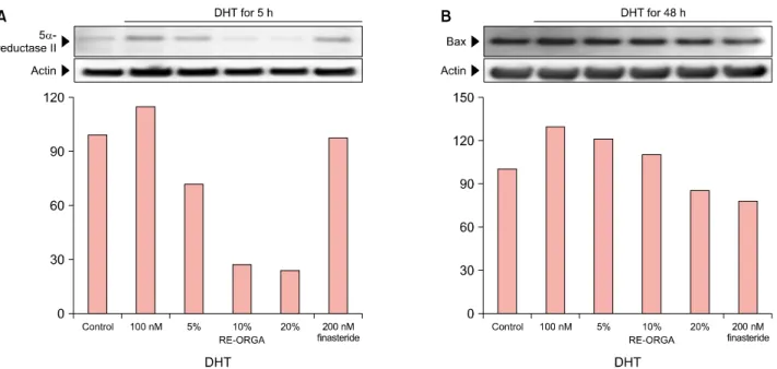
Fig. 3. (A, B) Western blot analysis of 5-α-reductase type II and Bax in human dermal papilla cells. Expression levels of 5-α-reductase type II decreased after addition of RE-ORGA. After treatment with 20% RE-ORGA, the expression level of 5-α-reductase type II dropped to 21.3% of that treated with 100 nM dihydrotestosterone (DHT) and 34.0% of that treated with 200 nM finasteride. Reduction in expression level of Bax was not higher than that of 5-α-reductase type II. However, treatment with 20% RE-ORGA resulted in decreased expression level of the Bax gene (86%) compared to treatment with 200 nM finasteride (78%).
Fig. 2. Cell proliferation in human dermal papilla (DP) cells after treatment with RE-ORGA at various concentrations. The highest number of proliferated DP cells was measured on day 5. Human DP cells proliferated most efficiently in groups treated with RE-ORGA at 1% and 5% on day 1 and in groups treated with RE-ORGA at 1%, 5%, and 10% on day 5. Growth rates of human DP cells after treatment with 10% RE-ORGA were similar to those after treatment with 20 ng/ml epidermal growth factor (EGF) on all days.
10%. DP cells treated with 20 ng/ml EGF showed sig- nificantly higher cell proliferation rate only on day 5 com- pared to control. When RE-ORGA treated cells were com-
pared with EGF-treated cells, the cells treated with 1%
RE-ORGA showed a significantly higher proliferation rate only on day 2 (p-value=0.00005), whereas the cells treat- ed with 5% RE-ORGA showed a significantly higher cell proliferation rate on day 2 (p-value=0.00003) and on day 5 (p-value=0.004). However, cells treated with 10% RE- ORGA showed similar cell proliferation rates as cells treat- ed with EGF at 20 ng/ml.
Western blot analysis on 5α-reductase type II and Bax Western blotting analysis was performed to examine the expression levels of 5α-reductase type II and Bax in DHT- stimulated DP cells treated with RE-ORGA (Fig. 3). The ex- pression levels of 5α-reductase type II were decreased af- ter treatment with RE-ORGA. After the DHT-stimulated cells were treated with RE-ORGA for 5 hours, the expression levels of 5α-reductase type II decreased to 62.6% in the 5% RE-ORGA treatment group, to 24.1% in the 10% RE- ORGA treatment group, and to 21.3% in the 20% RE-ORGA treatment group compared to their levels in the only DHT- treated cells. Protein levels of Bax (one of the late genes induced by DHT) also decreased after treatment with RE- ORGA. RE-ORGA treatment decreased the expression of 5 α-reductase type II and Bax in a concentration dependent manner. RE-ORGA at 20% resulted in an 86% reduction in the expression of Bax, comparable to the 78% reduc-
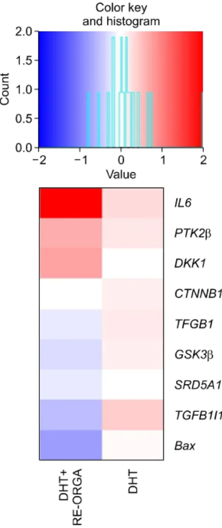
Fig. 4. RNA-seq analysis of dihydro- testosterone (DHT)-stimulated der- mal papilla (DP) cells treated with or without RE-ORGA. In DHT-stimu- lated cells, transforming growth factor-beta 1 (TGF-β1), glycogen synthatase kinase 3 beta (GSK3β), TGF-β1 induced transcript 1 (TGF β1I1), and Bax genes were highly expressed. However, these genes are downregulated in DHT-stimu- lated DP cells treated with RE- ORGA. On the other hand, protein tyrosine kinase 2 beta (PTK2β), a negative regulator of androgen re- ceptor, was upregulated, whereas 5-α-reductase type I and Bax were downregulated in DHT-stimulated DP cells treated with RE-ORGA.
IL6: interleukin 6, DKK1: Dickkopf- 1, CTNNB1: catenin beta-1.
tion observed after treatment with 200 nM finasteride.
RNA-seq results with RE-ORGA
RNA-seq was performed to investigate changes in expres- sion patterns of AR-induced genes in DHT stimulated DP cells treated with RE-ORGA (Fig. 4). RNA-seq results show- ed down-regulation of the Bax gene in mRNA levels after treatment with 20% RE-ORGA (log fc: −0.786). However, the mRNA levels for 5α-reductase type II were below the detection limit, even in the cells treated with only DHT. It was found that mRNA transcripts of 5α-reductase type I and 5-α-reductase type III were decreased after RE-ORGA treatment (log fc: −0.158 for 5α-reductase type I, −0.253 for 5-α-reductase type III). RNA-seq revealed that mRNA levels of ARs (log fc: −0.575) and TGF-β1 (log fc: −0.182) were downregulated. In contrast, mRNA levels of DKK1 (log fc: 0.735) and IL6 (log fc: 2.04) were highly increased.
It has been reported that two molecules, TGFβ1I1 and protein tyrosine kinase 2 beta (PTK2β), can regulate the transcriptional activity of the AR. RNA seq showed that RE-ORGA induced the expression of PTK2β mRNA (log fc: 0.659) but downregulated the expression of TGFβ1I1
mRNA (log fc: −0.542), indicating that RE-ORGA has a negative effect on the activation of ARs. RE-ORGA also downregulated the mRNA level (log fc: −0.298) of glyco- gen synthatase kinase 3 beta (GSK3β), which inhibits Wnt signaling via the phosphorylation of β-catenin. How- ever, mRNA level of the β-catenin was almost unaffected by RE-ORGA (log fc: −0.0405).
Anti-inflammation activity of RE-ORGA
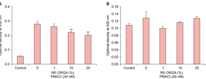
We evaluated the anti-inflammatory activities of RE-ORGA by measuring protein levels of TNF-α and IL6 (Fig. 5).
Treatment with 40 nM PMACI induced a 4.7-fold (0.28±
0.015) increase in the expression levels of TNF-α in hu- man Mast cell-1 when compared with the control (0.06±
0.001). When different concentrations (0%∼20%) of RE- ORGA were added to cells following PMACI treatment, the levels of TNF-α decreased with increasing concen- tration of RE-ORGA. The expression levels of TNF-α were reduced to 72.4% (0.20±0.008) in the 20% RE-ORGA treat- ment group compared to its levels in the 40-nM PMACI- only treatment group. RE-ORGA also decreased the ex- pression of IL6 compared to control, however the de-
Fig. 5. Anti-inflammatory activity of RE-ORGA in Mast cell-1. Tumor necrosis factor-alpha (TNF-α) (A) and interleukin 6 (IL6) (B) were chosen as biomarkers for inflammation. Levels of TNF-α (0.28±0.015) that were high after treatment with 40 nM phorbol-12-myristate 13-acetate plus calcium ionophore A23187 (PMACI) showed RE-ORGA-concentration dependent reduction. They decreased to 0.22±
0.004 (79.2%) in the 10% RE-ORGA treatment group and 0.20±0.008 (72.4%) in the 20% RE-ORGA treatment group, showing a statistically significant difference compared to their levels in the 0% RE-ORGA treatment group. However, the maximum inhibition of IL6 level was observed in the 1% RE-ORGA treatment group (p-value=0.053). **p-value<0.01 compared with 0% Re-ORGA.
crease in IL6 expression was not dependent on RE-ORGA concentration.
DISCUSSION
RE-ORGA was extracted from a mixture of various Korean herbs by steam distillation. It was prepared to help hair growth in men with AGA. From the results of cytotoxicity tests, 20% of RE-ORGA was chosen as the maximum non-toxic concentration for DP cells. At lower concen- trations than 20%, RE-ORGA helped the proliferation of DP cells and its activity was comparable to that of 20 ng/ml EGF. However, RNA-seq was performed with DP cells treated with 20% RE-ORGA to find as many genes that will be influenced by RE-ORGA as possible.
Actually, mRNA levels of 5α-reductase type II were not quantified in RNA-seq analysis. However Western blot analysis showed that RE-ORGA decreased expression of 5 α-reductase type II in DHT-stimulated DP cells by RE- ORGA (Fig. 3). Moreover, RNA-seq analyzed that the tran- script levels of 5α-reductase type I were reduced by RE- ORGA. A report suggested that AR activation would lead to repress the mRNA expression of 5α-reductase type II but induce the mRNA expression of 5α-reductase type I in prostate cancer cell lines16. Therefore, we assumed that DHT-activated AR had already decreased mRNA levels of 5α-reductase type II in all DP cells.
We had also investigated some candidate genes for AGA and AR-driven genes from RNA-seq results. TGFβ1I1, a gene for co-activator of ARs, was slightly upregulated by
DHT but was down-regulated by RE-ORGA. The PTK2β, mRNAs, which act as a negative regulator of ARs, were more activated with RE-ORGA than with DHT. Moreover, only mRNA levels of TGF-β1, among the three main hair-loss effectors (TGF-β1, DKK1, and IL6), was down- regulated with RE-ORGA, while the other two genes were overexpressed. The suppression of TGF-β1 expression with RE-ORGA could help hair loss induced by oxidative stress, because it was reported that DP cells from AGA pa- tients are more sensitive to ROS and that TGF-β1 can be induced by ROS16,17. The transcript levels of Bax, a pro- apoptotic gene, were also down-regulated, which means that RE-ORGA could protect DP cells from apoptosis fol- lowing shortened anagen phases.
Cytokines may play an important negative role in hair loss by influencing hair growth18. Addition of TNF-α to human hair follicles can dose-dependently inhibit cell growth19. Serum levels of TNF-α in patients with alopecia areata are significantly elevated compared to those in healthy subjects20. IL6 can inhibit hair shaft elongation and sup- press proliferation of matrix cells in cultured human hair follicles12. Therefore, we evaluated the anti-inflammatory activity of RE-ORGA in human Mast cell-1 using ELISA assays. The results showed that RE-ORGA could decrease TNF-α expression in dose-dependent manner but not IL6, although these results were not statistically significant. Con- sequently, it seems that further studies are required to con- firm anti-inflammatory activities of RE-ORGA.
Although RE-ORGA can be a new material to help hair loss, we had not yet verified results from RNA-seq analysis
in translational levels and we had not revealed the main target proteins of RE-ORGA. Because RE-ORGA is not a single chemical, it is expected to interact with multiple proteins. If all these targets would be revealed through fur- ther studies, we could understand the mechanisms for RE- ORGA to lessen hair loss in AGA patients.
In summary, our study showed that RE-ORGA could pro- mote the proliferation of hair follicle DP cells by altering the androgen sensitivity of AR through the transcriptional regulation of TGFβ1I1 gene and PTK2β gene, which might negatively affect the expression of 5α-reductase type I, Bax, TGF-β1, and GSK3β. Anti-inflammatory ac- tivities of RE-ORGA might also contribute to the decrease in cytokine-induced cell death. Therefore, we expect that RE-ORGA could be a useful candidate to inhibit hair loss by alleviating progressive AGA induced by male sex hormones.
ACKNOWLEDGMENT
This research was supported by the Ministry of Trade, Industry & Energy (MOTIE), Korea Institute for Advance- ment of Technology (KIAT) through the Encouragement Program for The Industries of Economic Cooperation Re- gion (R0005729).
CONFLICTS OF INTEREST
The authors have nothing to disclose.
ORCID
Myung-Gyun Kang, https://orcid.org/0000-0002-9499-7571 Daeui Park, https://orcid.org/0000-0002-9452-5849 Hyoung-Yun Han, https://orcid.org/0000-0002-7957-7448 Hyeeun Shim, https://orcid.org/0000-0002-2940-4362 Yoonjung Hong, https://orcid.org/0000-0001-5449-549X Jiyeon Moon, https://orcid.org/0000-0002-6988-6714 Seokjoo Yoon, https://orcid.org/0000-0003-1884-2591 Bosun Kwon, https://orcid.org/0000-0002-8585-2194
REFERENCES
1. Courtois M, Loussouarn G, Hourseau C, Grollier JF. Hair cycle and alopecia. Skin Pharmacol 1994;7:84-89.
2. Choudhry R, Hodgins MB, Van der Kwast TH, Brinkmann AO, Boersma WJ. Localization of androgen receptors in human skin by immunohistochemistry: implications for the hormonal regulation of hair growth, sebaceous glands and sweat glands. J Endocrinol 1992;133:467-475.
3. Hibberts NA, Howell AE, Randall VA. Balding hair follicle
dermal papilla cells contain higher levels of androgen receptors than those from non-balding scalp. J Endocrinol 1998;156:59-65.
4. Sawaya ME, Price VH. Different levels of 5alpha-reductase type I and II, aromatase, and androgen receptor in hair follicles of women and men with androgenetic alopecia. J Invest Dermatol 1997;109:296-300.
5. Kaufman KD, Olsen EA, Whiting D, Savin R, DeVillez R, Bergfeld W, et al. Finasteride in the treatment of men with androgenetic alopecia. Finasteride Male Pattern Hair Loss Study Group. J Am Acad Dermatol 1998;39(4 Pt 1):578- 589.
6. Schweikert HU, Wilson JD. Regulation of human hair growth by steroid hormones. II. Androstenedione metabolism in isolated hairs. J Clin Endocrinol Metab 1974;39:1012-1019.
7. Inui S, Fukuzato Y, Nakajima T, Yoshikawa K, Itami S. Iden- tification of androgen-inducible TGF-beta1 derived from der- mal papilla cells as a key mediator in androgenetic alopecia.
J Investig Dermatol Symp Proc 2003;8:69-71.
8. Kwack MH, Kim MK, Kim JC, Sung YK. Dickkopf 1 promotes regression of hair follicles. J Invest Dermatol 2012;132:1554- 1560.
9. Yu M, Kissling S, Freyschmidt-Paul P, Hoffmann R, Shapiro J, McElwee KJ. Interleukin-6 cytokine family member oncos- tatin M is a hair-follicle-expressed factor with hair growth inhibitory properties. Exp Dermatol 2008;17:12-19.
10. Inui S, Fukuzato Y, Nakajima T, Kurata S, Itami S. Androgen receptor co-activator Hic-5/ARA55 as a molecular regulator of androgen sensitivity in dermal papilla cells of human hair follicles. J Invest Dermatol 2007;127:2302-2306.
11. Kaliyadan F, Nambiar A, Vijayaraghavan S. Androgenetic alo- pecia: an update. Indian J Dermatol Venereol Leprol 2013;
79:613-625.
12. Kwack MH, Ahn JS, Kim MK, Kim JC, Sung YK. Dihydro- testosterone-inducible IL-6 inhibits elongation of human hair shafts by suppressing matrix cell proliferation and pro- motes regression of hair follicles in mice. J Invest Dermatol 2012;132:43-49.
13. Inui S, Itami S. Molecular basis of androgenetic alopecia:
from androgen to paracrine mediators through dermal pa- pilla. J Dermatol Sci 2011;61:1-6.
14. Uchiyama C, Ishida K, Tsutsui T, Naito A, Kurita K, Hanihara H, et al. Effects of Hura crepitans and its active ingredient, daphne factor F3, on dihydrotestosterone-induced neuro- trophin-4 activation and hair retardation. Biol Pharm Bull 2012;35:42-47.
15. Upton JH, Hannen RF, Bahta AW, Farjo N, Farjo B, Philpott MP. Oxidative stress-associated senescence in dermal pa- pilla cells of men with androgenetic alopecia. J Invest Der- matol 2015;135:1244-1252.
16. Audet-Walsh É, Yee T, Tam IS, Giguère V. Inverse regu- lation of DHT synthesis enzymes 5α-reductase types 1 and 2 by the androgen receptor in prostate cancer. Endocrino- logy 2017;158:1015-1021.
17. Shin H, Yoo HG, Inui S, Itami S, Kim IG, Cho AR, et al.
Induction of transforming growth factor-beta 1 by androgen is mediated by reactive oxygen species in hair follicle
dermal papilla cells. BMB Rep 2013;46:460-464.
18. Hoffmann R. The potential role of cytokines and T cells in alopecia areata. J Investig Dermatol Symp Proc 1999;4:235- 238.
19. Philpott MP, Sanders DA, Bowen J, Kealey T. Effects of interleukins, colony-stimulating factor and tumour necrosis
factor on human hair follicle growth in vitro: a possible role for interleukin-1 and tumour necrosis factor-alpha in alopecia areata. Br J Dermatol 1996;135:942-948.
20. Kasumagic-Halilovic E, Prohic A, Cavaljuga S. Tumor necrosis factor-alpha in patients with alopecia areata. Indian J Der- matol 2011;56:494-496.