The Role of Interventional Radiology in Treatment
of Patients with Acute Trauma:
A Pictorial Essay
급성 외상 환자 치료에서 인터벤션 영상의 역할: 임상화보
Kyung Sik Kang, MD1 , Mu Sook Lee, MD2* , Doo Ri Kim, MD1 , Young Hwan Kim, MD2
1Department of Radiology, Jeju National University Hospital, Jeju National University School of Medicine, Jeju, Korea
2Department of Radiology, Keimyung University Dongsan Hospital, Keimyung University, School of Medicine, Daegu, Korea
Acute trauma is a common cause of mortality in individuals aged < 40 years. As organ preservation has become important in treating trauma patients, the treatment is shifting from surgical manage- ment to non-operative management. A multidisciplinary team approach, including interventional radiology (IR), is essential for the optimal management of trauma patients, as IR plays an impor- tant role in injury evaluation and management. IR also contributes significantly to achieving the best clinical outcomes in critically ill trauma patients. This pictorial essay aims to present and sum- marize various interventional treatments in trauma patients requiring critical care.
Index terms Trauma; Injuries; Angiography; Interventional Radiology
INTRODUCTION
Acute trauma is a common mortality cause in individuals aged < 40 years. Proper di- agnosis and appropriate treatments are crucial due to residual disabilities and sequelae (1, 2). Previously, surgeries, including surgical exploration and vessel ligation, were pre- ferred for trauma-related vascular injuries (3, 4). However, management of hemody- namically stable patients has changed from surgical to non-operative management (NOM), as organ preservation became increasingly important. Recently, NOM has also been conducted in hemodynamically unstable patients (3, 4). Interventional treatment, equipment, and agents have developed dramatically since the introduction of endovas- cular techniques in the 1970s. Therefore, interventional radiology (IR) plays an essen-
Received May 18, 2020 Revised July 1, 2020 Accepted July 11, 2020
*Corresponding author Mu Sook Lee, MD Department of Radiology, Keimyung University Dongsan Hospital, Keimyung University, School of Medicine, 1035 Dalgubeol-daero, Dalseo-gu, Daegu 42601, Korea.
Tel 82-53-258-7862 Fax 82-53-258-4153
E-mail [email protected] This is an Open Access article distributed under the terms of the Creative Commons Attribu- tion Non-Commercial License (https://creativecommons.org/
licenses/by-nc/4.0) which permits unrestricted non-commercial use, distribution, and reproduc- tion in any medium, provided the original work is properly cited.
ORCID iDs Kyung Sik Kang https://
orcid.org/0000-0002-4711-8458 Mu Sook Lee
https://
orcid.org/0000-0002-0382-5564 Doo Ri Kim
https://
orcid.org/0000-0001-5263-7693 Young Hwan Kim
https://
orcid.org/0000-0002-2715-358X
tial role in treating trauma patients (3, 4).
This pictorial essay aims to present and summarize various cases that the IR plays an es- sential role in acute trauma management to achieve the best clinical outcomes of trauma pa- tients.
LIVER INJURY
The liver is among the most frequently injured organs in blunt abdominal trauma. Liver injuries are detected in approximately 25% of severely injured patients with whole-body computed tomography (CT) (5).
The American Association for the Surgery of Trauma (AAST) liver injury scale is used to grade livery injury on CT. However, surgeries are determined based on hemodynamic stabil- ity rather than on grade severity (6).
Most hemodynamically stable patients with liver injury undergo conservative manage- ment. However, embolization can be considered when active bleeding is present on CT, or hemorrhage control is not achieved by laparotomy with perihepatic packing alone (Fig. 1) (7).
SPLENIC INJURY
The spleen is another organ frequently injured. The extent of injury is measured using the AAST splenic injury scale. Treatment decision is made based on CT results and various clini- cal factors (8).
Laparotomy with splenectomy or splenic salvage is performed for hemodynamically un- stable patients, whereas conservative management is considered initially in hemodynami- cally stable patients. Splenic injury in patients with high AAST grades is less likely to be treat-
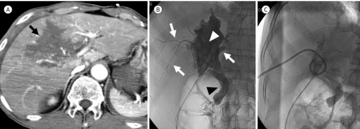
Fig. 1. Liver injury in a 32-year-old man who had an in-car traffic accident.
A. CT shows liver laceration (grade IV, American Association for the Surgery of Trauma) and active contrast extravasation (arrows) in the right hepatic lobe.
B. Angiography confirms the extravasation (arrow) from a branch of the right hepatic artery, which was successfully treated with transcatheter arterial embolization using N-butyl cyanoacrylate (arrowhead).
A B
ed by conservative management alone (9). However, recent studies show that splenic arterial embolization meaningfully decreased the NOM failure rates in patient with high AAST grade splenic injury (10). Splenic artery embolization is indicated when vascular injuries, including active arterial bleeding, pseudoaneurysm, and arteriovenous fistula, are evident on CT of he- modynamically stable patients (Fig. 2) (11).
KIDNEY INJURY
Traumatic renal injury develops in approximately 10% of abdominal trauma patients and is the third most common abdominal organ injured. It mostly occurs after a blunt trauma and is considered non-life-threatening (6, 12). Traumatic renal bleeding is managed conser- vatively due to spontaneous cessation in most cases owing to the tamponade effect of the ret- roperitoneal fascia. However, endovascular or surgical intervention is required in cases with massive hemorrhage, pseudoaneurysm formation, continuous hematuria, and hemodynam- ic instability (12, 13). Recently, selective renal angioembolization has been preferred over surgery in hemodynamically stable patients with high-grade injury (grade III or higher) on the AAST kidney injury scale (Fig. 3) (6, 12).
PANCREATIC INJURY
Pancreas injury is rare, accounting for approximately 1% of blunt abdominal trauma cases.
It usually accompanies other abdominal organ injuries (14).
Its management depends on the AAST pancreatic injury scale, and the presence of ductal injury determines the prognosis and treatment methods. In AAST grade III and higher inju- ries accompanied with ductal injury, surgery is performed. Recently, NOM has shown posi- tive results in liver, splenic, and kidney injuries. Thus, NOM, including embolization and Fig. 2. Splenic injury in a 64-year-old woman who fell from a height.
A. CT scan shows splenic rupture (grade IV, American Association for the Surgery of Trauma) with contrast extravasation (arrow) and hemo- peritoneum (arrowhead).
B. Angiography confirms the contrast extravasation (arrows) from the splenic artery, which was successfully treated with transcatheter arterial embolization using coils (arrowhead) superselectively deployed in the bleeding vessel with preservation of the surrounding branches.
A B
percutaneous drainage, has been performed gradually in pancreatic injury (Fig. 4) (14). Per- cutaneous drainage can reduce symptoms and complications of enzyme leakage in pancre- atic duct injury (14).
BILIARY TRACT INJURY
The biliary system remains relatively uninjured. because it is protected by the ribs, liver, Fig. 3. Kidney injury in a 50-year-old woman who fell from a height.
A. CT scan shows renal laceration (grade III, American Association for the Surgery of Trauma) with active extravasation (arrow) and perirenal hematoma (arrowhead).
B. Angiography confirms the contrast extravasation (arrow) from the left anterior renal artery, which was successfully treated with transcathe- ter arterial embolization using coils (arrowheads).
Fig. 4. Pancreatic injury in a 19-year-old man who had a pedestrian accident.
A. CT scan shows active extravasation (arrows) and pancreatic disruption (grade IV, American Association for the Surgery of Trauma) (arrow- head).
B. Angiography confirms the contrast extravasation (arrow) from the pancreaticoduodenal arcade of the gastroduodenal artery, which was successfully treated with transcatheter arterial embolization using coils (arrowhead).
A B
A B
and mesentery. According to the locations, biliary tract injury is classified as gallbladder, which is the most common, intrahepatic, and extrahepatic bile duct injuries. Gallbladder in- jury can be accompanied with cystic artery transection, causing major blood loss. Bile leak- age leads to combined infection and sepsis (15).
Treatment options include endoscopic or percutaneous biliary drainage, and surgical bili- ary reconstruction. Interventional treatment involves percutaneous transhepatic biliary drain placement for biliary decompression and diversion (Fig. 5). Biloma, resulting from bile leakage, can lead to complications, including abscess, cholangitis, and sepsis, and may be treated with percutaneous catheter drainage (16).
MESENTERIC INJURY
Mesenteric injury is relatively rare and difficult to diagnose. It frequently results from seat- belt injury in motor vehicular accidents. Its rapid diagnosis and appropriate treatment are essential to avoid life-threatening conditions, including intraperitoneal and gastrointestinal bleeding, intestinal ischemia, and/or perforation from mesenteric vessel interruption (17).
Mesenteric injuries are traditionally managed surgically. However, isolated mesenteric ves- sel injury, without gastrointestinal perforation, can be treated with angiography and trans- catheter arterial embolization (TAE) (Fig. 6) (17).
URETHRAL INJURY
Traumatic urethral injury results from iatrogenic, blunt, or penetrating trauma. Its man- agement depends on injury location. Anterior urethral contusions are observed without treatment, but incomplete or complete anterior urethral injury is treated with suprapubic di-
Fig. 5. Biliary tract injury in a 51-year-old man who had a car accident.
A. CT scan shows liver laceration (grade IV, American Association for the Surgery of Trauma) (arrow).
B, C. A tubogram (B) through the Jackson-Pratt drain shows a collection (white arrowhead) of contrast material, communicating with the in- trahepatic bile duct (white arrows) and common bile duct (black arrowhead), which disappearred after percutaneous transhepatic biliary drain placement (C).
A B C
version and delayed repair (18).
Posterior urethral injury management includes primary surgical repair, suprapubic cystos- tomy with delayed urethroplasty, and primary interventional urethral realignment (PIUR).
PIUR can accurately and rapidly evaluate injury severity through retrograde urethrography, and immediate realignment can also be performed (Fig. 7) (18).
PENILE INJURY
Priapism is a persistent erection regardless of sexual stimulation. Appropriate treatment is crucial to prevent complications, including structural damage or permanent erectile dys- function. Its two major types are ischemic and non-ischemic (19).
Fig. 6. Mesenteric injury in a 72-year-old man who had a car accident.
A. CT scan shows contrast extravasation (arrows) and mesenteric hematoma (arrowhead).
B. Angiography confirms the contrast extravasation (arrows) from the branch of the ileocecal artery, which was successfully treated with transcatheter arterial embolization using N-butyl cyanoacrylate (arrowhead).
Fig. 7. Urethral injury in a 41-year-old man who had a straddle accident.
A. Complete disruption of the bulbous urethra (arrow) is seen on a retrograde urethrogram.
B. Retrograde primary interventional urethral realignment was performed using a 5-F catheter (black arrow) and guidewire (white arrow), and a 14-F Foley catheter (arrowhead) was placed transurethrally over the wire.
A B
A B
Ischemic priapism develops from little or no cavernous arterial inflow and corpora caver- nosa rigidity. Contrarily, non-ischemic priapism results from an increased corpora cavernosa inflow, occurring from the arteriolar sinusoidal fistula as a result of the trauma. Most non- ischemic priapism do not develop permanent injuries, but may require proper treatment be- cause patients may experience decreased function (19).
The first-line treatment for non-ischemic priapism is clinical observation, as more than two-thirds of cases could achieve spontaneous resolution. If symptoms persist, treatment is required, and interventional treatment, including cavernous arterial embolization, can be performed (Fig. 8) (19).
PELVIC INJURY
Most pelvic fractures occur from high-energy crush injuries and are often accompanied with organ damages. Mortality rate with pelvic fracture alone ranges from 6% to 15%, in- creasing to 36–54% when accompanied with hemorrhage. Mortality rate can further increase with delayed diagnosis. Most pelvic injury-associated hemorrhages occur in fractured bones or disrupted pelvic veins, and 10–20% of cases involve hemorrhage from arterial injury, most commonly the internal iliac artery branch. Hemodynamically unstable patients require ag- gressive treatment, as pelvic bone fracture can lead to massive bleeding (11, 20). Treatment includes external fixation of the unstable fracture, pelvic packing, and TAE. Interventional treatment is preferred due to the risks of large volume loss or uncontrolled vessel bleeding with surgeries. When hemodynamically unstable, nonselective proximal gelfoam emboliza- tion is performed on both internal iliac arteries as life-saving procedures, followed by addi- tional selective embolization after stabilization (Fig. 9) (11).
Evaluation for collateral vessel hemorrhage or additional site bleeding in the contralateral internal iliac artery is necessary after embolization. Moreover, thorough evaluations are criti-
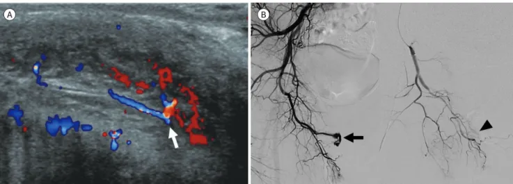
Fig. 8. Penile injury in a 35-year-old man who had a straddle accident.
A. Color Doppler ultrasonogram shows aliasing phenomena (arrow) due to the turbulent high velocity flow in the cavernous artery.
B. Angiography confirms the presence of an arteriocavernous fistula (arrow), which was successfully treated with transcatheter arterial embo- lization using an autologous blood clot and a gelfoam (arrowhead).
A B
cal post-procedure to prevent complications, including tissue necrosis, abscess formation, re-bleeding, and sepsis (11).
ABDOMINAL WALL INJURY
Abdominal wall hematoma rarely occurs in blunt trauma. Conservative management is mostly sufficient, but it can cause life-threatening conditions as well. Traumatic abdominal wall hematoma develops mainly from injuries to the deep circumflex iliac or inferior epigas- tric arteries (Fig. 10). Treatment for hemodynamically unstable patients is not well estab- lished. However, TAE can accurately assess bleeding sites through angiography and provide Fig. 9. Pelvic injury in a 74-year-old man who had a pedestrian accident.
A. CT scan shows contrast extravasation (arrow) and right pelvic bone fracture with hematoma.
B. Angiography confirms the pseudoaneurysm (arrow) from the right medial circumflex femoral artery, which was successfully treated with transcatheter arterial embolization using coils (arrowhead).
Fig. 10. Abdominal wall injury in an 83-year-old man who hit his abdomen against a chair.
A. CT scan shows active extravasation (arrow) and right rectus sheath hematoma.
B. Angiography confirms the small pseudoaneurysms (arrows) from the right inferior epigastric artery, which was successfully treated with transcatheter arterial embolization using N-butyl cyanoacrylate (arrowheads).
A B
A B
rapid bleeding control through embolization (21).
THORACIC INJURY
Intrathoracic hemorrhage in blunt thoracic trauma is caused by rib fracture-associated in- tercostal artery injuries. Self-limited bleeding can be managed conservatively, but persistent bleeding requires active treatment due to life-threatening complications (22).
Although exploratory thoracotomy was the treatment of choice in the past, interventional treatment is preferred for those not eligible for surgeries or have difficulty for surgical access due to deep posterior intercostal artery bleeding (Fig. 11). TAE of the intercostal artery is a minimally invasive, safe, and reliable treatment for intra-thoracic hemorrhage. However, cautions are required due to spinal cord ischemia, a severe complication of intercostal artery embolization (22).
EXTREMITY INJURY
Extremity vascular injury mainly occurs in blunt or penetrating trauma. Clinical practice has shifted from performing immediate surgeries in all suspected patients to those with dis- tal pulse loss during physical examination, limb ischemia, expanding hematoma, thrill or bruit, pulsatile bleeding, significant external bleeding, or compartment syndrome (11).
Interventional treatments include embolization, balloon occlusion, and stent-graft place- ment. Large proximal extremity vessel injuries, such as subclavian or femoral arteries, are rare but may cause life-threatening conditions and require stent graft or balloon occlusion (11). Small vessel injury requires evaluation for collateral circulation of distal extremity be- fore embolization. In the upper extremity, embolization can be performed safely in radial,
Fig. 11. Thoracic injury in a 43-year-old man who had an out-of-car traffic accident.
A. CT scan shows active extravasation (arrow) and multiple rib fractures with hematoma (arrowhead).
B. Angiography confirms the extravasation (arrow) from the 10th right intercostal artery, which was successfully treated with transcatheter ar- terial embolization using coils (arrowheads).
A B
ulnar, or interosseous arteries, as their collateral vessels are well developed. However, embo- lization in the iliac and femoral arteries in the lower extremity should be performed with caution, as they are vital vessels that cannot be sacrificed (Fig. 12) (23).
CONCLUSION
A multidisciplinary team approach among surgeons and interventional radiologists with use of advanced equipment is necessary for optimal management of trauma patients requir- ing critical care. Although emergency laparotomy remains the standard treatment for hemo- dynamically unstable patients, IR plays an essential role in acute trauma management to achieve the best clinical outcomes of trauma critical care patients.
Author Contributions
Conceptualization, L.M.S.; data curation, K.K.S., L.M.S.; investigation, K.K.S., L.M.S.; project ad- ministration, L.M.S.; supervision, L.M.S., K.D.R., K.Y.H.; visualization, all authors; writing—original draft, K.K.S., L.M.S.; and writing—review & editing, L.M.S., K.D.R., K.Y.H.
Conflicts of Interest
The authors have no potential conflicts of interest to disclose.
Funding None
Fig. 12. Extremity injury in a 55-year-old man who had a pedestrian traffic accident.
A. CT scan shows active (arrow) in the tibialis muscle.
B. Angiography confirms the extravasation (arrow) from the muscular branch of the right anterior tibial artery, which was successfully treated with transcatheter arterial embolization using N-butyl cyanoacrylate (arrowhead).
A B
REFERENCES
1. Artigas Martín JM, Martí de Gracia M, Claraco Vega LM, Parrilla Herranz P. Radiology and imaging tech- niques in severe trauma. Med Intensiva 2015;39:49-59
2. Lee J, Ahn JH. Multidetector CT in blunt abdominal trauma: imaging findings and pitfalls. J Korean Soc Radiol 2019;80:445-465
3. Bauer JR, Ray CE. Transcatheter arterial embolization in the trauma patient: a review. Semin Intervent Ra- diol 2004;21:11-22
4. Gilyard S, Shinn K, Nezami N, Findeiss LK, Dariushnia S, Grant AA, et al. Contemporary management of he- patic trauma: what IRs need to know. Semin Intervent Radiol 2020;37:35-43
5. Yoon W, Jeong YY, Kim JK, Seo JJ, Lim HS, Shin SS, et al. CT in blunt liver trauma. Radiographics 2005;
25:87-104
6. Kozar RA, Crandall M, Shanmuganathan K, Zarzaur BL, Coburn M, Cribari C, et al. Organ injury scaling 2018 update: spleen, liver, and kidney. J Trauma Acute Care Surg 2018;85:1119-1122
7. Letoublon C, Morra I, Chen Y, Monnin V, Voirin D, Arvieux C. Hepatic arterial embolization in the manage- ment of blunt hepatic trauma: indications and complications. J Trauma 2011;70:1032-1036; discussion 1036-1037
8. Patil MS, Goodin SZ, Findeiss LK. Update: splenic artery embolization in blunt abdominal trauma. Semin Intervent Radiol 2020;37:97-102
9. Imbrogno BF, Ray CE. Splenic artery embolization in blunt trauma. Semin Intervent Radiol 2012;29:147- 149
10. Crichton JCI, Naidoo K, Yet B, Brundage SI, Perkins Z. The role of splenic angioembolization as an adjunct to nonoperative management of blunt splenic injuries: a systematic review and meta-analysis. J Trauma Acute Care Surg 2017;83:934-943
11. Gould JE, Vedantham S. The role of interventional radiology in trauma. Semin Intervent Radiol 2006;23:
270-278
12. Mani NB, Kim L. The role of interventional radiology in urologic tract trauma. Semin Intervent Radiol 2011;
28:415-423
13. Kwon H, Jeon CH, Kim CW. Intervention for urologic trauma. J Korean Soc Radiol 2019;80:667-683 14. Chan KV, Cheung MT. Towards a non-operative strategy for severe blunt pancreatic injury–Case report. In-
jury Extra 2014;45:35-39
15. Gupta A, Stuhlfaut JW, Fleming KW, Lucey BC, Soto JA. Blunt trauma of the pancreas and biliary tract: a multimodality imaging approach to diagnosis. Radiographics 2004;24:1381-1395
16. Thompson CM, Saad NE, Quazi RR, Darcy MD, Picus DD, Menias CO. Management of iatrogenic bile duct injuries: role of the interventional radiologist. Radiographics 2013;33:117-134
17. Shin JS, Shin JH, Ko HK, Kim JW, Yoon HK. Transcatheter arterial embolization for traumatic mesenteric bleeding: a 15-year, single-center experience. Diagn Interv Radiol 2016;22:385-389
18. Lee MS, Kim SH, Kim BS, Choi GM, Huh JS. The efficacy of primary interventional urethral realignment for the treatment of traumatic urethral injuries. J Vasc Interv Radiol 2016;27:226-231
19. Levey HR, Segal RL, Bivalacqua TJ. Management of priapism: an update for clinicians. Ther Adv Urol 2014;6:230-244
20. Franco DF, Zangan SM. Interventional radiology in pelvic trauma. Semin Intervent Radiol 2020;37:44-54 21. Lefere P, Gryspeerdt S, Van Holsbeeck B, Baekelandt M. Diagnosis and treatment of expanding haemato-
ma of the lateral abdominal wall after blunt abdominal trauma. Eur Radiol 1999;9:1553-1555
22. Chemelli AP, Thauerer M, Wiedermann F, Strasak A, Klocker J, Chemelli-Steingruber IE. Transcatheter arte- rial embolization for the management of iatrogenic and blunt traumatic intercostal artery injuries. J Vasc Surg 2009;49:1505-1513
23. Lopera JE. Embolization in trauma: principles and techniques. Semin Intervent Radiol 2010;27:14-28
급성 외상 환자 치료에서 인터벤션 영상의 역할: 임상화보
강경식1 · 이무숙2* · 김두리1 · 김영환2
외상은 40세 이하에서 가장 흔한 사망의 원인 중에 하나이다. 과거에는 외상 환자의 치료로 대부분 수술적 치료가 우선 되었다. 하지만 점차 외상 환자의 치료에서 장기 보존이 중요하 게 되었으며, 외상 후 환자의 치료는 수술적 치료에서 비수술적 치료로 바뀌고 있다. 외상 환 자의 적절한 치료를 위해서는 다양한 분야의 협력이 필요하다. 그중 인터벤션 영상 의학은 외상 환자를 평가하고 치료하는데 중요한 역할을 담당하고 있다. 이에 본 논문은 인터벤션 치료가 급성 외상 환자에서 중요한 역할을 했던 다양한 증례들에 대해 소개해 보고자 한다.
1제주대학교 의과대학 제주대학교병원 영상의학과,
2계명대학교 의과대학 동산의료원 영상의학과