Endocrinol Metab 2018;33:296-304 https://doi.org/10.3803/EnM.2018.33.2.296 pISSN 2093-596X · eISSN 2093-5978
Original Article
Influence of Vitamin D Deficiency on Progression of Experimental Otitis Media in Rats
Hee-Bok Kim1, So Hyun Lim2, Chang Gun Cho3, Han Seok Choi2
1Department of Otorhinolaryngology-Head and Neck Surgery, BK21plus, Korea University College of Medicine, Seoul;
2Division of Endocrinology and Metabolism, Department of Internal Medicine, 3Department of Otorhinolaryngology-Head and Neck Surgery, Dongguk University Ilsan Hospital, Dongguk University College of Medicine, Goyang, Korea
Background: Vitamin D plays an important role in the immune response against infection. The purpose of the present study was to investigate the influence of vitamin D deficiency on the progression of otitis media (OM) using an experimental rat model.
Methods: Four-week-old male Sprague-Dawley rats (n=72) were divided into two groups based on their diet: a control diet group (n=36) and a vitamin D-deficient diet group (n=36). After 8 weeks of diet, experimental OM was induced by inoculation of non- typeable Haemophilus influenzae in the middle ear cavity. The rats were evaluated with otomicroscopy to determine the inflamma- tion in the middle ear mucosa on days 1, 2, 4, 7, and 14 post-inoculation. Bullae from sacrificed rats were collected and analyzed his- tologically.
Results: The middle ear mucosa from rats with vitamin D deficiency showed a significantly higher thickness than that of controls during the course of OM. The maximum mucosal thickness was 56.0±9.1 μm in the vitamin D deficiency group, and 43.9±9.8 μm in the control group, although there was no significant difference in the tympanic membrane score between the two groups evaluated with otomicroscopy. An immunohistochemical study showed increased expression of interleukin 6 (IL-6) and tumor necrosis factor α in rats manifesting vitamin D deficiency and decreased expression of IL-10 compared with controls.
Conclusion: Our results showed that vitamin D deficiency may exacerbate the pathophysiological changes of OM via altered cyto- kine production. Therefore, maintaining vitamin D status in the optimal range may be beneficial for proper management of OM.
Keywords: Vitamin D; Infection; Vitamin D deficiency; Otitis media; Rats
INTRODUCTION
Vitamin D is well known for its crucial role in bone and mineral
metabolism. Besides, its role in immune response to infection has been an interesting topic in vitamin D research [1-3]. In the pre-antibiotic era, sunlight exposure or cod liver oil, which pre-
Received: 15 March 2018, Revised: 11 April 2018, Accepted: 20 April 2018 Corresponding authors: Chang Gun Cho
Department of Otorhinolaryngology-Head and Neck Surgery, Dongguk University Ilsan Hospital, Dongguk University College of Medicine, 27 Dongguk-ro, Ilsandong-gu, Goyang 10326, Korea
Tel: +82-31-961-7434, Fax: +82-31-961-7429, E-mail: cho69@dumc.or.kr Han Seok Choi
Division of Endocrinology and Metabolism, Department of Internal Medicine, Dongguk University Ilsan Hospital, Dongguk University College of Medicine, 27 Dongguk-ro, Ilsandong-gu, Goyang 10326, Korea
Tel: +82-31-961-7137, Fax: +82-31-961-7147, E-mail: hschoi402@dumc.or.kr
Copyright © 2018 Korean Endocrine Society
This is an Open Access article distributed under the terms of the Creative Com- mons Attribution Non-Commercial License (http://creativecommons.org/
licenses/by-nc/4.0/) which permits unrestricted non-commercial use, distribu- tion, and reproduction in any medium, provided the original work is properly cited.
Vitamin D Deficiency and Otitis Media Endocrinol Metab 2018;33:296-304
https://doi.org/10.3803/EnM.2018.33.2.296 pISSN 2093-596X · eISSN 2093-5978
sumably raised the vitamin D level of individuals, was adopted as an effective treatment for tuberculosis infection [3]. Recently, accumulating evidence based on scientific studies suggests that vitamin D plays an important role in the immune system. Vita- min D receptor is expressed in most cells of the immune system and its genetic polymorphisms have been associated with sus- ceptibility to infection [4-6]. The vitamin D-activating enzyme, 25-hydroxyvitamin D (25(OH)D)-1α-hydroxylase, is also ex- pressed in immune cells including macrophages, dendritic cells, and T and B lymphocytes [4]. In addition, the mechanisms un- derlying the immunoregulatory function of vitamin D including regulation of cytokine profiles or antimicrobial peptides have been reported [1-3].
Otitis media (OM) is one of the most common inflammatory diseases associated with childhood. Approximately 80% of chil- dren report at least one episode of acute OM and more than 40% experience three or more episodes by the age of 3 years [7]. OM is caused by the interaction between multiple factors including Eustachian tube dysfunction, viral and bacterial infec- tion, exposure to smoke, and impaired host immunity [8,9].
Pathologically, OM is characterized by transformation and hy- perplasia of the middle ear mucosa following infiltration of var- ious inflammatory cells [10]. A variety of animal models have been developed to explore and understand the pathophysiologi- cal mechanisms of OM [11], although the mechanism underly- ing hyperplastic changes has not been fully identified.
Several clinical studies reported a significant association be- tween vitamin D status and OM [12-15]. However, little is known about the role of vitamin D status in the pathophysiology of OM. In the present study, we investigated the influence of vi- tamin D deficiency on the progression of OM using an experi- mental rat model.
METHODS
Animal and experimental design
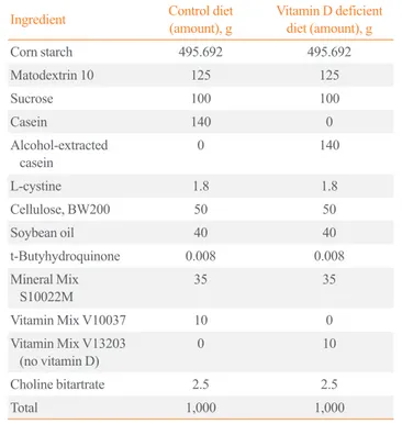
A total of 72 male Sprague-Dawley rats (Harlan CPB, Horst, the Netherlands) aged 4 weeks, and each weighing 200 to 230 g at the beginning of the study were used. The rats underwent 1-week acclimatization to the animal facility prior to study. The breeding room was maintained at a temperature of 21°C to 23°C with 40% to 60% humidity and a 12-hour light/darkness cycle. The rats were divided into two groups based on their diet (Rearch Diets Inc., New Brunswick, NJ, USA): control diet group (n=36) and vitamin D-deficient diet group (n=36) as shown in Table 1. Starting with day 1 of the experiment, the rats
were fed a control diet or a vitamin D-deficient diet for 8 weeks.
Subsequently, experimental OM was induced by inoculation of nontypeable Haemophilus influenzae (NTHi) 3,655 in the mid- dle ear of the rats. Rats were sacrificed on days 1, 2, 4, 7, and 14 post-inoculation for evaluation. All care and experiments were performed in accordance with the regulations of the Animal Re- search Institute of Medical Science, Dongguk University, and the study protocol was approved by the Animal Institutional Re- view Board (AIRB no. 2016-02114).
Bacterial strain
NTHi 3655 was preserved in a deep freezer at –80°C. It was used to induce OM with effusion. The bacteria were streaked onto BD BBLTM chocolate II agar dish (prod. no. 221169, Becton Dickinson, Franklin Lakes, NJ, USA). The dish was incubated overnight at 37°C and 5% CO2. Two colonies were selected and blended with 25 mL Brain Heart Infusion Broth (prod. no.
B9500, Teknova Inc., Burlington, ON, Canada) and 1 mL of RemelTM Fildes enrichment (prod. no. R45037, Thermo Fisher Scientific, Waltham, MA, USA). The mixture was incubated overnight at 37°C in the rotator, and centrifuged at 1,000 ×g for 5 minutes. Finally, the solution contained a bacterial suspension (105 cells/mL).
Table 1. Contents of Diet Provided to Experimental Rats
Ingredient Control diet
(amount), g Vitamin D deficient diet (amount), g
Corn starch 495.692 495.692
Matodextrin 10 125 125
Sucrose 100 100
Casein 140 0
Alcohol-extracted
casein 0 140
L-cystine 1.8 1.8
Cellulose, BW200 50 50
Soybean oil 40 40
t-Butyhydroquinone 0.008 0.008
Mineral Mix
S10022M 35 35
Vitamin Mix V10037 10 0
Vitamin Mix V13203
(no vitamin D) 0 10
Choline bitartrate 2.5 2.5
Total 1,000 1,000
Kim HB, et al.
Induction of otitis media
The tympanic membrane of rats was observed for abnormalities such as middle ear effusion, using otomicroscopy. The anesthet- ic solution used for the animals contained 0.1 mL/kg of ket- amine (Bayer, Leverkusen, Germany) and 0.1 mL/kg of Zoletile (Virbac, Carros, France). Under anesthesia, the rat was held in the supine position. Anterior neck was disinfected with betadine and a vertical incision was made. The soft tissue was dissected and both bullae were exposed by pushing aside the strap muscle and the submandibular gland. A 25 G needle tip was used to pierce a hole in the bulla, and 0.05 mL of bacterial suspension was injected into the hole.
Otomicroscopic examination
The anesthetic solution was injected on days 1, 2, 4, 7, and 14 post-inoculation to obtain a picture of the tympanic membrane and to monitor the progress of OM. Inflammation of the middle ear mucosa and middle ear effusion in all the rats was ensured using a digital microscope-USB (Dino-Lite, New Taipei City, Taiwan). Scoring of tympanic membrane was made based on the otomicroscopic findings (0: normal; 1: 1/2 or less exudate, congestion; 2: more than 1/2 exudate, severe congestion; 3:
high-pitched whole effervescent fluid, severe congestion).
Measurement of serum 25(OH)D concentration
The rats were anesthetized and 0.5 to 1 mL of blood was drawn by cardiac puncture at the time of sacrifice. Blood samples were collected in BD Microtainer SSTTM (prod. no. REF 365967, Becton Dickinson) and centrifuged. The collected serum sam- ples were left in a –80°C deep freezer. For determination of vi- tamin D levels, 25-OH vitamin D total ELISA kits (ALPCO, Salem, NH, USA) were used. Each vitamin D concentration was determined using spectrophotometry.
Immunohistochemistry of middle ear mucosa
The 72 bullae were fixed in 4% neutral buffered formalin for 24 hours, decalcified for over 3 days, using a decalcifying solution (prod. no. D0818-1L, Sigma-Aldrich, St. Louis, MO, USA).
The tissue was embedded in paraffin and sliced to a thickness of 4 μm on a microtome (prod. no. RM2235, Leica, Nussloch, Germany). For the analysis of mucosal thickness, hematoxylin (prod. no. HHS32, Sigma-Aldrich) and eosin Y (prod. no.
E6003, Sigma-Aldrich) (H&E) stains were used on the prepared slides. Photographs of each slide at 400× magnification were obtained. To determine the expression of cytokines and antimi- crobial peptides, all slides were immunostained with interleukin
6 (IL-6) (prod. no. orb303667, Biorbyt Ltd., Cambridge, UK), tumor necrosis factor α (TNF-α) (prod. no. ab6671, Abcam, Cambridge, MA, USA), IL-10 (prod. no. P29456, Cloud-Clone Corp., Huston, TX, USA), cathelicidin-related antimicrobial peptide (CRAMP) (prod. no. sc-374218, Santa Cruz Biotech- nology Inc., Dallas, TX, USA), and β-defensin 2 (prod. no.
251659, Abbiotec LLC, San Diego, CA, USA) and coupled with 3, 3´-diaminobenzidine (prod. no. K3468, Dako North America Inc., Carpinteria, CA, USA) for color development.
We obtained microscopic images and photographs at 400×
magnification of each slide using Olympus BX53F (Olympus, Tokyo, Japan). All the detected antibodies were collected using LEICA Qwin V3 (Leica Microsystems Imaging Solutions Ltd., Cambridge, UK) digital image processing and analysis soft- ware.
Statistical analysis
Each subgroup was analyzed with the GraphPad Prism version 5.0 (GraphPad Software, San Diego, CA, USA) program using the t test. The results were considered statistically significant at P<0.05.
RESULTS
Induction of otitis media with effusion
A summary of otomicroscopic findings of OM in the control and vitamin D deficiency groups is presented in Fig. 1A. In- flammation of each middle ear was graded according to the oto- microscopic findings. The mean value of the tympanic mem- brane scores in both groups are listed in Fig. 1B. The maximum increase in OM with effusion in both groups occurred on days 1 and 2, which decreased subsequently. After day 7, the tympanic membrane has almost returned to normal. However, there was no statistical difference of the scores between control group and vitamin D deficiency group.
Middle ear mucosal thickness
Bullae from control and vitamin D deficiency groups were stained with H&E (Fig. 2A). The mucosal thickness of both groups increased gradually from day 1. The middle ear mucosal thickness of the vitamin D deficiency group was significantly higher than that of the control group from day 2 to day 7 (Fig.
2B). On day 2, the maximum thickness was 56.0±9.1 μm in the vitamin D deficiency group and 43.9±9.8 μm in the control group, which represents 28.2% higher thickness in the vitamin D deficiency group. On days 4 and 7, the mucosal thickness of
Vitamin D Deficiency and Otitis Media
the vitamin D deficiency group was 47% to 50% higher than that of the control group. Most of the middle ear mucosa was restored by day 14.
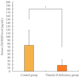
Measurement of serum 25(OH)D concentration
The serum 25(OH)D concentration was 75.19±43.43 ng/mL in the control group and 17.43±15.10 ng/mL in the vitamin D de- ficiency group (P<0.05) (Fig. 3).
Expression of cytokines in the middle ear mucosa
Immunohistochemical staining showed that proinflammatory cytokine IL-6 detection area gradually increased from day 0 (0.27%±0.10%) to day 7 (3.29%±1.21%) in the vitamin D de- ficiency group, while its detection area was slightly altered be- tween day 1 and 7 in the control group. This difference of the detection area made a statistical significance on day 7 (P<0.05) (Fig. 4A, B). TNF-α detection areas also showed a gradual in- crease from day 0 (0.29%±0.22%) to day 4 (3.12%±1.37%) in
the vitamin D deficiency group, while its detection area re- mained almost unchanged between day 1 and 4 in the control group. There was a statistically significant difference of the TNF-α detection area between the control group and the vitamin D deficiency group on day 2 (P<0.05) and day 4 (P<0.005) (Fig. 4C, D). Anti-inflammatory cytokine IL-10 detection in- creased gradually from day 0 (3.13%±1.23%) to day 7 (12.50%±4.11%) in the control group. Its expression also in- creased from day 0 (3.02%±1.02%) to day 2 (8.83%±2.49%), but decreased from day 4 (8.83%±3.93%) to day 14 (7.28%±
2.30%) in the vitamin D deficiency group, which made a statis- tically significant difference on day 7 (P<0.05) and day 14 (P<0.05) (Fig. 4E, F).
Expression of antimicrobial peptide in the middle ear mucosa
The expression of innate immune defense factor CRAMP was increased in the control group compared with the vitamin D de- Fig. 1. (A) Serial otomicroscopic images of middle ear in bacterially induced otitis media were taken for 14 days (×90 magnification). (B) The mean value of the tympanic membrane scores in the control group and vitamin D deficiency group (0: normal; 1: 1/2 or less exudate;
congestion; 2: more than 1/2 exudate, severe congestion; 3: high-pitched whole effervescent fluid, severe congestion).
Day 0 Day 1 Day 2 Day 4 Day 7 Day 14
Control group
Vitamin D deficiency group
Figure 1A.
Control group
Day 0 Day 1 Day 2 Day 4 Day 7 Day 14
Vitamin D deficiency group
A 3
2
1
0
Score of tympanic membrane (0–3)
Post-inoculation day
Day 1 Day 2 Day 4 Day 7 Day 14 B Control group Vitamin D deficiency group
Kim HB, et al.
ficiency group on day 1, but its expression was decreased in the control group compared with the vitamin D deficiency group on day 2 (Supplemental Fig. S1A, B). β-Defensin 2 was highly ex- pressed in the control group than in the vitamin D deficiency group on day 7 (Supplemental Fig. S1C, D). Most of the CRAMP and β-defensin 2 expression in both groups was recov- ered by day 7.
DISCUSSION
Several clinical studies indicate that low vitamin D status is sig- nificantly associated with a high risk or severity of OM [12-15].
A recent case-control study by Cayir et al. [12] showed that the mean serum 25(OH)D level was significantly lower in children with recurrent OM compared with controls (28.5 nmol/L vs.
72.9 nmol/L). Another case-control study by Walker et al. [13]
also showed that children with a higher serum 25(OH)D level Day 1 Day 2 Day 4 Day 7
Control group
Vitamin D deficiency group
Figure 2A.
Day 1 Day 2 Day 4 Day 7
Control group
Vitamin D deficiency group
Figure 2A.
Control group
Day 1 Day 2 Day 4 Day 7
Vitamin D deficiency group
A
Fig. 2. (A) H&E stain of middle ear mucosa (×200 magnification). (B) Middle ear mucosa thickness. aP<0.005.
100 80 60 40 20 0
Meddle ear mucosa thickness (μm)
Post-inoculation day
Day 0 Day 1 Day 2a Day 4a Day 7a Day 14 B Control group Vitamin D deficiency group
Fig. 3. Serum 25-hydroxyvitamin D (25(OH)D) levels in the con- trol group and the vitamin D deficiency group. aP<0.05.
200190 180170 160150 140130 120110 10090 8070 6050 4030 2010 0
Serum 25(OH)D level (ng/mL)
Control group Vitamin D deficiency group
a
Vitamin D Deficiency and Otitis Media
A IL-6
Figure 4A.
Day 0 Day 1 Day 4 Day 7 Day 14
Control group
Vitamin D deficiency group
IL-6 Figure 4A.
Day 0 Day 1 Day 4 Day 7 Day 14
Control group
Vitamin D deficiency group
Control group
Day 0 Day 1 Day 4 Day 7 Day 14
Vitamin D deficiency group
IL-6
C Day 0 Day 1 Day 4 Day 7 Day 14
Control group
Vitamin D deficiency group
Figure 4C.
TNF-α
Day 0 Day 1 Day 4 Day 7 Day 14
Control group
Vitamin D deficiency group
Figure 4C.
TNF-α
Control group
Day 0 Day 1 Day 4 Day 7 Day 14
Vitamin D deficiency group
TNF-α 5
4 3 2 1
2IL-6 detection area (%)/1.75 mm 0
Post-inoculation day
Day 0 Day 1 Day 2 Day 4 Day 7a Day 14 B
Control group
Vitamin D deficiency group IL-6
5 4 3 2 1 0
TNF-α detection area (%)/1.75 mm2
Post-inoculation day
Day 0 Day 1 Day 2a Day 4b Day 7 Day 14 D
Control group
Vitamin D deficiency group TNF-α
Fig. 4. (Continued to the next page)
Kim HB, et al.
carried a lower risk of chronic OM with effusion (odds ratio, 0.86 per 10 nmol/L; 95% confidence interval, 0.77 to 0.97). In a randomized controlled trial, administration of vitamin D 1,000 IU per day for 4 months significantly reduced the risk of un- complicated acute OM in children with a history of recurrent acute OM (hazard ratio, 0.23; 95% confidence interval, 0.12 to 0.46) [14]. In addition, a meta-analysis of five clinical studies involving 16,689 subjects concluded that the lower vitamin D level might play an important role in increasing the occurrence of acute OM [15].
Despite the aforementioned clinical evidence showing a sig- nificant association between vitamin D status and OM, little is known about the pathophysiological changes underlying this association. The middle ear mucosa normally consists of a sim- ple squamous epithelium ranging from 15 to 20 μm in thick- ness. However, bacterial infection or trauma can trigger infiltra- tion of inflammatory cells leading to mucosal growth and pro- liferation into a pseudostratified columnar epithelium that may exceed 1,000 μm in thickness [10]. In the present study, the rats showed a thickening of middle ear mucosa after induction of
experimental OM. We found that rats with vitamin D deficiency showed significantly higher mucosal thickness than controls during the course of OM, particularly from day 2 to 7. This finding indicates that vitamin D deficiency might be a factor that exacerbates the pathological changes of OM.
Vitamin D appears to influence the host immune system via multiple ways. One of the most important mechanisms involves T-cell–mediated immunity and cytokine production. By activat- ing the vitamin D receptors in T-cell, vitamin D promotes the proliferation of type 2 helper T (TH2) cells and related cytokine production while suppressing type 1 helper T (TH1) cell prolif- eration and associated cytokine expression [1-3]. TH1 cyto- kines, which include IL-2, interferon γ, and TNF-α, are general- ly considered proinflammatory. By contrast, TH2 cytokines in- cluding IL-4, IL-5, and IL-10 appear to exhibit anti-inflamma- tory effects [2]. Therefore, optimal vitamin D status leads to a healthy and tolerogenic immune response, whereas vitamin D deficiency may lead to a more proinflammatory state. Vitamin D also appears to promote regulatory T-cell production, which limits the extent of tissue damage [2,3]. In addition, it was Fig. 4. Immunohistochemical stain and the average detection area (%) of (A, B) interleukin 6 (IL-6), (C, D) tumor necrosis factor α (TNF-α), and (E, F) IL-10 for bacterially induced middle ear mucosa. aP<0.05; bP<0.005.
20 15 10 5
2IL-10 detection area (%)/1.75 mm 0
Post-inoculation day
Day 0 Day 1 Day 2 Day 4 Day 7a Day 14a F
Control group
Vitamin D deficiency group IL-10
E Control
group
Day 0 Day 1 Day 4 Day 7 Day 14
Vitamin D deficiency group
IL-10
Vitamin D Deficiency and Otitis Media
shown that vitamin D inhibits the production of proinflammato- ry cytokines in the respiratory epithelium by modulating the nu- clear factor κB pathway [16,17]. In the present study, our results also showed that the expression of IL-6 and TNF-α in the mid- dle ear mucosa was increased, while IL-10 expression was de- creased in the vitamin D-deficient rats, which indicates that vi- tamin D deficiency may alter the host immune response to a more proinflammatory state in OM.
It was also suggested that vitamin D upregulated the expres- sion of antimicrobial peptide in phagocytes and epithelial cells.
The vitamin D receptor-binding site, the so-called vitamin D re- sponse element, was discovered in the promoter regions of the genes encoding antimicrobial peptides such as cathelicidin and β-defensin 2, which have a lethal effect on microsomes [18-20].
However, this mechanism was shown to be conserved in only humans and primates, but not in nonprimate mammals includ- ing mice and rats [19,20]. Immunohistochemistry of our study showed an inconsistent result. The expression of CRAMP on day 1 after induction of OM was decreased in the vitamin D de- ficiency group compared with controls, but its expression on day 2 was increased in the vitamin D deficiency group. The ex- pression of β-defensin 2 was decreased in the vitamin D defi- ciency group compared with controls on day 7. In addition, oth- er antimicrobial factors such as reactive oxygen species and ni- tric oxide synthase were also known to be stimulated by vitamin D [3,21].
The present study has a few limitations. First, the role of vita- min D in the OM pathophysiology was investigated only in rats with vitamin D deficiency. To elucidate the effects of vitamin D on the development or progression of OM, further studies in- vestigating the role of vitamin D supplementation are needed.
Second, the altered expression of cytokines in the middle ear mucosa resulting from vitamin D deficiency was demonstrated immunohistochemically. Serum levels of cytokines were not in- vestigated in the present study. Therefore, it is not clear whether the role of vitamin D in the progression of OM is mediated via local or systemic mechanisms.
In conclusion, our study showed that pathological changes in the middle ear mucosa derived from OM were more severe in rats with vitamin D deficiency compared with controls, al- though the duration of OM was not different between the two groups. Our results also showed that vitamin D deficiency is as- sociated with a proinflammatory state due to the increased pro- duction of proinflammatory cytokines and decreased expression of anti-inflammatory cytokines. Based on these findings, we suggest that vitamin D deficiency may exacerbate the patho-
physiological changes of OM via altered cytokine production.
Therefore, maintenance of vitamin D status in the optimal range may be critical to successful clinical management of OM. The influence of vitamin D supplementation on the pathophysiology of OM requires additional investigation.
CONFLICTS OF INTEREST
No potential conflict of interest relevant to this article was re- ported.
ACKNOWLEDGMENTS
This research was funded by Basic Science Research Program through the National Research Foundation of Korea (NRF) funded by the Ministry of Science, ICT & Future Planning (NRF-2015R1C1A1A01054333).
AUTHOR CONTRIBUTIONS
Conception or design: C.G.C., H.S.C. Acquisition, analysis, or interpretation of data: H.B.K., S.H.L. Drafting the work or re- vising: H.B.K., H.S.C. Final approval of the manuscript: C.
G.C., H.S.C.
ORCID
Han Seok Choi https://orcid.org/0000-0002-6506-4342
REFERENCES
1. Prietl B, Treiber G, Pieber TR, Amrein K. Vitamin D and immune function. Nutrients 2013;5:2502-21.
2. Gunville CF, Mourani PM, Ginde AA. The role of vitamin D in prevention and treatment of infection. Inflamm Allergy Drug Targets 2013;12:239-45.
3. Borella E, Nesher G, Israeli E, Shoenfeld Y. Vitamin D: a new anti-infective agent? Ann N Y Acad Sci 2014;1317:76- 83.
4. Bouillon R, Carmeliet G, Verlinden L, van Etten E, Verstuyf A, Luderer HF, et al. Vitamin D and human health: lessons from vitamin D receptor null mice. Endocr Rev 2008;29:
726-76.
5. Rathored J, Sharma SK, Singh B, Banavaliker JN, Sreenivas V, Srivastava AK, et al. Risk and outcome of multidrug-re- sistant tuberculosis: vitamin D receptor polymorphisms and
Kim HB, et al.
serum 25(OH)D. Int J Tuberc Lung Dis 2012;16:1522-8.
6. Aslan S, Akil I, Aslan G, Onay H, Ozyut BC, Ozkinay F. Vi- tamin D receptor gene polymorphism in children with uri- nary tract infection. Pediatr Nephrol 2012;27:417-21.
7. Teele DW, Klein JO, Rosner B. Epidemiology of otitis me- dia during the first seven years of life in children in greater Boston: a prospective, cohort study. J Infect Dis 1989;160:
83-94.
8. Mittal R, Kodiyan J, Gerring R, Mathee K, Li JD, Grati M, et al. Role of innate immunity in the pathogenesis of otitis media. Int J Infect Dis 2014;29:259-67.
9. Cho CG, Gong SH, Kim HB, Song JJ, Park JH, Lim YS, et al. Role of group 3 innate lymphoid cells during experimen- tal otitis media in a rat model. Int J Pediatr Otorhinolaryngol 2016;88:146-52.
10. Lim DJ, Birck H. Ultrastructural pathology of the middle ear mucosa in serous otitis media. Ann Otol Rhinol Laryngol 1971;80:838-53.
11. Cho CG. Animal models of otitis media. Korean J Otorhino- laryngol-Head Neck Surg 2015;58:371-7.
12. Cayir A, Turan MI, Ozkan O, Cayir Y, Kaya A, Davutoglu S, et al. Serum vitamin D levels in children with recurrent otitis media. Eur Arch Otorhinolaryngol 2014;271:689-93.
13. Walker RE, Bartley J, Camargo CA Jr, Flint D, Thompson JM, Mitchell EA. Higher serum 25(OH)D concentration is associated with lower risk of chronic otitis media with effu- sion: a case-control study. Acta Paediatr 2017;106:1487-92.
14. Marchisio P, Consonni D, Baggi E, Zampiero A, Bianchini S, Terranova L, et al. Vitamin D supplementation reduces the risk of acute otitis media in otitis-prone children. Pediatr In- fect Dis J 2013;32:1055-60.
15. Li HB, Tai XH, Sang YH, Jia JP, Xu ZM, Cui XF, et al. As- sociation between vitamin D and development of otitis me- dia: a PRISMA-compliant meta-analysis and systematic re- view. Medicine (Baltimore) 2016;95:e4739.
16. Hansdottir S, Monick MM, Lovan N, Powers L, Gerke A, Hunninghake GW. Vitamin D decreases respiratory syncy- tial virus induction of NF-kappaB-linked chemokines and cytokines in airway epithelium while maintaining the antivi- ral state. J Immunol 2010;184:965-74.
17. McNally P, Coughlan C, Bergsson G, Doyle M, Taggart C, Adorini L, et al. Vitamin D receptor agonists inhibit proin- flammatory cytokine production from the respiratory epithe- lium in cystic fibrosis. J Cyst Fibros 2011;10:428-34.
18. Wang TT, Nestel FP, Bourdeau V, Nagai Y, Wang Q, Liao J, et al. Cutting edge: 1,25-dihydroxyvitamin D3 is a direct in- ducer of antimicrobial peptide gene expression. J Immunol 2004;173:2909-12.
19. Gombart, AF, Borregaard N, Koeffler HP. Human cathelici- din antimicrobial peptide (CAMP) gene is a direct target of the vitamin D receptor and is strongly up-regulated in my- eloid cells by 1,25-dihydroxyvitamin D3. FASEB J 2005;19:
1067-77.
20. Liu PT, Stenger S, Li H, Wenzel L, Tan BH, Krutzik SR, et al. Toll-like receptor triggering of a vitamin D-mediated hu- man antimicrobial response. Science 2006;311:1770-3.
21. Rockett KA, Brookes R, Udalova I, Vidal V, Hill AV, Kwiat- kowski D. 1,25-DihydroxyvitaminD3 induces nitric oxide synthase and suppresses growth of Mycobacterium tubercu- losis in a human macrophage-like cell line. Infect Immun 1998;66:5314-21.