Fragility hip fractures are considered amongst the most prognostically poor consequences of age-related osteo- porosis owing to their propensity for complications such as chronic pain, reduced mobility, poor quality of life and
mortality. In the year 2000, there were an estimated 1.6 million hip fractures worldwide.1) With increasing life ex- pectancy and rising numbers of elderly individuals in all geographic populations, the incidence of fragility hip frac- tures is estimated to reach 6.3 million by the year 20502) and will certainly pose a significant health burden world- wide. Death within the first month following fragility hip fracture injuries has been shown to be as high as 13.3%
in previous studies.3) Furthermore, this risk of mortality tends to cluster in the month following surgery but dimin- ishes thereafter.4)
A Comprehensive Analysis of the Causes and Predictors of 30-Day Mortality Following Hip
Fracture Surgery
Hassaan Qaiser Sheikh, MRCS, Fahad Siddique Hossain, FRCS*, Adeel Aqil, MRCS, Babawande Akinbamijo, MBBS, Vhaid Mushtaq, MUDr, Harish Kapoor, FRCS
Department of Trauma and Orthopaedics, Leeds General Infirmary, Leeds,
*Department of Trauma and Orthopaedics, University College London, London, UK
Background: A fracture neck of femur is the leading cause of injury-related mortality in the elderly population. The 30-day mortal- ity figure is a well utilised marker of clinical outcome following a fracture neck of femur. Current studies fail to analyse all patient demographic, biochemical and comorbid parameters associated with increased 30-day mortality. We aimed to assess medical risk factors for mortality, which are easily identifiable on admission for patients presenting with a fractured neck of femur.
Methods: A retrospective review of a prospectively populated database was undertaken to identify all consecutive patients with a fracture neck of femur between October 2008 and March 2011. All factors related to the patient, injury and surgery were identi- fied. The primary outcome of interest was 30-day mortality. Univariate and subsequent multivariate analyses using a backward stepwise likelihood ratio Cox regression model were performed in order to establish all parameters that significantly increased the risk of death.
Results: A total of 1,356 patients were included in the study. The 30-day mortality was 8.7%. The most common causes of death included pneumonia, sepsis and acute myocardial infarction. Multiple regression analysis revealed male gender, increasing age, admission source other than the patient’s own home, admission haemoglobin of less than 10 g/dL, a history of myocardial infarc- tion, concomitant chest infection during admission, increasing Charlson comorbidity score and liver disease to be significant pre- dictors of mortality.
Conclusions: This study has elucidated risk factors for mortality using clinical and biochemical information which are easily gath- ered at the point of hospitalization. These results allow for identification of vulnerable patients who may benefit from a prioritisa- tion of resources.
Keywords: Hip fractures, Mortality, Risk factors, Epidemiology
Copyright © 2017 by The Korean Orthopaedic Association
This is an Open Access article distributed under the terms of the Creative Commons Attribution Non-Commercial License (http://creativecommons.org/licenses/by-nc/4.0) which permits unrestricted non-commercial use, distribution, and reproduction in any medium, provided the original work is properly cited.
Clinics in Orthopedic Surgery • pISSN 2005-291X eISSN 2005-4408 Received August 16, 2016; Accepted November 8, 2016
Correspondence to: Hassaan Qaiser Sheikh, MRCS
Department of Trauma and Orthopaedics, Leeds General Infirmary, 10 Oldroyd Way, Dewsbury, Wakefield 13 2JJ, UK
Tel: +44-113-243-2799, Fax: +44-113-392-3290 E-mail: hqsheikh@doctors.org.uk
Mortality incidence at 30 days has long been rec- ognised as an important care quality indicator for health- care institutions and has been in longstanding use by the National Hip Fracture Database (NHFD) of the English healthcare system.5) Identifying modifiable risk factors contributing to mortality can, therefore, play a crucial role in reducing it.4,6,7) Current literature has identified that age, gender, pre-existing cardiovascular, cerebrovascular or respiratory disease and admission source predispose to mortality.8-11) A commonly used and validated hip fracture score12) has also identified several variables contributing to mortality. However, it stratifies mortality risk accord- ing to the number of comorbidities rather than severity or type. It also attributes risk of death based on the presence of anaemia to the exclusion of other commonly collected blood parameters. Similarly, other studies have also failed to consider all comorbid and biochemical parameters identifiable at admission in their analysis of risk factors for mortality.4,6-11) Despite advances in medical and surgical treatments for patients along with the implementation of national evidence-based guidelines for the management of such hip fracture injuries, 30-day mortality rates have shown only a trivial improvement nationally. According to the most recent NHFD reports, the 30-day mortality rates have only improved from 8.1% in 2013 to 8.02% in 2014 and then 7.5% in 2015. It is hence entirely possible that a more thorough and exhaustive analysis of predictors of early mortality may aid in our understanding resulting in improved care.
We, therefore, aimed to perform a comprehensive evaluation of all risk factors for 30-day mortality that are easily obtainable at the point of hospital admission.
METHODS
Hip fracture patient data is prospectively input into the NHFD at our level 1 trauma centre institution by trained personnel. We used this database to identify all consecu- tive patients admitted over a 3-year period. The clinical data for each patient were then identified and cross refer- enced with hospital electronic patient records to ensure accuracy.
This project was approved by Leeds General In- firmary Institutional Review Board (Project 668). We collected variables including demographic (age, gender, admission source, and walking ability), biochemical (hae- moglobin level, white blood cell count, platelet count, coagulation profile and urea and electrolyte levels) and medical comorbidity data. Patient’s preoperative admis- sion source was grouped as one of four categories: patients
who were independent in their own home or sheltered ac- commodation; those who were in a nursing or residential home; those who were already in hospital and had a fall as an inpatient; and those transferred from another hos- pital. Mobility was categorised into independent walkers;
walkers with a single aid (stick or crutch); walkers with two aids (two sticks/crutches or frame); and those who were wheelchair-bound. All patient comorbidity data was identified from hospital records using International Clas- sification of Disease, 10th revision (ICD-10), codes and these were used to calculate the Charlson Comorbidity Index for each patient.13) The American Society of Anes- thesiologists (ASA) grade recorded at the time of surgery was also noted. Other factors analysed included the type of fracture and the time from presentation to hospital to receiving surgical intervention. Fractures were classified as intracapsular undisplaced, intracapsular displaced, intertrochanteric, subtrochanteric or other (if the injury couldn’t be distinctively classified). The primary outcome of interest was 30-day mortality following surgery. Causes of death were collected from death certificate entries as well as from coroner’s autopsy records and the primary cause of death was used for analysis. Patients that had in- complete datasets or that were managed nonoperatively were excluded from analysis.
All patients were treated on a standardised hip frac- ture pathway. On presentation, a clinical assessment was performed allowing identification of medical comorbidity data. Blood tests ensured haematological, biochemical and coagulation profiling. A chest radiography and electrocar- diography completed the basic cardio-respiratory assess- ment. Our unit aimed to perform surgery on the day of or the day after presentation of injury in accordance with national health care guidelines.14)
Following review of imaging by multiple orthopae- dic consultants and anaesthetic team at a multidisciplinary trauma meeting, operative management was decided and performed in a theatre with laminar air flow. All opera- tions were performed by or under direct supervision of an orthopaedic consultant. The majority of displaced intra- capsular fractures were treated with a cemented hip hemi- arthroplasty and undisplaced intracapsular fractures were fixed in situ with screw fixation. Intertrochanteric fractures were fixed with a sliding hip screw, while subtrochanteric fractures were managed with a cephalomedullary device.
In accordance with national guidelines, patients that are not cognitively impaired and can mobilise independently or with a single stick were treated with a total hip arthro- plasty. After the operation, patients were mobilised fully weight bearing with physiotherapy support.
Statistical Analysis
To aid clinical interpretation, admission haemoglobin levels and white blood cell counts were categorized as di- chotomous variables. Haemoglobin levels were grouped
as being less than or more than 10 g/dL—this was done following evidence from a recent study which found haemoglobin < 10 g/dL to be an independent predictor of mortality.12) White blood cell count levels were grouped
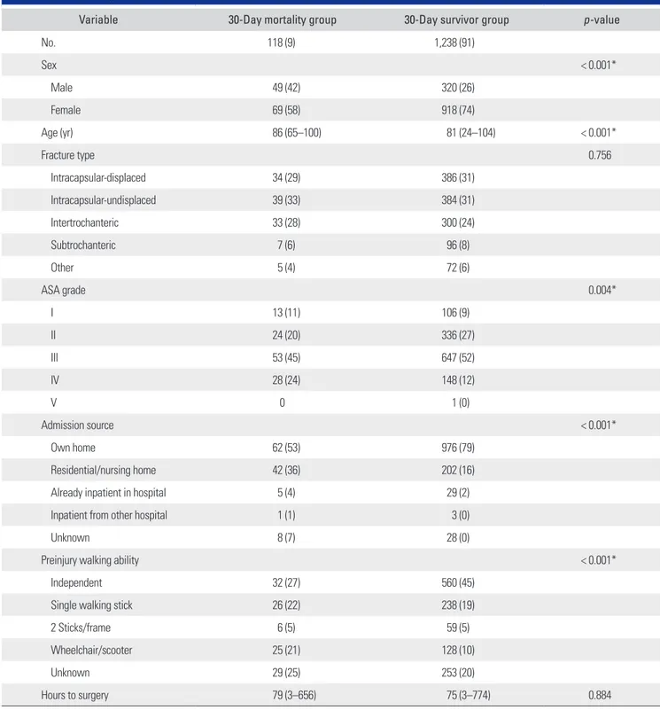
Table 1. Patient Demographics
Variable 30-Day mortality group 30-Day survivor group p-value
No. 118 (9) 1,238 (91)
Sex < 0.001*
Male 49 (42) 320 (26)
Female 69 (58) 918 (74)
Age (yr) 86 (65–100) 81 (24–104) < 0.001*
Fracture type 0.756
Intracapsular-displaced 34 (29) 386 (31)
Intracapsular-undisplaced 39 (33) 384 (31)
Intertrochanteric 33 (28) 300 (24)
Subtrochanteric 7 (6) 96 (8)
Other 5 (4) 72 (6)
ASA grade 0.004*
I 13 (11) 106 (9)
II 24 (20) 336 (27)
III 53 (45) 647 (52)
IV 28 (24) 148 (12)
V 0 1 (0)
Admission source < 0.001*
Own home 62 (53) 976 (79)
Residential/nursing home 42 (36) 202 (16)
Already inpatient in hospital 5 (4) 29 (2)
Inpatient from other hospital 1 (1) 3 (0)
Unknown 8 (7) 28 (0)
Preinjury walking ability < 0.001*
Independent 32 (27) 560 (45)
Single walking stick 26 (22) 238 (19)
2 Sticks/frame 6 (5) 59 (5)
Wheelchair/scooter 25 (21) 128 (10)
Unknown 29 (25) 253 (20)
Hours to surgery 79 (3–656) 75 (3–774) 0.884
Values are presented as number (%) or mean (range).
ASA: American Society of Anesthesiologists.
*Variables have a p-value < 0.15 and were included in the multivariate analysis.
as being normal (4 × 10⁹/L–12 × 109/L) or abnormal (less than 4 × 10⁹/L or more than 12 × 109/L), in keeping with the widely accepted definition of systemic inflammatory response syndrome.
All factors were initially analysed for risk of mortali- ty with univariate analysis. This assessment involved either using a chi-squared/Fisher exact test for categorical data or the independent t/Mann-Whitney U-test for continuous variables. The impact of these variables were then analysed using a backward stepwise likelihood ratio Cox regres- sion analysis whilst adjusting for covariates. Covariates were included in the multivariate analysis if the univariate analysis resulted in a p-value of < 0.15, in accordance with accepted and published statistical methods.15) Results were displayed as hazard ratios to aid clinical interpretation. All statistical calculations were performed using IBM SPSS ver. 21 (IBM Co., Armonk, NY, USA).
RESULTS
A total of 1,673 patients were initially identified. After exclusion of 132 duplicates, 44 patients that did not have an operation and 141 incomplete datasets, a total of 1,356 patients were included in the study. The 30-day mortality rate was 8.7% (118/1,356). Of these patients, 89 died dur- ing their index admission episode following hip fracture surgery. There were no on-table deaths during surgery.
The clinical and demographic data for these patients are summarised in Table 1, grouped by their 30-day mortality status. The commonest cause of death was pneumonia fol- lowed by myocardial infarction (MI) (Fig. 1).
On univariate analysis, patients that died within 30 days of surgery were likely to be older, male, functionally less independent and living in a care institution rather that their own home. The mortality group also had a signifi- cantly higher incidence of hypertension, chronic obstruc-
tive airways disease (COAD), previous stroke, previous MI, chronic liver disease and chest infection on admission (Table 2). Analysis of admission blood parameters re- vealed that the mortality group was more likely to have an admission haemoglobin level of less than 10 g/dL, an ab- normal white blood cell count, a raised serum potassium level (although the mean potassium level was still within normal range), deranged urea and creatinine levels and a higher international normalized ratio (Table 2).
Following multivariate analysis, the risk factors sig- nificant for 30-day mortality included increasing age, male gender, admission source other than patient’s own home, admission haemoglobin levels less than 10 g/dL and an increasing Charlson comorbidity score. A history of previ- ous MI, concomitant chest infection during admission and chronic liver disease were the strongest predictors of early mortality with hazard ratios of 2.191, 3.738, and 3.945, re- spectively (Table 3).
Identification of Risk Factors for Mortality
Table 2 summarises the comorbidities and admission blood parameters that were significant on univariate anal- ysis. Table 3 summarises the outcomes of the multivariate analysis analysing risk factors for 30-day mortality. The causes of death for the 118 patients that died within 30 days of hip fracture surgery are summarised in Fig. 1.
DISCUSSION
Patients who died within 30 days of hip fracture surgery were likely to have admission haemoglobin levels less than 10 g/dL, be older, be male, have a history of previous myocardial infarction and/or have suffered a concomitant chest infection during their index admission. Chronic liver disease was the strongest risk factor for early death within 30 days of surgery. The overall 30-day mortality rate was
60 0
Pneumonia Acute myocardial infarction Sepsis Carcinomatosis Upper GI haemorrhage Cerebral infarction Pulmonary embolism Chronic cardiac failure Bowel infarction Acute renal failure Acute exacerbation of COAD Osteopenic fracture neck of femur Old age Chronic liver disease
10 20 30 40 50
52 22
12 7 4 4 3 3 3 3 2 1 1 1
Fig. 1. 30-Day morality causes of death.
GI: gastrointestinal, COAD: chronic ob- structive airways disease.
Table 2. Univariate Analysis of Risk Factors for 30-Day Mortality
Variable 30-Day mortality group
(n = 118) 30-Day survivor group
(n = 1,238) p-value
Comorbidities on admission
Charlson score 5 (3–9) 4 (0–9) < 0.001*
Hypertension 13 (11) 30 (2) < 0.001*
COAD 15 (13) 77 (6) 0.012*
Previous stroke 11 (9) 38 (3) 0.002*
Previous myocardial infarction 12 (10) 27 (22) < 0.001*
Chest infection during admission 48 (41) 113 (9) < 0.001*
Chronic heart failure 7 (6) 32 (3) 0.046*
Chronic liver disease 3 (3) 4 (0) 0.017*
Dementia 9 (7) 88 (7) 0.851
Diabetes mellitus 11 (9) 128 (10) 0.874
Neurological disease 2 (2) 26 (2) 1.000
Thyroid disease 5 (4) 71 (6) 0.675
Malignancy 15 (13) 114 (9) 0.248
Alcohol excess 4 (3) 39 (3) 0.785
Urinary tract infection 14 (12) 206 (17) 0.194
Peripheral vascular disease 0 19 (2) 0.400
Cardiovascular disease 7 (6) 25 (2) 0.017*
Connective tissue disorder 0 1 (0) 1.000
Peptic ulcer disease 0 9 (1) 1.000
Chronic kidney disease 33 (28) 87 (7) < 0.001*
Hemiplegia 2 (2) 15 (1) 0.654
Leukaemia 0 4 (0) 1.000
Blood parameters on admission
Haemoglobin < 10 g/dL 25 (21) 141 (11) 0.003*
Abnormal white blood cell count 51 (43) 382 (31) 0.007*
Platelet count 119 (93–843) 282 (43–938) 0.435
Sodium 137.8 (123–151) 137.1 (115–151) 0.076*
Potassium 4.5 (2.9–7.0) 4.3 (2.4–7.1) < 0.001*
Urea 11.5 (2.6–33.8) 8.1 (0.9–35.7) < 0.001*
Creatinine 139 (58–817) 102 (42–598) < 0.001*
INR 1.2 (0.9–4.2) 1.1 (0.8–6.3) 0.002*
APTT 30.7 (24–51) 30.5 (19–195) 0.749
Values are presented as mean (range) or number (%).
COAD: chronic obstructive airways disease, INR: international normalized ratio, APTT: activated partial thromboplastin time.
*Variables have a p-value < 0.15 and were included in the multivariate analysis.
8.7% (118/1,356 patients). The commonest causes of death were pneumonia and acute myocardial infarction followed by sepsis from other sources. Mortality risk was reduced in patients if they were admitted from their own home. In our cohort, other comorbidities such as diabetes mellitus, admission electrolyte disturbances and malignancy did not increase the risk of 30-day mortality. While increasing ASA grade did not affect early death after hip fracture sur- gery, an increasing Charlson score did.
Our 30-day mortality rate of 8.7% is comparable with the figure of 7.5% across England and Wales as re- ported by the NHFD (this figure is case-mix adjusted for multiple variables including age, ASA grade and source of admission, whereas our cohort is an unselected and unadjusted group).5) Furthermore, our institutional find- ings compare favourably to those of other developed na- tions. A study of over 38,000 Danish patients with hip fractures showed the 30-day mortality to be between 9.2%
and 10.9%.16) Elsewhere in the world, this figure is higher, reaching up to 13.3% worldwide as reported by a recent meta-analysis.3)
Over 60% of patients that died within 30 days after surgery did so as a result of a chest infection and/or an acute MI in our study. In fact, there were four mortalities within 24 hours of surgery resulting from overwhelming chest infection or an acute myocardial infarction. Such patients already have a higher incidence of pre-existing cardio-respiratory disease and the reduced mobility fol- lowing a hip fracture is known to increase the risk of pneumonia.17) A chest infection is an independent risk factor for early readmission after hip fracture surgery and is a common postoperative complication.6,18) The develop- ment of a chest infection following hip fracture surgery in our cohort was one of the strongest predictors of 30-day
death after hip fracture surgery with a hazard ratio of 3.738.
These findings are in complete agreement with those of a similar study from Japan showing a fivefold increase in early death in hip fracture patients who develop a postop- erative pneumonia.17)
A hip fracture in itself is an independent risk fac- tor for acute myocardial infarction.19) A recent prospec- tive study of 200 hip fracture patients undergoing surgery found that the incidence of acute myocardial infarction in the perioperative period may in fact be as high as 35.5%–
some of these may be completely asymptomatic.4,20) Due to the high incidence of perioperative myocardial infarction in this cohort, previous authors have made recommenda- tions to routinely measure postoperative cardiac biochem- ical markers to reduce cardiac-related deaths.21)
Increasing age, male gender and presentation with injury from a source other than patients’ own home were also statistically significant predictors of early mortality in our cohort. Similar findings have also been demonstrated in cohort studies of hip fracture patients looking at early mortality where increasing age, male gender, dementia and residence in an institution were identified as risk fac- tors.3,22) Mortality amongst men is generally higher than in women23) and perhaps multifactorial. Factors such as dementia and increasing age likely reflect the patients’
poor physiological reserve in withstanding the stress of surgery and its sequelae. Patients admitted from sources other than their own homes are likely to be care-depen- dent within nursing or care homes with poor mobility and health. In contrast, patients who live in their own homes are more likely to be independent and active resulting in better postoperative outcomes.24)
Interestingly, in our cohort, the presence of diabetes mellitus, malignancy and admission electrolyte imbalances Table 3. Multivariate Analysis of Risk Factors for 30-Day Mortality
Variable p-value Hazard ratio 95% Confidence interval
Increasing age < 0.001 1.057 1.030–1.084
Gender (male) 0.005 1.760 1.185–2.615
Admission source other than home 0.002 1.295 1.102–1.521
Admission hemoglobin < 10 g/dL 0.012 1.765 1.131–2.755
Increasing Charlson comorbidity score 0.004 1.249 1.075–1.452
Previous myocardial infarction 0.014 2.191 1.174–4.087
Chest infection during admission < 0.001 3.738 2.549–5.480
Chronic liver disease 0.024 3.945 1.203–12.938
did not significantly increase the risk of 30-day mortality.
In our centre, patients with diabetes mellitus are assessed on admission for current anti-hyperglycaemic treatment and prescribed the appropriate inpatient anti-hyperglycae- mic treatment immediately. This includes adjustable rate intravenous insulin therapy if the patient usually requires insulin or if the blood glucose readings are unusually high. The patient’s glucose levels are monitored regularly and they are placed back on their usual treatment once they are eating and drinking normally after their opera- tion. Acute electrolyte imbalances, if severe enough, are similarly treated in an aggressive manner on admission and the hip fracture surgery is delayed until the patient is deemed anaesthetically fit to undergo the operation. We believe this strict inpatient diabetic and electrolyte control is responsible for the lack of association between diabetes mellitus and 30-day mortality in this cohort.
Increasing ASA grade also did not significantly in- crease the risk of mortality. However, a higher Charlson score was associated with increased mortality. This likely reflects the fact that the ASA grade may be more subject to higher interobserver variability.25) The Charlson comorbid- ity scoring system, however, scores specific diagnoses and is a more robust and reproducible system.26) Our results are in accordance with a previously pooled analysis of over 500,000 hip fracture patients which also demonstrated that a high admission Charlson score was a preinjury predictor of mortality.27)
Chronic liver disease was the strongest predictor of 30-day mortality in our cohort with a hazard ratio of 3.98.
Although only one patient died directly from chronic liver disease (and, therefore, hepatorenal failure), the statistical analysis concluded that patients with long-term liver dis- ease are at much higher risk of 30-day mortality from any cause. It is known that chronic liver disease also increases the risk of cardiovascular disease. These results are echoed by the findings of a similar Australian study which showed liver disease to be an overwhelmingly strong predictor of inpatient mortality amongst hip fracture patients with a hazard ratio of 4.75.28) It has previously been suggested that such patients have an increased susceptibility to infection which has also been demonstrated in the elective setting following arthroplasty surgery.29) The mechanisms behind the increased surgical risk in patients with liver disease are poorly understood; however, dysregulation of metabolic homeostasis and the imbalance of oxidative and antioxida- tive processes have been implicated as underlying mecha- nisms leading to morbidity and mortality.30)
Our study is strengthened by the inclusion of a large number of patients and is unique in that it provides risk
factors not previously found in literature for a number of reasons. Previous studies have omitted blood parameters as risk factors. The fact that anaemia is a risk factor for myocardial infarction and that chest infection may result in hyponatremia strengthens our reasons for including blood results as variables in our analysis. We also exam- ined specific comorbidities for association with early mortality as well as Charlson score which is a weighted score based on specific diagnoses and has previously been used in similar patient cohorts. Other studies looking at mortality following hip fractures have previously used co- morbidity counts which can be vague and have high inter- observer errors.
This study is limited by its retrospective design and, therefore, does not offer the robustness of data offered by prospective data collection. However, we are confident that due to the cross-referencing of data between the NHFD and multiple local hospital records, we obtained an accurate dataset. The NHFD is a prospectively populated audit database with accurate data input by trained trauma coordinators at our centre. In addition, we used multivari- ate analysis to investigate a large cohort of patients analys- ing many parameters that can influence mortality–this minimises the confounding effect of covariates.
A specific limitation that we faced was that we did not have complete body mass index data on all patients and, therefore, this was not included in the final analysis.
Although this could impact mortality, any such effect is likely to be indirect due to the sequelae of a high body mass index such as diabetes mellitus and ischaemic heart disease. These factors were independently analysed in this study for association with mortality.
In conclusion, increasing age, male gender, admis- sion source and admission haemoglobin of less than 10 g/dL predispose to early mortality. A previous history of myocardial infarction, concomitant chest infection during index admission and chronic liver disease are the strongest predictors of 30-day mortality in hip fracture patients. The Charlson comorbidity score allows indexing of patient comorbidities in a more robust manner than a simple co- morbidity count and is also an independent predictor of early mortality. A multidisciplinary approach to the care of these patients should be undertaken from the point of ad- mission with physicians supporting early medical optimi- sation of cardiorespiratory, metabolic and liver functions.
CONFLICT OF INTEREST
No potential conflict of interest relevant to this article was reported.
REFERENCES
1. Cooper C, Cole ZA, Holroyd CR, et al. Secular trends in the incidence of hip and other osteoporotic fractures. Osteopo- ros Int. 2011;22(5):1277-88.
2. Cooper C, Campion G, Melton LJ 3rd. Hip fractures in the elderly: a world-wide projection. Osteoporos Int.
1992;2(6):285-9.
3. Hu F, Jiang C, Shen J, Tang P, Wang Y. Preoperative predic- tors for mortality following hip fracture surgery: a system- atic review and meta-analysis. Injury. 2012;43(6):676-85.
4. Khan MA, Hossain FS, Ahmed I, Muthukumar N, Mohsen A. Predictors of early mortality after hip fracture surgery.
Int Orthop. 2013;37(11):2119-24.
5. Royal College of Physicians. National Hip Fracture Data- base (NHFD) annual report 2014. London: Royal College of Physicians; 2014.
6. Roche JJ, Wenn RT, Sahota O, Moran CG. Effect of comor- bidities and postoperative complications on mortality after hip fracture in elderly people: prospective observational cohort study. BMJ. 2005;331(7529):1374.
7. Moran CG, Wenn RT, Sikand M, Taylor AM. Early mortal- ity after hip fracture: is delay before surgery important? J Bone Joint Surg Am. 2005;87(3):483-9.
8. Elliott J, Beringer T, Kee F, Marsh D, Willis C, Stevenson M.
Predicting survival after treatment for fracture of the proxi- mal femur and the effect of delays to surgery. J Clin Epide- miol. 2003;56(8):788-95.
9. Gruson KI, Aharonoff GB, Egol KA, Zuckerman JD, Koval KJ. The relationship between admission hemoglobin level and outcome after hip fracture. J Orthop Trauma.
2002;16(1):39-44.
10. Ramnemark A, Nilsson M, Borssen B, Gustafson Y. Stroke, a major and increasing risk factor for femoral neck fracture.
Stroke. 2000;31(7):1572-7.
11. Eiskjaer S, Ostgard SE. Risk factors influencing mortality after bipolar hemiarthroplasty in the treatment of fracture of the femoral neck. Clin Orthop Relat Res. 1991;(270):295- 300.
12. Maxwell MJ, Moran CG, Moppett IK. Development and validation of a preoperative scoring system to predict 30 day mortality in patients undergoing hip fracture surgery. Br J Anaesth. 2008;101(4):511-7.
13. Charlson ME, Pompei P, Ales KL, MacKenzie CR. A new method of classifying prognostic comorbidity in longitu- dinal studies: development and validation. J Chronic Dis.
1987;40(5):373-83.
14. National Clinical Guideline Centre. The management of hip fracture in adults. London: National Clinical Guideline Centre; 2011.
15. Bursac Z, Gauss CH, Williams DK, Hosmer DW. Purpose- ful selection of variables in logistic regression. Source Code Biol Med. 2008;3:17.
16. Daugaard CL, Jorgensen HL, Riis T, Lauritzen JB, Duus BR, van der Mark S. Is mortality after hip fracture associated with surgical delay or admission during weekends and pub- lic holidays? A retrospective study of 38,020 patients. Acta Orthop. 2012;83(6):609-13.
17. Muraki S, Yamamoto S, Ishibashi H, Nakamura K. Factors associated with mortality following hip fracture in Japan. J Bone Miner Metab. 2006;24(2):100-4.
18. Khan MA, Hossain FS, Dashti Z, Muthukumar N. Causes and predictors of early re-admission after surgery for a frac- ture of the hip. J Bone Joint Surg Br. 2012;94(5):690-7.
19. Chiang CH, Liu CJ, Chen PJ, et al. Hip fracture and risk of acute myocardial infarction: a nationwide study. J Bone Miner Res. 2013;28(2):404-11.
20. Hietala P, Strandberg M, Strandberg N, Gullichsen E, Airak- sinen KE. Perioperative myocardial infarctions are common and often unrecognized in patients undergoing hip fracture surgery. J Trauma Acute Care Surg. 2013;74(4):1087-91.
21. Gupta BP, Huddleston JM, Kirkland LL, et al. Clinical pre- sentation and outcome of perioperative myocardial infarc- tion in the very elderly following hip fracture surgery. J Hosp Med. 2012;7(9):713-6.
22. Hasegawa Y, Suzuki S, Wingstrand H. Risk of mortality fol- lowing hip fracture in Japan. J Orthop Sci. 2007;12(2):113-7.
23. Singh-Manoux A, Gueguen A, Ferrie J, et al. Gender differ- ences in the association between morbidity and mortality among middle-aged men and women. Am J Public Health.
2008;98(12):2251-7.
24. Vochteloo AJ, Tuinebreijer WE, Maier AB, Nelissen RG, Bloem RM, Pilot P. Predicting discharge location of hip fracture patients; the new discharge of hip fracture patients score. Int Orthop. 2012;36(8):1709-14.
25. Mak PH, Campbell RC, Irwin MG; American Society of Anesthesiologists. The ASA Physical Status Classification:
inter-observer consistency: American Society of Anesthesi- ologists. Anaesth Intensive Care. 2002;30(5):633-40.
26. Bernardini J, Callen S, Fried L, Piraino B. Inter-rater reliabil- ity and annual rescoring of the Charlson comorbidity index.
Adv Perit Dial. 2004;20:125-7.
27. Smith T, Pelpola K, Ball M, Ong A, Myint PK. Pre-oper- ative indicators for mortality following hip fracture sur- gery: a systematic review and meta-analysis. Age Ageing.
2014;43(4):464-71.
28. Frost SA, Nguyen ND, Black DA, Eisman JA, Nguyen TV.
Risk factors for in-hospital post-hip fracture mortality.
Bone. 2011;49(3):553-8.
29. Deleuran T, Vilstrup H, Overgaard S, Jepsen P. Cirrhosis pa- tients have increased risk of complications after hip or knee arthroplasty. Acta Orthop. 2015;86(1):108-13.
30. Fisher L, Srikusalanukul W, Fisher A, Smith P. Liver func- tion parameters in hip fracture patients: relations to age, adipokines, comorbidities and outcomes. Int J Med Sci.
2015;12(2):100-15.