ARTICLE
https://doi.org/10.11106/ijt.2016.9.2.168Received October 31, 2015 / Revised 1st May 12, 2016, 2nd May 26, 2016 / Accepted May 26, 2016
Correspondence: Changhoon Lee, MD, PhD, Department of Surgery, Mirae Woman’s Hospital, 459 Gaya-daero, Busanjin-gu, Busan 47268, Korea
Tel: 82-51-890-9955, Fax: 82-51-890-9987, E-mail: gschlee@naver.com
Copyright ⓒ 2016, the Korean Thyroid Association. All rights reserved.
This is an open-access article distributed under the terms of the Creative Commons Attribution Non-Commercial License (http://creative- commons.org/licenses/by-nc/4.0/), which permits unrestricted non-commercial use, distribution, and reproduction in any medium, provided the original work is properly cited.
임신 중 진단된 갑상선유두암의 외과적 치료 시기
미래여성병원 외과1, 산부인과2
김윤석
1, 이창훈
1, 제은애
2, 이영진
2, 정주은
2, 김수선
2, 김미향
2, 이은숙
2, 박천숙
2, 박재묵
2, 정현우
2, 박무실
2, 이재준
2, 안준모
2, 이 수
2Timing of Surgical Management of Papillary Thyroid Cancer Diagnosed during Pregnancy
Yoonseok Kim1, Changhoon Lee1, Eunae Jae2, Youngjin Lee2, Jueun Jung2, Susun Kim2, Mihyang Kim2, Eunsuk Lee2, Chunsuk Park2, Jaemook Park2, Hyunwoo Jung2, Musil Park2, Jaejun Lee2, Junmo Ahn2 and Soo Lee2
Departments of Surgery1 and Obstetrics & Gynecology2, Mirae Woman’s Hospital, Busan, Korea
Background and Objectives: Although the thyroid cancer occurs in every one of 1000 pregnant women, the optimal timing of surgery is still uncertain. The aim of this study is to propose the timing of surgical management of papillary thyroid cancer in pregnant woman. Materials and Methods: The authors reviewed the medical records of papillary thyroid cancer patients diagnosed during pregnancy in our hospital from May 1st, 2013 to April 30th, 2015. We analyzed the changes of radiologic and pathologic findings during prenatal and postpartum period. Results: 17 of 4978 patients were diagnosed with papillary thyroid cancer. 10 of 17 patients enrolled in this study. Each size of thyroid cancer in 1st trimester, in 2nd trimester, in 3rd trimester, and after delivery was 11.30±6.01 mm, 12.74±7.79 mm, 13.82±9.93 mm, and 13.82±8.19 mm, respectively. No patient showed the recurrence or death after surgery. Conclusion: There was no statistical significance on the prognosis of papillary thyroid cancer during prenatal and postpartum period. The authors propose that the surgical treatment of papillary thyroid cancer diagnosed during pregnancy could be delayed after delivery.
Key Words: Pregnancy, Thyroid neoplasms, Timing of surgery
서 론
최근 한국인의 암 발생 통계에서 갑상선암은 전체 암 중 19.6%를 차지하여 우리나라에서 가장 호발하는 암으로 알려졌으며, 특히 여성에서 가장 높은 빈도를 차지하는 암으로 보고되고 있다.1) 2013년 Cho 등2)의 보고에 의하면 한국인의 갑상선암은 대부분 분화갑상 선암이 차지하고 있으며, 이는 예후가 매우 양호하여 10년 생존율이 96% 이상에 이르는 것으로 알려져 있
다.2,3) 가임기 여성에서 발견되는 암은 대부분 분화갑 상선암이며, 임산부 약 1000명당 1명꼴로 발생한다고 알려져 있다.4,5) 임신 중 갑상선암의 수술적 치료는 임 신2기에 해야 한다는 주장과 출산 후 수술을 해야 한다 는 의견으로 대비되고 있다.6-9) 이러한 논란은 임신 중 갑상선암 수술에 명확한 방침이 없다는 것 이외에도 모체와 태아에 발생할 수 있는 합병증의 위험에 의한 것이라고 할 수 있다.9,10)
이에 저자들은 최근 2년간 본원에서 출산한 산모들 중 외과에서 임신 중 갑상선유두암을 진단받고, 수술
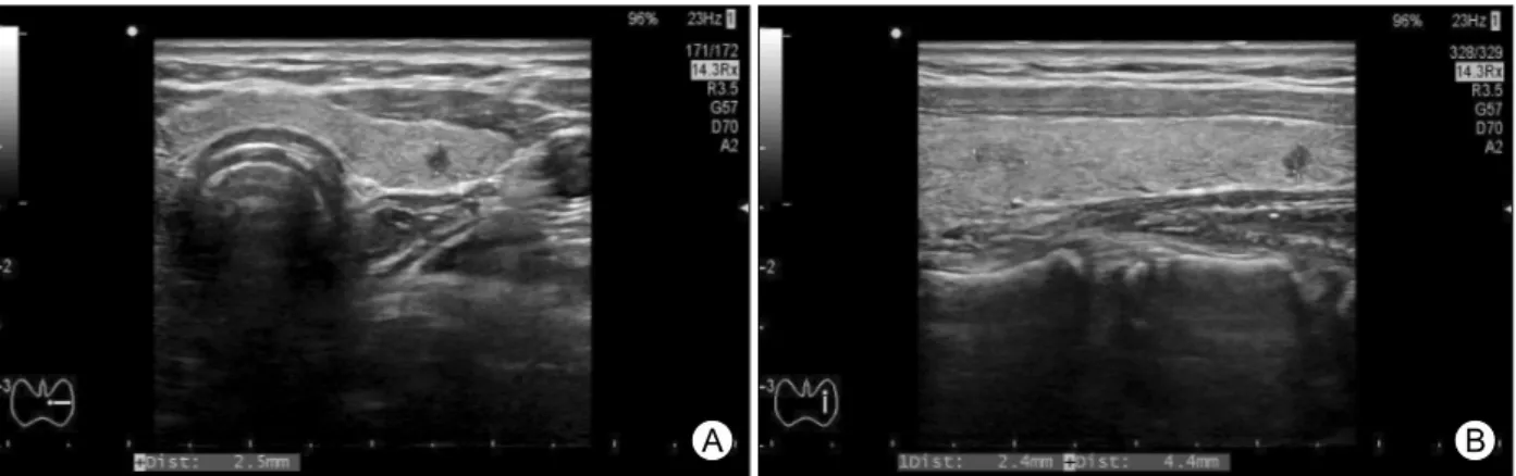
Fig. 1. TIRADS 4a mass which sized less than 0.5 centimeters. (A) Transverse view, (B) Longitudinal view. TIRADS: Thyroid Image Reporting and Data System.
한 환자를 대상으로 후향적 분석을 통해 임산부에서 갑상선유두암의 수술적 치료를 임상적으로 고찰하여, 적합한 외과적 치료 시기를 알아보고자 하였다.
대상 및 방법
2013년 5월 1일부터 2015년 4월 30일까지 만 2년간 미래여성병원 산부인과에서 시행한 총 분만 4978건 중, 임신진단을 위해 본원 외래를 첫 방문하여 스크리닝 유방/갑상선 초음파를 시행한 1863예의 환자를 대상으 로 하였다. 스크리닝 초음파를 한 환자들의 갑상선 초 음파 소견은 TIRADS (Thyroid Image Reporting and Data System) 분류11)에 따랐으며, 이 중 TIRADS 4a 이 상인 환자를 대상으로 하였다. TIRADS 4a 이상이지만 종양의 크기가 0.5 cm 미만인 경우는 추적관찰을 하였 으며(Fig. 1), 0.5 cm 이상의 경우에 한해 초음파유도하 세침흡인세포검사를 시행하였다. 이 중 병리학적 소견 상 갑상선유두암으로 진단받은 환자 17명을 대상으로 후향적 의무기록 조사를 시행하였으며, 갑상선암 진단 환자 중 임신 또는 분만 후에 갑상선절제술을 시행한 10명을 최종 대상으로 선정하였다.
환자의 의무기록을 통해 갑상선암 진단 시 임신 주 수 및 환자의 나이, 진단초음파상 크기와 경부임파선 전이 여부, 수술 시기, 진단 시기와 수술 시기의 시간 간격, 수술방법 및 술 후 방사성요오드 치료 여부, 수술 후 최종조직 결과, 임신 분기당 초음파상 병변의 크기 변화를 분석하였고, 술 후 재발 여부를 확인하였다.
환자의 임신 주수 평가는 규칙적인 월경력을 가진 경우 최종 월경일을 기준으로 하였고, 불규칙적인 월 경력을 가진 환자는 임신 초기 혹은 초진 초음파 소견 을 참고로 교정한 예정일을 기준으로 하였다.
통계분석은 SPSS 19 (SPSS Inc., Chicago, IL, USA) 를 이용하여 양측검정을 시행하였으며, Student t-test 를 이용하여 95% 신뢰구간을 기준으로 p값이 0.05 미 만인 경우를 통계적으로 유의하다고 판정하였다.
결 과
환자군의 특징
연구기간 중 미래여성병원 산부인과에서 시행한 분 만 건수는 총 4978건이었으며, 이 중 임신 확인을 위해 첫 외래 방문 시 스크리닝 유방/갑상선 초음파를 시행 한 경우는 1863예였다.
이 중 병리학적 소견상 갑상선유두암으로 진단받은 환자는 17명이었고, 갑상선절제술을 시행한 경우는 최 종 10명으로, 본원에서 7명, 타원에서 3명을 시행하였으 며 나머지 7명 중 6명은 연구기간 내에 분만 전이었고 1명은 분만 후 수술받지 않고 추적조사만 하고 있었다.
수술 환자 10명의 수술 시기는 임신 2분기 1예를 제외 한 9명의 환자가 출산 후 6개월 이내에 수술을 받았으 며, 진단 시기와 수술 시기의 시간 간격은 평균 5.87±
4.91개월이었다. 17명의 갑상선암 진단 환자의 나이는 평균 34.80±3.01세였고, 진단 당시 임신 태령은 임신 1분기 16예, 3분기 1예로 평균 임신 9.06±8.04주였다 (Table 1). 전체 17명의 환자 중 수술을 시행한 환자는 10명이었으며, 본 연구에 최종 대상으로 선정되었다.
10명의 수술 환자 모두가 갑상선전절제 및 중앙경부 림프절곽청술을 시행하였으며, 2명의 환자에서 변형근 치적경부곽청술이 추가되었다. 수술 후 방사성요오드 치료를 시행한 경우는 4예였으며, 모든 환자의 수술 전 세침흡인검사와 술 후 조직검사결과 갑상선유두암으
Table 3. Data of patients who enrolled in this study (n=10)
No. Age
(yr) Location Size*
(mm)
Node metastasis status†
Time of surgery
Surgical
procedure RAI‡
1 32 Right 28.0 Negative S§ TT c CCND∥ No
2 39 Both 26.0 Negative P¶ TT c CCND Yes
3 35 Both 11.9 Central
Lateral
P TT c mRND** Yes
4 32 Left 10.7 Negative P TT c CCND No
5 39 Left 10.0 Negative P TT c CCND No
6 34 Left 3.0 Central P TT c CCND No
7 34 Both 10.0 Negative P TT c CCND No
8 36 Right 9.0 Negative P TT c CCND No
9 37 Both 16.0 Negative P TT c CCND Yes
10 30 Both 35.0 Central
Lateral
P TT c mRND Yes
*Largest mass size at ultrasonographic diagnosis, †Pathologic finding, ‡Radioactive iodine, §Second trimester, ∥Bilateral total thyroidectomy with central compartment neck dissection, ¶Postpartum period, **Bilateral total thyroidectomy with modified radical neck dissection
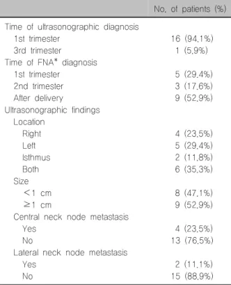
Table 1. Characteristics of patients (n=17)
No. of patients (%) Time of ultrasonographic diagnosis
1st trimester 16 (94.1%)
3rd trimester 1 (5.9%)
Time of FNA* diagnosis
1st trimester 5 (29.4%)
2nd trimester 3 (17.6%)
After delivery 9 (52.9%)
Ultrasonographic findings Location
Right 4 (23.5%)
Left 5 (29.4%)
Isthmus 2 (11.8%)
Both 6 (35.3%)
Size
<1 cm 8 (47.1%)
≥1 cm 9 (52.9%)
Central neck node metastasis
Yes 4 (23.5%)
No 13 (76.5%)
Lateral neck node metastasis
Yes 2 (11.1%)
No 15 (88.9%)
*FNA: fine needle aspiration
Table 2. Characteristics of patients who received thyroid surgery (n=10)
No. of patients (%) Timing of surgery
2nd trimester 1 (10.0%)
After delivery 9 (90.0%)
Surgical procedure
TT c CCND* 8 (80.0%)
TT c mRND† 2 (20.0%)
RAI‡
Yes 4 (40.0%)
No 6 (60.0%)
*Bilateral total thyroidectomy with central compartment neck dissection; †Bilateral total thyroidectomy with modified radical neck dissection; ‡Radioactive iodine
로 확인되었다(Table 2). 최종 선정된 환자 각각의 데이 터는 Table 3과 같다.
수술 전후 종괴 크기의 변화 및 술 후 경과
전체 수술환자 10명 중 1명은 임신 3분기에 초음파 진단되어 총 9명의 환자에서 임신 중 크기 변화를 확인
할 수 있었다. 1분기 초음파상 종괴의 크기는 평균 11.30±6.01 mm였으며, 임신 2분기와 3분기 종괴의 크 기는 각각 평균 12.74±7.79 mm, 13.82±9.93 mm였다.
수술 후 최종 병리검사상 종괴의 크기는 평균 13.84±
8.19 mm로 약간의 크기증가가 있었으나 진단 당시와 비교하면 통계적으로 차이는 없었다(Table 4, Fig. 2).
전체 수술 환자에서 수술 후 합병증은 관찰되지 않았 으며, 특별한 징후 또한 보이지 않았다. 수술 후 경과관 찰 기간은 평균 16.50±4.48개월이었고, 이 기간 동안 병변의 재발 및 환자사망은 관찰되지 않았다.
고 찰
한국인의 갑상선암은 인구 10만 명당 120.4명으로
Table 4. Change in tumor size during prenatal and postpartum period (n=9)
1st trimester 2nd trimester 3rd trimester Final pathology
Size* (mm) 11.30±6.01 12.74±7.79 13.82±9.93 13.84±8.19
p value† Reference standard 0.12 0.13 0.08
*Mean±standard deviation, †Statistical value compared with 1st trimester
Fig. 2. Ultrasonographic findings which shows an increase in the size of tumor. (A) 1st trimester, (B) 2nd trimester and (C) 3rd trimester.
알려져 있고, 일반적으로 임산부 1000명당 한 명꼴로 발견되는 것으로 보고되고 있다.1,4,5) 본원에서 진단된 임산부 갑상선암의 경우 만 2년간의 분만 환자 4978명 중 17명이 진단되어 외국사례의 유병률보다 약 3배 정 도 높은 것으로 확인되었다.
일반적인 갑상선암의 첫 번째 치료는 수술적 치료와 방사성 동위원소를 이용한 치료로 나뉜다. 이런 치료 원칙하에 수술이 임산부의 경우 임신 시 정상적인 생 리적 변화 이외에도 수술 합병증이 산모뿐 아니라 태 아에도 치명적인 영향을 줄 수 있다는 점에서 좀 더 신중한 치료 결정이 필요하다.12-15) 특히 방사성 동위원 소 치료는 일반인의 경우와 달리 임신 시 태아에 많은 양의 방사선을 노출시켜, 그로 인한 후유증으로 임산 부의 갑상선암 치료에는 금기이며, 모유에 다량의 방 사선이 축적되어 분비되므로 수유부에게도 금기되는 치료법이다. 따라서 임산부 갑상선암 치료는 수술적 치료가 유일한 치료법으로 알려져 있다.16-18)
임산부의 갑상선암 수술 시기에 대한 의견은 크게 임신 중과 출산 후로 나뉜다. 임신 중 수술을 해야 한다 는 측의 주장은 임신에 의한 여러 가지 생리학적 변화 가 갑상선에 자극을 주기 때문이다. 임신 중 갑상선자 극호르몬과 구조적으로 유사한 사람융모성 고나도트 로핀의 증가로 인해 갑상선암 조직의 자극이 증가되고 이는 임신 중 갑상선암의 급격한 크기 증가와 관련이 있다고 주장한다.19) 반대로 출산 후로 수술시기를 연기 해야 한다는 주장의 근거는 다음과 같다. 첫째, 임신 2분기 때의 수술적 치료는 모체의 갑상선기능저하를
가져오고, 이는 저체중아 및 기형아 출산과 신생아의 인지능력 저하 등을 유도하기 때문이다. 둘째, 전신 마 취제에 의한 태아의 기관형성(organogenesis) 장애유발 및 자연유산, 조산 등의 위험성이 증가되기 때문이 다.14) 이상과 같은 이유로 임신 중 갑상선암의 수술은 그 시기에 있어 많은 논란이 있다.6-9,20,21)
본원에서는 임신 중 분화갑상선암이 발견된 환자에 서 경부림프절 전이가 없거나 공격적인 병의 진행을 보이지 않는 경우는 수술적 치료를 분만 후로 연기하 고 있다. 이에 본 연구는 임신 중과 출산 후 기간 동안 의 갑상선 병변 크기의 변화 및 술 후 최종 병변의 크 기를 비교 분석하여 임신이 병변의 변화에 어떠한 영 향을 미치는지 알아보고, 수술 후 추적검사를 통해 예 후를 분석하여 그 수술 시기를 출산 후로 연기하는 것 이 옳은 것인지에 대한 방침을 제시하고자 한다.
이전의 여러 연구를 통하여 보면 Messuti 등22)은 임 신이 분화갑상선암의 예후에 부정적인 영향을 준다고 하였으나, 에스트로겐 수용체, 프로게스테론 수용체, 아로마타제, NIS유전자, BRAFV600E 변이가 모두 병변 의 병태생리에 관여하지 않아 그에 대한 연구가 필요 하다 하였고, Hirsch 등23)은 출산 후 갑상선암 수술 기 간과 임신과의 상관관계를 연구한 결과 임신 시 병변 의 진행을 의심할 수 있는 소견이 관찰되나 임신 자체 가 갑상선암의 재발을 유도하지 않는다고 하였다. 그 들이 관찰한 생물학적/방사선학적으로 병변의 진행을 보이는 환자들은 이미 임신 전부터 공격적인 성향을 지닌 종양이었다고 하며, 질병 자체의 과거력이 더욱
중요하다 하였다. Yasmeen 등24)의 연구에서는 임신 자 체가 갑상선암의 예후에 영향을 주지 않는다고 하며, 임신 중 갑상선절제술이 산모/태아에 악영향을 주지 않는다고 하였다. Vini 등7)은 평균 28세의 산모를 대상 으로 한 연구에서 갑상선암이 진단된 지 1년 이후에 수술을 하는 경우 1년 이내에 하는 것에 비해 나쁜 예 후를 가진다고 하였으며, 초기임신에 진단된 경우 임 신 2분기, 임신 후반기에 진단된 경우 출산 후로 그 수 술 시기를 정해야 한다고 하였다. 그에 반해, Moosa 등9) 은 동일한 연령대 환자들의 경우 임산부와 비임산부의 예후는 차이가 없어 수술 시기를 출산 후로 연기하는 것이 옳다고 하였다. 또한 Shindo 등25)은 미세유두암 임산부 환자 중 44%에서 임신 중 크기의 증가가 관찰 되어 비임산부 환자에 비해 통계적으로 의미 있는 차 이를 나타내었으나, 예후에는 관련이 없다는 보고를 하였으며, Nam 등26)도 임신 2분기 때나 유산 후 수술 하는 경우에 비해 출산 후로 수술을 연기하는 경우가 종양의 크기를 약간 증가시키지만 수술 시간, 수술관 련 합병증, 치료결과 등이 차이를 나타내지 않아 출산 후 수술을 권장한다고 하였다. 본 연구에서도 마찬가 지로 임신 중 종양의 크기는 증가되는 양상을 보였으 나, 통계적으로 유의하지 않아 임신 자체가 병의 예후 에 영향을 주지 않는다고 할 수 있었다.
본 연구에서 관찰된 임상적 변화와 수술 결과의 비 교분석을 통해 임신 중 갑상선유두암의 변화는 통계적 인 의미를 나타내지 않았다. 또한 수술 후 추적검사에 서 전신 재발 혹은 갑상선암에 의한 사망은 단 한 예도 나타나지 않아 전체 생존 기간에는 영향을 미치지 않 았다. 따라서, 임신 중 발견된 갑상선유두암의 수술적 치료는 모체와 태아에 합병증을 발생 시킬 수 있다는 점에서 볼 때 수술 시기를 출산 후로 연기하는 것을 고려할 수 있겠으며, 좀 더 많은 환자를 대상으로 한 추가적인 연구가 필요할 것으로 생각된다.
결 론
본 연구를 통해 임신 중 진단된 갑상선유두암의 수 술을 출산 후로 연기한 예에서 종양의 크기는 약간 증 가하였지만 예후에는 변화가 없었고, 임신 중 수술에 의한 합병증을 피할 수 있으므로, 출산 이후로 수술을 연기하는 것이 좋을 것으로 생각된다.
중심 단어: 임신, 갑상선암, 수술시기.
References
1) Jung KW, Won YJ, Kong HJ, Oh CM, Cho H, Lee DH, et al. Cancer statistics in Korea: incidence, mortality, survival, and prevalence in 2012. Cancer Res Treat 2015;47(2):127-41.
2) Cho BY, Choi HS, Park YJ, Lim JA, Ahn HY, Lee EK, et al. Changes in the clinicopathological characteristics and outcomes of thyroid cancer in Korea over the past four decades.
Thyroid 2013;23(7):797-804.
3) Machens A, Holzhausen HJ, Dralle H. The prognostic value of primary tumor size in papillary and follicular thyroid carcinoma. Cancer 2005;103(11):2269-73.
4) Donegan WL. Cancer and pregnancy. CA Cancer J Clin 1983;33(4):194-214.
5) Akslen LA, Haldorsen T, Thoresen SO, Glattre E. Incidence of thyroid cancer in Norway 1970-1985. Population review on time trend, sex, age, histological type and tumour stage in 2625 cases. APMIS 1990;98(6):549-58.
6) Hod M, Sharony R, Friedman S, Ovadia J. Pregnancy and thyroid carcinoma: a review of incidence, course, and prognosis.
Obstet Gynecol Surv 1989;44(11):774-9.
7) Vini L, Hyer S, Pratt B, Harmer C. Management of differentiated thyroid cancer diagnosed during pregnancy. Eur J Endocrinol 1999;140(5):404-6.
8) Herzon FS, Morris DM, Segal MN, Rauch G, Parnell T.
Coexistent thyroid cancer and pregnancy. Arch Otolaryngol Head Neck Surg 1994;120(11):1191-3.
9) Moosa M, Mazzaferri EL. Outcome of differentiated thyroid cancer diagnosed in pregnant women. J Clin Endocrinol Metab 1997;82(9):2862-6.
10) Yoshimura M, Hershman JM. Thyrotropic action of human chorionic gonadotropin. Thyroid 1995;5(5):425-34.
11) Horvath E, Majlis S, Rossi R, Franco C, Niedmann JP, Castro A, et al. An ultrasonogram reporting system for thyroid nodules stratifying cancer risk for clinical management. J Clin Endocrinol Metab 2009;94(5):1748-51.
12) Mazzaferri EL. Approach to the pregnant patient with thyroid cancer. J Clin Endocrinol Metab 2011;96(2):265-72.
13) Owen RP, Chou KJ, Silver CE, Beilin Y, Tang JJ, Yanagisawa RT, et al. Thyroid and parathyroid surgery in pregnancy. Eur Arch Otorhinolaryngol 2010;267(12):1825-35.
14) Fanarjian N, Athavale SM, Herrero N, Fiorica J, Padhya TA.
Thyroid cancer in pregnancy. Laryngoscope 2007;117(10):
1777-81.
15) Gibelli B, Zamperini P, Proh M, Giugliano G. Management and follow-up of thyroid cancer in pregnant women. Acta Otorhinolaryngol Ital 2011;31(6):358-65.
16) American Thyroid Association (ATA) Guidelines Taskforce on Thyroid Nodules and Differentiated Thyroid Cancer, Cooper DS, Doherty GM, Haugen BR, Kloos RT, Lee SL, et al. Revised American Thyroid Association management guidelines for patients with thyroid nodules and differentiated thyroid cancer. Thyroid 2009;19(11):1167-214.
17) Gorman CA. Radioiodine and pregnancy. Thyroid 1999;9(7):
721-6.
18) Imran SA, Rajaraman M. Management of differentiated thyroid cancer in pregnancy. J Thyroid Res 2011;2011:549609.
19) Kung AW, Chau MT, Lao TT, Tam SC, Low LC. The effect of pregnancy on thyroid nodule formation. J Clin Endocrinol Metab 2002;87(3):1010-4.
20) Kobayashi K, Tanaka Y, Ishiguro S, Mori T. Rapidly growing thyroid carcinoma during pregnancy. J Surg Oncol 1994;55(1):
61-4.
21) Rosen IB, Walfish PG. Pregnancy as a predisposing factor in thyroid neoplasia. Arch Surg 1986;121(11):1287-90.
22) Messuti I, Corvisieri S, Bardesono F, Rapa I, Giorcelli J, Pellerito R, et al. Impact of pregnancy on prognosis of differentiated thyroid cancer: clinical and molecular features. Eur J Endocrinol 2014;170(5):659-66.
23) Hirsch D, Levy S, Tsvetov G, Weinstein R, Lifshitz A, Singer J, et al. Impact of pregnancy on outcome and prognosis of survivors of papillary thyroid cancer. Thyroid 2010;20(10):
1179-85.
24) Yasmeen S, Cress R, Romano PS, Xing G, Berger-Chen S, Danielsen B, et al. Thyroid cancer in pregnancy. Int J Gynaecol Obstet 2005;91(1):15-20.
25) Shindo H, Amino N, Ito Y, Kihara M, Kobayashi K, Miya A, et al. Papillary thyroid microcarcinoma might progress during pregnancy. Thyroid 2014;24(5):840-4.
26) Nam KH, Yoon JH, Chang HS, Park CS. Optimal timing of surgery in well-differentiated thyroid carcinoma detected during pregnancy. J Surg Oncol 2005;91(3):199-203.