bloodresearch.or.kr Blood Res 2013;48:292-303.
Letters to the Editor 293
to promptly diagnose any recurrence of her hematological complaints. Thus, we report a rare and unexplained associa- tion of primary mixed AIHA with acute SVT, both of idio- pathic origin that simultaneously occurred in a young patient.
Laura Scaramucci, Marco Giovannini, Pasquale Niscola, Alessio Perrotti, Paolo de Fabritiis
Haematology Division, S. Eugenio Hospital, Rome, Italy Correspondence to: Laura Scaramucci Hematology Division, S. Eugenio Hospital, Piazzale dell’Umanesimo 10, 00144, Rome, Italy
E-mail: scaramucci.laura@gmail.com
Received on Jul. 26, 2013; Revised on Sep. 9, 2013; Accepted on Nov. 21, 2013 http://dx.doi.org/10.5045/br.2013.48.4.292
AuthorsÊ Disclosures of Potential Conflicts of Interest No potential conflicts of interest relevant to this article were reported.
REFERENCES
1. Mayer B, Yurek S, Kiesewetter H, Salama A. Mixed-type auto- immune hemolytic anemia: differential diagnosis and a critical review of reported cases. Transfusion 2008;48:2229-34.
2. Sudha Reddy VR, Samayam P, Ravichander B, Bai U. Autoim- mune hemolytic anemia: mixed type-a case report. Indian J Hematol Blood Transfus 2011;27:107-10.
3. Hoffman PC. Immune hemolytic anemia-selected topics.
Hematology Am Soc Hematol Educ Program 2009:80-6.
4. Tsiopoulos FD, Manolakis AC, Kapsoritakis AN, Psychos AK, Potamianos SP. Autoimmune hemolytic anemia and ophthalmic artery thrombosis preceding the intestinal manifestations of Crohn's disease. Inflamm Bowel Dis 2009;15:487-8.
5. Kowal-Vern A, Radhakrishnan J, Goldman J, Hutchins W, Blank J. Mesenteric and portal vein thrombosis after splenectomy for autoimmune hemolytic anemia. J Clin Gastroenterol 1988;10:
108-10.
Successful treatment of steroid resistant hypereosinophilic syn- drome with low-dose CsA
TO THE EDITOR: Although much is known about hyper- eosinophilic syndrome (HES), the notable discovery of a few genetic rearrangements, such as FIP1L1/PDGFRA (F/P) and TEL/ PDGFRB, and the identification of a phenotypi- cally aberrant clonal T lymphocyte brought a new paradigm to HES. As a result, multidisciplinary groups of hematologists
and scientists have created more stratified treatment guide- lines for patients with HES.
However, more than half of patients with HES are still classified as undefined under the current diagnostic criteria and the best course of treatment for these patients remains unclear, especially after initial steroid treatments fail. Here, we present two cases of undefined HES who were success- fully treated with cyclosporine A (CsA) after corticosteroid treatment failed.
CASE 1
A 41-year-old man visited our emergency room with rashes on both legs and his trunk area.
On physical examination, only a skin lesion was found.
His complete blood count (CBC) showed hypereosinophilia (eosinophils 4,730/mm3). Although he had no history of allergies, a previous CBC reports showed persistent hyper- eosinophilia over the previous 6 months.
Subsequent analyses did not reveal any evidence of secon- dary HES including any autoimmune disease or parasitic infestation. Bone marrow aspirate and biopsy showed marked eosinophilia without dyspoiesis. FIP1L1-PDGFRA, TEL-PDGFRB, or BCR-ABL rearrangement was not detected on fluorescence in situ hybridization. JAK2 V617F mutation analysis performed with a reverse transcription polymerase chain reaction technique was negative. Serum immunoglob- ulin E (IgE) level (1,599 mg/dL) and eosinophilic cationic protein level (163.35 ng/mL) were elevated.
Skin biopsy of the leg rash showed perivascular lympho- histiocytic infiltration and many eosinophils. Flow cyto- metric analysis to evaluate the associated aberrant T-lym- phocyte found no abnormal phenotypes such as CD3-CD4+
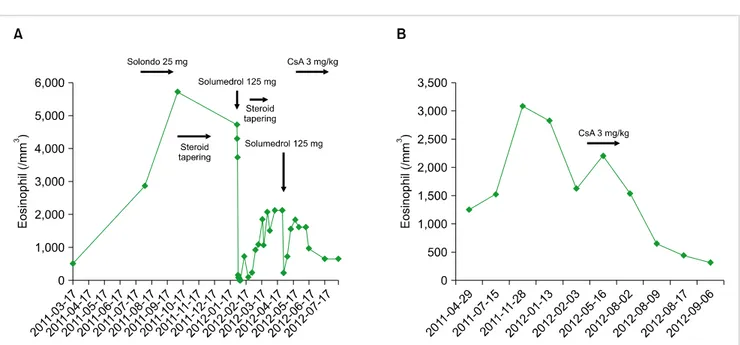
or CD3+CD4-CD8-, but the T cell receptor (TCR) gene rearrangement was not checked. Although we could not confirm the subclass of HES, clinical findings indicated a lymphocytic variant of HES (L-HES). The patient initially responded well to a high-dose glucocorticoid treatment, but after reducing prednisone, the eosinophil count and IgE level rebounded. Thus, we administered a low dose of CsA (100 mg bid), and his eosinophil count returned to normal after just 1 week of treatment. Prednisone was then tapered to 10 mg per day (Fig. 1A).
CASE 2
A 41-year-old man who had been suffering from intract- able eosinophilic pustular folliculitis for 5 years was referred to our hospital. Skin biopsy from the lesion showed peri- vascular and periadnexal eosinophilic infiltration. He had no history of allergies. Although he had been taking dapsone, prednisone, and an antihistamine since 2008, his skin lesion had waxed and waned and the CBC showed persistent hyper- eosinophilia for over 1 year. The laboratory results showed an eosinophil count of 1,790/mm3 and a serum IgE level of 119 IU/mL. Nevertheless, there was no evidence of secon- dary HES. Sequentially performed bone-marrow aspiration and biopsy showed hyperplasia of the eosinophilic lineage,
Blood Res2013;48:292-303. bloodresearch.or.kr
294 Letters to the Editor
Fig. 1. Changes in the peripheral blood eosinophil count of case 1 (A) and case 2 (B) over time in relation to the treatment.
but the molecular analysis did not show any specific aberrant genetic mutations consistent with the myelocytic variant of HES (M-HES). Unfortunately, we did not analyze the T cell phenotype and T cell clonality. On physical examina- tion, only skin manifestations were found. The patient could not be categorized as L-HES according to the laboratory results; however, considering the relatively gentle clinical course and absence of cardiac involvement, we selected an immune-modulating drug as a second line treatment rather than a cytotoxic agent or imatinib. We administered a low dose of CsA (100 mg bid), and his eosinophil count returned to normal after 1 week of treatment. In addition, his skin lesion disappeared (Fig. 1B).
DISCUSSION
Previously, the diagnosis of HES was usually based on exclusion and treatment included mostly steroids and cyto- toxic agents. With the improved understanding of the patho- genesis of HES, distinguishing M-HES from non-M-HES and analyzing the F/P rearrangement have become pre- requisites for optimizing HES treatment. However, steroids still play a major role as the first line treatment for HES, except for F/P positive HES, which should be treated with imatinib.
Corticosteroids are known to interfere with the tran- scription of pro-inflammatory genes necessary for eosinophil maturation, proliferation, migration, and chemo-attraction.
Because these mechanisms perform diverse actions, cortico- steroids have been used for decades to treat HES, irrespective of the subtype. However, the accompanying side effects of systemic corticosteroids preclude long-term usage and some patients with HES respond only to a high dose of steroids or are resistant to steroid treatment. Nevertheless, treatment guidelines after failure or intolerance of gluco-
corticoid therapy for HES have not been established.
In a review of the literature on HES treatment, previous reports mention various second lines of treatment including hydroxyurea, leukapheresis, vincristine, 6-mercaptopurine, IFN-α, or allogenic bone marrow transplantation, but most were case studies or retrospective analyses.
According to the largest study, a retrospective analysis on HES treatment, 163 patients of 188 (81%) without FP rearrangement received corticosteroids as the initial ther- apy; 85% of these patients experienced a favorable response [1]. However, 42% of these patients eventually had to dis- continue steroid use or add another drug because of treat- ment failure or steroid toxicity. Furthermore, the most fre- quently combined agent was hydroxyurea. Therefore, con- sidering the different prognoses and natural course of M-HES and L-HES, different approaches to treatment are needed to avoid unnecessary exposure to cytotoxic drugs.
Since Cogan et al. discovered the aberrant T lymphocyte clone in one patient with HES with a high interleukin 5 level, L-HES was defined as a lymphoid disorder caused by a sustained over-production of eosinopoietic cytokines such as interleukin 5 from T-lymphocytes that have under- gone clonal expansion or aberrant T-lymphocytes [2].
Furthermore, these discoveries of a lymphocyte-based path- ogenetic mechanism aroused attention regarding an im- mune-mediated treatment approach targeting lymphocytes for the treatment of L-HES. Compared with other im- mune-modulating drugs such as mepolizumab or IFN-α, CsA has not received much attention for the treatment of HES. However, systemic toxicity and a possible inhibitory effect on apoptosis of aberrant CD3-CD4+ T cells became an obstacle for the widespread application of IFN-α in the treatment of L-HES [3-5]. Thus, further studies are needed to link the anti-apoptotic effect on aberrant T cells with
bloodresearch.or.kr Blood Res 2013;48:292-303.
Letters to the Editor 295
subsequent lymphoma. Although a few prospective studies on mepolizumab recently showed positive results, high cost can be a limitation in widespread use and the results of long-term use along with the toxicity profile are still un- known [6, 7]. CsA, a calcineurine inhibitor, has been used to prevent rejection after organ transplant because of its inhibitory effect on interleukin 2 production of T cells.
The feasible toxicity profile and effective T-lymphocyte sup- pression properties of CsA made this drug applicable to many T lymphocyte-related diseases. However, not many studies have investigated the use of CsA for the treatment of HES; the first report was in 1996 [8]. A 7-year-old girl presented with a maculopapular rash with erosion over her entire body with hypereosinophilia and hyper IgE. Because she could not maintain a high dose of steroids and had a cushingoid appearance, 3 mg/kg of CsA was administered.
Just one week after CsA administration, the eosinophil count returned to normal and she was weaned off corticosteroid use at an increment dose of up to 8 mg/kg. In 2009, the first adult case was reported. A 63-year-old man was diag- nosed with L-HES by performing TCR gene rearrangement [9]. The patient did not respond to initial prednisone therapy or subsequent hydroxyurea. However, a low dose of CsA (100 mg/day) was attempted and a complete resolution was achieved. In our two cases, both patients also showed a favorable response 1 week after CsA administration and a 3–4 mg/kg dose was enough for to control the disease.
Although we could not categorize our two cases into an HES subtype, a number of clinical features caused us to treat them as L-HES cases; thus, CsA was selected as the second line of treatment.
Firstly, both cases did not show an F/P rearrangement and were partially responsive to the initial steroid treatment.
Secondly, neither patient had M-HES features such as hep- atosplenomegaly, cardiac eosinophilic involvement, or a high vitamin B12 level. Lastly, both cases showed dermato- logic manifestations. While an effort to find reliable clinical indicators that predict treatment response and help classify the subtype of HES has continued, a few parameters were suggested and some were accepted as minor criteria in differ- entiating between the subclasses of HES, such as serum thymus and regulated chemokine, IgE, interleukin 5, tryp- tase level, and organ involvement patterns [10, 11]. Even if these factors are not absolute indicators, a combination of these clinical parameters is expected to help predict treat- ment outcomes and select optimal drugs in patients with undefined HES.
Moreover, further prospective investigation is needed to clarify the effectiveness of CsA in non-M-HES patients;
however, CsA may be a feasible and effective second-line drug for L-HES and patients with undefined HES with sim- ilar features.
Yun Hwa Jung1, Sang Bong Han2, Young Jae Park1, In Sook Woo1, Baik Kee Cho3, Chi Wha Han1
1Division of Hematology-Oncology, Department of Internal Medicine, 2Department of Laboratory Medicine,
3Department of Dermatology, St. Mary’s Hospital, The Catholic University of Korea College of Medicine, Seoul, Korea Correspondence to: Yun Hwa Jung Division of Hematology-Oncology, Department of
Internal Medicine, Yeouido St. Mary’s Hospital, 10, 63-ro, Yeongdeungpo-gu, Seoul 150-713, Korea
E-mail: ksdmz@hanmail.net
Received on Jul. 23, 2013; Revised on Nov. 1, 2013; Accepted on Nov. 21, 2013 http://dx.doi.org/10.5045/br.2013.48.4.293
AuthorsÊ Disclosures of Potential Conflicts of Interest No potential conflicts of interest relevant to this article were reported.
REFERENCES
1. Ogbogu PU, Bochner BS, Butterfield JH, et al. Hypereosinophilic syndrome: a multicenter, retrospective analysis of clinical char- acteristics and response to therapy. J Allergy Clin Immunol 2009;124:1319-25.
2. Cogan E, Schandene L, Crusiaux A, Cochaux P, Velu T, Goldman M. Brief report: clonal proliferation of type 2 helper T cells in a man with the hypereosinophilic syndrome. N Engl J Med 1994;330:535-8.
3. Butterfield JH. Interferon treatment for hypereosinophilic syn- dromes and systemic mastocytosis. Acta Haematol 2005;114:
26-40.
4. Schandene L, Roufosse F, de Lavareille A, et al. Interferon alpha prevents spontaneous apoptosis of clonal Th2 cells associated with chronic hypereosinophilia. Blood 2000;96:4285-92.
5. Klion AD, Bochner BS, Gleich GJ, et al. Approaches to the treat- ment of hypereosinophilic syndromes: a workshop summary report. J Allergy Clin Immunol 2006;117:1292-302.
6. Rothenberg ME, Klion AD, Roufosse FE, et al. Treatment of pa- tients with the hypereosinophilic syndrome with mepolizumab.
N Engl J Med 2008;358:1215-28.
7. Roufosse F, de Lavareille A, Schandene L, et al. Mepolizumab as a corticosteroid-sparing agent in lymphocytic variant hyper- eosinophilic syndrome. J Allergy Clin Immunol 2010;126:828- 35.
8. Nadarajah S, Krafchik B, Roifman C, Horgan-Bell C. Treatment of hypereosinophilic syndrome in a child using cyclosporine: im- plication for a primary T-cell abnormality. Pediatrics 1997;99:
630-3.
9. Donald CE, Kahn MJ. Successful treatment of hypereosinophilic syndrome with cyclosporine. Am J Med Sci 2009;337:65-6.
10. Roufosse F. Hypereosinophilic syndrome variants: diagnostic and therapeutic considerations. Haematologica 2009;94:1188- 93.
11. Klion AD. Recent advances in the diagnosis and treatment of hy- pereosinophilic syndromes. Hematology Am Soc Hematol Educ Program 2005:209-14.