Recurrent Pneumomediastinum and Subcutaneous Emphysema Complicating Chronic Graft versus Host Disease after Allogeneic Bone Marrow Transplantation
Na-Ri Lee, M.D., Tae-Hwan Lee, M.D., Eun-Kee Song, M.D., Jae-Yong Kwak, M.D. and Chang-Yeol Yim, M.D.
Department of Internal Medicine, Chonbuk National University Medical School, Jeonju, Korea
Several noninfectious pulmonary complications can be associated with chronic graft versus host disease (GVHD). Obstructive airway disease can be a clinical feature of chronic GVHD and the histopathology reveals characteristic lesions of bronchiolitis obliterans. Bronchiolitis obliterans is an obstructive pulmonary disorder affecting the small airways, and it was first described as a late complication of allogeneic bone marrow transplantation (BMT). Spontaneous pneumomediastinum and subcutaneous emphysema can occur in the setting of severe bronchiolitis obliterans and only rarely are they the first sign of such disease. We describe here a case of a 27-year old woman who developed recurrent pneu- momediastinum and subcutaneous emphysema that were secondary to the bronchiolitis obliterans that complicated chronic GVHD after allogeneic BMT. (Korean J Hematol 2006;41:61-65.)
Key Words: Pneumomediastinum, Subcutaneous emphysema, Bronchiolitis obliterans, Chronic graft versus host disease, Allogeneic bone marrow transplantation
61
Correspondence to: Chang-Yeol Yim, M.D., Ph.D.
Department of Internal Medicine, Chonbuk National University Medical School
San 2-20, Geumam-dong, Deokjin-gu, Jeonju 561-180, Korea Tel: +82-63-250-1682, Fax: +82-63-254-1609
E-mail: cyyim@chonbuk.ac.kr
접수:2006년 1월 4일, 수정:2006년 2월 1일 승인:2006년 2월 9일
교신저자:임창열, 전북 전주시 덕진구 금암동 산 2-20
561-180, 전북대학교 의과대학 내과학교실 Tel: 063-250-1682, Fax: 063-254-1609
E-mail: cyyim@chonbuk.ac.kr INTRODUCTION
Half of all patients who undergo allogeneic bone marrow transplantation (BMT) achieve long- term disease free survival, but a similar number develop significant complications. Pulmonary com- plications remain frequent cause of post-tran- splantation mortality.1,2)
Bronchiolitis obliterans (BO), a nonspecific in- flammatory injury affecting the small airways primarily, has been reported to occur between 5~
11% in long term survivors of BMT with chronic
graft versus host disease (GVHD).3) It can com- prise a spectrum of pathology ranging from mild histological changes to severe pulmonary function abnormalities and cripping airway obstruction leading to death. Pneumothorax, pneumomedias- tinum, and subcutaneous emphysema are consi- dered as rare complications of BO.4)
Here we describe a patient with BO presenting with recurrent spontaneous pneumomediastinum, and subcutaneous and emphysema complicating chronic GVHD after allogeneic BMT.
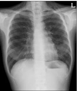
Fig. 1. Plain chest radiograph showing subcutaneous em- physema in the neck and upper trunk and pneumomed- iastinum.
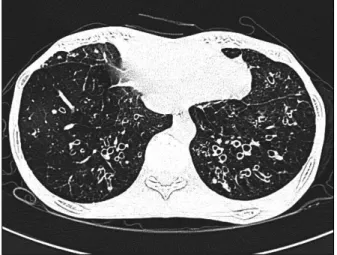
Fig. 2. High resolution CT of the chest showing the sub- cutaneous emphysema and pneumomediastinum at first event.
CASE REPORT
A 27-year old woman underwent related allo- geneic BMT for chronic phase of chronic myeloid leukemia in October 2003. She was diagnosed with chronic phase of chronic myeloid leukemia three months before, when she was presented with abdominal discomfort and headache. She was initially treated with imatinib mesylate for two months. She underwent allogeneic BMT from her brother (HLA full matched) after conditio- ning with intravenous busulfan (from days -7 to -4; 0.8mg/kg q 6hours per day) and cyclophos- phamide (from days -3 to -2; 60mg/kg per day).
Post-transplant GVHD prophylaxis was treated with cyclosporine and short-course methotrexate.
Pulmonary function tests that were made as a routine before the transplantation showed normal test parameters including spirometry, lung volu- mes, diffusion, and airway resistance. The course of the transplantation was uneventful, with good engraftment, and the patient was sent home after three weeks of hospitalization. Five weeks after
BMT, she developed jaundice and skin rash on her face and trunk which was proven as acute GVHD and were treated high dose steroids. Since then, she continued to have chronic GVHD in- volving skin, gut, liver and was treated steroids, cyclosporine and PUVA. Five months after BMT, she developed dyspnea, nonproductive cough and chest pain abruptly. Physical examination at that times showed crepitus involving the neck and anterior chest due to subcutaneous emphysema.
Pulmonary function tests showed severe obstruc- tive pattern of small airway. Chest radiographs demonstrated subcutaneous emphysema and pn- eumomediastinum (Fig. 1). A High resolution CT scan of the chest demonstrated pneumomedias- tinum and subcutaneous emphysema (Fig. 2). We could not find other causes of air-leak syndrome and the diagnosis of BO was made. Further more, chronic GVHD of the skin continued. Subsequ- ently she was added mycofenolate mofetil and treated conservatively including high-flow oxy- gen. Subcutaneous emphysema and pneumomed- iastinum was near completely resolved. After three weeks, subcutaneous emphysema and pneu- momediastinum was relapsed more extensively (Fig. 3). She was treated with tacrolimus instead
Fig. 3. High resolution CT of the chest showing the recurred subcutaneous emphysema and pneumomediastinum.
Fig. 4. High resolution CT of the chest showing bronchial wall thickening and dilatation in the both lung field at one year later.
of cyclosporine. After two weeks, her pulmonary condition was improved and subcutaneous em- physema and pneumomediastinum was comp- letely resolved. Her pulmonary condition con- tinued to mildly worsen with immunosuppressive therapy but air leak syndrome did not recur during a follow-up period. One year later, fol- lowing a high resolution CT scan of the chest demonstrated typical pattern of BO; There was striking dilatation of subsegmental airways in mainly both lower lung field (Fig. 4). She died at home due to sudden respiratory arrest approxi- mately 21 months after transplantation.
DISCUSSION
Chronic airflow obstruction is the most com- mon late complication of allogeneic hemato- poietic stem cell transplantation, typically occur- ring beyond the third month and associated with underlying chronic GVHD in the majority of cases.5,6) The incidence in the allogeneic popula- tion varies between 6 and 26% in published series. The underlying process accounting for airflow obstruction in the majority of cases is BO.6)
BO was first recognized in 1982 by Roca et al.,
who described a patient with chronic GVHD after allogeneic BMT for aplastic anemia, and who died of BO.7) In addition to chronic GVHD, risk factors for development of OB include recipient old age, low serum immunoglobulin levels, use of methotrexate, and history of a respiratory viral infection within the first 100 days.2,5) The etiology remains obscure. However, the strong association with chronic GVHD and the presence of un- derlying BO suggests that the bronchial epithe- lium may be the target of immune mediated injury induced by donor cytotoxic T cells.8) The onset of OB is often insidious. Presenting symptoms include nonproductive cough, dyspnea, and wheezing. Pulmonary function tests often reveal a moderate to severe airway obstruction as indicated by FEV1/FVC ratios close to 50%.9) The chest radiograph is commonly normal, but high-resolution CT often demonstrates evidence of air trapping, hypoattenuation, florid bronchial wall thickening and dilatation.10) The diagnosis is established by demonstration of persistent airflow obstruction on simple spirometry and exclusion of other causes. The yield of transbronchial biopsies is low and the utility of bronchoscopy is limited to its role in excluding infectious causes.
Surgical lung biopsy is rarely indicated.
Disease progression is variable, so some pa- tients experience a rapid decline in lung function whereas others have a more protracted course. In the study by Clark et al., overall mortality was 65% in patients with obstructive lung disease at 3 years after transplantation. There is no standar- dized approach for the treatment of BO in post BMT patients. The treatment has traditionally involved increased immunosuppression. Increased immunosuppression with cyclosporine or pre- dnisone has been shown to produce stabilization of the clinical condition in several studies.11) A number of other therapies have been suggested, such as antithymocyte globulin, extracorporeal photophoresis, hydroxychloroquine, mycofenylate, thalidomide and rapamycin.6,11) Lung transplanta- tion is an option for select patients with severe BO who are free of other significant comorbid conditions.12)
Spontaneous pneumomediastinum and subcu- taneous emphysema secondary to BO are unusual complications after allogeneic BMT. Multiple pleural bullae have been observed in some of these patients.13) Severe obstruction of the termi- nal bronchioles can lead to air trapping and de- velopment of the bullae. Infectious origin should be also considered on the differential diagnosis of those air leak syndromes.14)
Here we describe a patient with BO presenting with recurrent spontaneous pneumomediastinum, subcutaneous emphysema complicating chronic GVHD after allogeneic BMT for chronic phase of chronic myeloid leukemia. Unlikely the majority of patients presenting with BO, her symptomatic onset is abrupt and air leak syndrome is recurrent in spite of additional immunosuppressive therapy.
This case again highlights the irreversible nature of this disease and the poor response to con- ventional therapy. Avoidance of risk factors and the development of new modalities of therapy associated with OB after allogeneic BMT would help to decrease morbidity and mortality.
요 약
저자들은 만성골수성백혈병으로 동종골수이식을 시 행받은 환자에서 만성이식편대숙주질환에 합병해 발 생한 재발성 자발성 기종격동과 피하기종으로 인해 병 발한 폐색성 세기관지염을 진단했던 드문 1예를 경험 하여 보고하는 바이다.
REFERENCES
1) Krowka MJ, Rosenow EC 3rd, Hoagland HC. Pulmo- nary complications of bone marrow transplantation.
Chest 1985;87:237-46.
2) Dudek AZ, Mahaseth H, DeFor TE, Weisdorf DJ.
Bronchiolitis obliterans in chronic graft-versushost disease: analysis of risk factors and treatment out- comes. Biol Blood Marrow Transplant 2003;9:657- 66.
3) Crawford SW, Clark JG. Bronchiolitis associated with bone marrow transplantation. Clin Chest Med 1993;
14:741-9.
4) Galanis E, Litzow MR, Tefferi A, Scott JP. Spontane- ous pneumomediastinum in a patient with bron- chiolitis obliterans after bone marrow transplanta- tion. Bone Marrow Transplant 1997;20:695-6.
5) Chien JW, Martin PJ, Gooley TA, et al. Airflow obs- truction after myeloablative allogeneic hematopoietic stem cell transplantation. Am J Respir Crit Care Med 2003;168:208-14.
6) Kotloff RM, Ahya VN, Crawford SW. Pulmonary complications of solid organ and hematopoietic stem cell transplantation. Am J Respir Crit Care Med 2004;170:22-48.
7) Roca J, Granena A, Rodriguez-Roisin R, Alvarez P, Agusti-Vidal A, Rozman C. Fetal airway disease in an adult with chronic graft-versus-host disease. Thorax 1982;37:77-8.
8) Holland HK, Wingard JR, Beschorner WE, Saral R, Santos GW. Bronchiolitis obliterans in bone marrow transplantation and its relationship to chronic graft- v-host disease and low serum IgG. Blood 1988;72:
621-7.
9) Kumar S, Tefferi A. Spontaneous pneumomediast- inum and subcutaneous emphysema complicationing bronchiolitis obliterans after allogeneic bone marrow transplantation - case report and review of literature.
Ann Hematol 2001;80:430-5.
10) Yu DFQC, Desai SR. Lung complications in patients
undergoing bone marrow transplantation. Imaging 2002;14:272-7.
11) Sanchez J, Torres A, Serrano J, et al. Long-term follow-up of immunosuppressive treatment for obs- tructive airways disease after allogeneic bone marrow transplantation. Bone Marrow Transplant 1997;20:
403-8.
12) Rabitsch W, Deviatko E, Keil F, et al. Successful lung transplantation for bronchiolitis obliterans after allogeneic marrow transplantation. Transplantation 2001;71:1341-3.
13) Suzuki T, Saijo Y, Ebina M, et al. Bilateral pneu- mothoraces with multiple bullae in a patient with asymptomatic bronchiolitis obliterans 10 years after bone marrow transplantation. Bone Marrow Trans- plant 1999;23:829-31.
14) Merino JM, Diaz MA, Ramirez M, Ruano D, Madero L. Complicated pulmonary aspergillosis with pneu- mothorax and pneumopericardium in a child with acute lymphoblastic leukemia. Pediatr Hematol Oncol 1995;12:195-9.