다중절편나선식 CT : 흉부 영역에서의 적용
MDCT Application of Thoracic Imaging
정 명 진 | 성균관의대 영상의학과 |Myung Jin Chung, MD
Department of Radiology and Imaging Science, Sungkyunkwan University School of Medicine E-mail : [email protected]
J Korean Med Assoc 2007; 50(1): 57 - 64
M
ultidetector-row computed tomography (MDCT) provides new opportunities and poses challenges for medical imaging to radiologists and physicians. Isotropic imaging (similar resolution in three dimensional directions) allows in-depth views of anatomy and disease. Ultra-fast scan enables whole-body volume imaging within a single breath hold and thus the reduction of contrast medium consumption. CT volume data sets can be used for three- dimensional visualization of the whole body, with which the detailed and comprehensive interpretation of thoracic anatomy and specific disease location and extent is plausible. Moreover, four-dimensional CT imaging can be possible and therefore, we can observe and quantify cardiopulmonary functions without invasive procedures. The author reviews briefly the application of MDCT for the thoracic imaging.Keywords : Computed tomography; Helical technology computed tomography; Utilization thorax; CT
핵 심 용 어 : 전산화단층촬영; 나선식 기술; 전산화단층촬영; 응용; 흉부; 전산화단층촬영Abstract
서 론
19 99년 일본 Toshiba사의 Taguchi 등(1)이 기존의 나선식 CT(Single detector-row spiral CT, 이 하 SDCT로 약함)에 다중절편 형식의 개념을 소개한 이 래 다중절편 나선식 CT(Multi detector-row spiral CT, 이하 MDCT로 약함)는 다양한 분야의 진단에 효과적으 로 사용되고 있다. 특히 흉부 및 심장 영역의 CT 검사에
있어서 기존의 장비보다 유용한 특징이 있어 이를 소개 하고자 한다.
MDCT의 특징
1. MDCT의 장점
통상의 CT는 개념적으로 보았을 때 1회의 360도 회전
으로 한 장의 단면영상을 재구성하게 된다. MDCT가 기
존의 SDCT와 달라진 점은 이론적으로 1회의 회전시에
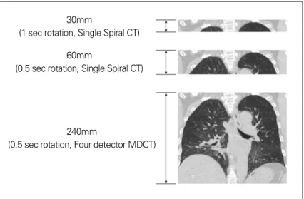
검출기 열의 수효만큼의 단면영상을 획득할 수 있다는 점이다(물론 실제적인 검사에서는 복수의 검출기열에서 얻어진 데이터가 복합되어 한 장의 단면영상을 구성하게 되며 회전당 얻어지는 단면영상의 수는 검출기열의 수보 다 적은 경우가 많다). 이에 의해 다음과 같은 세 가지의 장점을 갖게 된다. 첫째, 스캔 속도가 빠르다. 동일한 절 편 두께로 동일한 체적을 스캔할 경우 매 회전마다 MDCT는 SDCT에 비해 검출기열의 숫자만큼 많은 단 면영상을 획득할 수 있으므로 전체 스캔 시간은 검출기 열의 숫자 분율 대로 감소하게 된다. 근골격계나 두경부 와는 달리 흉부 및 복부의 CT 검사시엔 환자의 호흡이 개재되므로 단일 스캔시간은 30초 이내로 해야 하는 제 약이 있다. 특히 각종 호흡기 질환으로 호흡곤란을 겪는 환자들의 경우 스캔시간은 10초 이내로 줄여야 할 경우 도 있다. 따라서 빠른 스캔 속도는 흉부 영역의 CT 검사 시에 중요한 장점이 된다. 둘째, z-축 해상도가 높다. 동 일한 스캔 시간 동안 동일한 체적을 스캔할 경우, 검출기 열의 숫자의 분율만큼 얇은 절편 두께를 구현할 수 있다 (Figure 1). 기존의 SDCT의 해상도는 일반적으로 x-축
및 y- 축 상으로 0.5~1mm 의 pixel 크기를 갖는다고 보는데 이에 비해 z-축 크 기(두께)는 5 ~ 10mm로 가 로 해상도의 열 배에 달한다 (Figure 2). 이를 극복하기 위하여 세절편의 고해상 CT 가 적용되어 왔으나(2, 3) 기 존의 SDCT로는 절편두께 1.5mm의 조건에서 최대 초 당 3mm를 진행할 수 있는 데에 불과하여 제한된 호흡 정지 시간 동안 전폐야를 스 캔하는 것은 불가능하였다.
반면 MDCT는 단일 호흡정지간에 두께 1.25mm 내지 2.5mm로 전폐야를 스캔하는 것이 가능하다. 셋째, 더 넓은 체적을 포함할 수 있다. 동일한 스캔시간 동안 동일 한 절편두께로 스캔한다면 MDCT는 SDCT보다 검출기 열의 수만큼 넓은 범위를 스캔할 수 있다(Figure 3). 이 는 역시 제한된 호흡정지시간 동안 스캔을 완료하여야 하는 흉복부 영역에서 중요한 장점이라 할 수 있다. 실제 임상 적용시에는 이러한 세가지 장점을 적당히 혼합하여 필요한 체적을 최적의 검사시간 내에 스캔하도록 검사 프로토콜을 구성하게 된다.
2. MDCT의 단점
한편 MDCT는 몇 가지 단점도 가지게 되는데 첫째, 같 은 조건으로 검사 프로토콜을 구성하여도 SDCT에 비해 환자의 피폭선량은 증가하게 된다. 이는 초과피폭효과 (over-beaming effect)라고 하며 이는 경계영역(pe- numbra)의 조사 X-선이 SDCT에서는 가용한 데이터로 활용되는데 반해, MDCT에서는 검출기열간의 조건을 평준화시켜야 하므로 경계영역의 조사선은 폐기하여 야
Figure 1. Comparison of multiplanar image reformatted from conventional CT and MDCT(A) Coronal reformatted image from single spiral CT with 5mm thick axial image (B) Coronal reformatted image from sixteen multidetector-row CT with
1.25mm thick axial image
A B
하기 때문이다(Figure 4). 이는 검출기열 크기의 증가와 X- 선 발생기의 정밀화 등으로 경계영역을 최소화하도록 개선 되고 있으며 검출기 자체의 효율 향상 등으로 전반적인 피폭 선량 부분에서는 MDCT가 SDCT에 비해 높은 효율을 보인
다. 둘째, 방추형 X-선 조사에 따른 추상광선인공물(cone beam artifact) 이 발생한다. 이는 진정한 단면이 아 닌 방추형의 조사 X-선에 의해 투사된 영상을 가상의 단면에 재구성하는 과 정에서 발생하는 오류이다(Figure 5).
이로 인해 MDCT의 영상은 SDCT에 비해 불명확한 경계와 선상의 인공물 을 보이게 되나(4, 5) 다양한 후처리 알 고리듬을 적용하여 최신의 MDCT에 서는 상당히 극복되고 있다(6~8).
MDCT의 흉부 영역에서의 응용
앞서 언급한 장점들이 흉부 영역에 서의 MDCT의 유용성으로 사용되는 데 주요한 예는 다음과 같다.
1. 정입방(Isometric) 체적 CT 단면영상의 절편두께를 1mm 이 하로 줄이면 정입방체(isometric cubic) 혹은 근정입방체(near iso- metric)의 화소(voxel)를 만들 수 있다. 정입방 화소를 가진 영상은 다 중평면영상(multiplanar image)을 만들었을 때의 해상도가 단명영상 과 동일한 수준을 유지할 수 있어 진 단적 가치가 높다. 특히 관상면 (coronal plan)영상이 많이 이용되는데, 이는 첫째 관상 면이 흉부 단순 X-선 촬영과 같은 시야를 제공하여 임상 의사로 하여금 해부학적 이해를 쉽게 하고(9) 둘째, 인체 의 구조상 관상면으로 단면영상을 구성하였을 때가 축상 0.5mm
0.5mm
30mm
(1 sec rotation, Single Spiral CT) 60mm
(0.5 sec rotation, Single Spiral CT)
240mm
(0.5 sec rotation, Four detector MDCT)
0.5mm
10mm
Figure 2.Comparison of pixel and voxel. One pixel of axial CT has 0.5 x 0.5 mm size on XY plan. However Z axis length of this pixel on three dimensions is long to 10mm. This voxel shows unusually long hexa- hedron
Figure 3. How long scan with 1 mm slices thickness in 30 second scan time?
면단면영상으로 전폐야를 영상화 할 때보다 영상의 수가 적게 소요되는 편리함 때문이다(Figure 6)(10, 11) .
2. Dynamic Contract Enhancement
통상적으로 CT 조영제는 자동주입기를 사용할 경우
그 주입 속도가 1ml/second로부터 6ml/second까지 다양하게 사용된 다. 특히 CT 가상혈관조영술을 시 행할 경우 일시에 다량의 조영제를 주입하여(4ml/second 이상) 동맥 혈내 조영제 농도를 높게 유지하여 야 양질의 영상을 얻을 수 있다(12, 13). 그러나 주입가능한 조영제의 총량은 3.5ml/Kg을 초과해서는 안 되며(14 ~ 16) 통상 체중당 2ml/Kg 이하의 용량을 사용하게 되므로 자 동주입기를 이용한 고속주입시 조 영제 총 주입시간은 30초 이내이며 적절한 조영증강 평탄역(plateau) 은 이 중 15초의 전반기를 제외한 10 ~20초에 불과하다(17, 18). 또한 비교적 작은 직경을 갖는 혈관의 협 착, 관내혈전 등을 확인하기 위해서 는 z-축 해상도 역시 충분히 낮아야 한다(19). 따라서 짧은 평탄영 동안 에 전폐야를 2.5mm 이하의 절편두 께로 스캔하는 것은 기존의 SDCT로 는 불가능한 조건이었으나 MDCT 의 도래와 함께 현실화되어 특히 폐 동맥색전증의 조기진단에 널리 사 용되고 있다(Figure 7)(20, 21).
3. 3-Dimensional Demonstration
기관지 및 폐의 3차원 영상은 SDCT 당시부터 적극적 으로 활용되어온 기술이며 특히 기관지결핵과 이로 인한 협착이 흔한 우리나라의 상황에서 기관지내시경을 대치 할 수 있는 유용한 방법이었다(14, 22). 그러나 특히 z- 축 해상도의 한계로 주로 기관의 평가에만 사용되거나
Figure 5. Diagram of beam transmission with traditional axial scan vs. cone beam scan of MDCT
Traditional multislice assumption
Actal cone beam illumination Figure 4. Diagram of penumbra effect
주기관지 일부만을 표적 검사하여야 했다. MDCT의 도 래로 비로소 성대로부터 분엽기관지에 이르는 전장의 기 관 및 기관지 삼차원 영상이 실용화되었으며 가상 기관 지내시경도 유용하게 되었다. 이러한 기관지 삼차원 영 상을 이용하여 기관 협착 또는 종양의 정확한 위치를 영
상화하여 수술 계획에 도움을 줄 수 있고 치료 후 반응을 정확히 평가하는 데에도 유용하다(Figure 8).
4. 4-Dimensional study
MDCT는 z-축 해상도를 적절히 낮추면 전폐야를 불
Figure 7. Pulmonary thromboembolism on multplanar CT angiography. Filling defect (arrow) in right inferior pulmonary artery is conspicuous within highly enhanced vascular structure
(A) Axial image (B) Coronal image (C) Sagital image Figure 6. Comprehensive interpretation of anatomy using multiplanar image
(A) Plain radiography shows calcified granuloma in right upper lung zone (arrow)
(B) Coronal reformatted image of MDCT shows similar view with (A) It shows calcified granuloma also and fibrotic bands in right upper lobe as an additional finding (arrows)
A
A B C
B
과 수 초 내에 스캔할 수 있다. 이를 이용하여 이전 세대의 CT에서 시행되어온 단면 역동적 스캔(single level dy- namic scan)을 체적 역동적 스캔으로 확장할 수 있다.
이는 우선 조영증강 역동적 CT에 적용되고 있는데 (23~25), 두경부 CT에서와 달리 흉부 CT에서는 피사체 가 호흡에 따라 위치가 변동하기 때문에 단일 호흡정지 간에 시행한 스캔이 아니라면 매 스캔마다 피사체가 단 면에 포함되거나 벗어날 수 있다(respiratory misre- gistration). 나선식 CT는 체적 스캔을 통해 이러한 제한 점을 극복할 수 있으나(26, 27), 1회의 스캔에 10여 초 이 상의 시간이 소요되어 다중위상(multiphase) 스캔을 시 행하는 데에는 제약이 있었다. 그러나 MDCT를 이용하 여 호흡에 따른 피사체의 움직임을 보상할 만큼의 스캔 범위를 확보하고도 불과 수 초 이내에 대상 폐결절에 대 한 스캔을 완료할 수 있어 비로소 폐결절에 대한 다중위 상 스캔이 실용화되었다. 이를 이용하여 폐결절의 조영
증강 패턴을 평가, 양악성의 구분에 도움을 줄 수 있다 (Figure 9).
둘째, 전폐야에 걸친 체적스캔을 수 초 단위로 반복함 으로써 역동적 CT를 구현할 수 있다. 이를 조영제에 의 한 조영증강 작업과 함께 사용하면 폐관류영상 및 혈류 량 측정을 할 수 있으며(28, 29) 호기 및 흡기 운동중에 스 캔하면 폐 환기영상 및 폐기능 측정에 응용할 수 있으며 (30), 호기시 음압에 의한 기관이나 폐실질의 허탈 등을 역동적으로 관찰할 수도 있다(31).
결 론
MDCT에 이르러 비로소 인체에 대한 다중평면, 3차원 CT 영상이 가능해졌으며 특히 역동적 혈류와 호흡이 개 재되는 흉부 영역에서 단시간 내에 고해상도의 스캔이 가능한 장점이 두드러진다. 이를 이용하여 보다 구체적
Figure 8. Virtual bronchography using three dimensional volume rendering technique. Tubular structures with high (virtual) enhancement in right upper lobe and left lower lobe (arrows) are tubular bronchiec- tasis
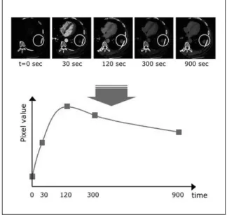
Figure 9. Enhancement dynamic CT of pulmonary nodule.
Serial images obtained at 30-second intervals and at similar levels show enhancement dynamics of nodule. We can extract objective attenuation of nodule and plot time-attenuation curve
인 해부학적 구조나 기능적인 면의 영상화가 진단적 가 치가 높을 것으로 사료된다.
참 고 문 헌
1. Taguchi K, Aradate H. Algorithm for image reconstruction in multi-slice helical CT. Med Phys 1998; 25: 550 - 561.
2. De Smedt E, Potvliege R, Pimontel-Appel B, Claus E, Vignaud J. High resolution CT-scan of the temporal bone; a preliminary report. J Belge Radiol 1980; 63: 205 - 212.
3. Todo G, Herman PG. High-resolution computed tomography of the pig lung. Invest Radiol 1986; 21: 689 - 696.
4. Kohler T, Proksa R, Bontus C, Grass M, Timmer J. Artifact analysis of approximate helical cone-beam CT reconstruc- tion algorithms. Med Phys 2002; 29: 51 - 64.
5. Zou Y, Sidky EY, Pan X. Partial volume and aliasing artefacts in helical cone-beam CT. Phys Med Biol 2004; 49: 65 - 75.
6. Yang K, Kwan AL, Miller DF, Boone JM. A geometric calibration method for cone beam CT systems. Med Phys 2006; 33: 1695 - 1706.
7. Mori S, Endo M, Kondo C, Tanada S. Physical evaluation of the weighted Feldkamp algorithms applied to the 256-detector row CT scanner for volumetric cine imaging. Acad Radiol 2006;
13: 701 - 712.
8. Schmidt TG, Fahrig R, Pelc NJ. A three-dimensional recon- struction algorithm for an inverse-geometry volumetric CT system. Med Phys 2005; 32: 3234 - 3245.
9. Remy J, Remy-Jardin M, Artaud D, Fribourg M. Multiplanar and three-dimensional reconstruction techniques in CT: im- pact on chest diseases. Eur Radiol 1998; 8: 335 - 351.
10. Studler U, Gluecker T, Bongartz G, Roth J, Steinbrich W.
Image quality from high-resolution CT of the lung: com- parison of axial scans and of sections reconstructed from volumetric data acquired using MDCT. AJR Am J Roentgenol 2005; 185: 602 - 607.
11. Sung YM, Lee KS, Yi CA, Yoon YC, Kim TS, Kim S. Additional coronal images using low-milliamperage multidetector-row computed tomography: effectiveness in the diagnosis of bron- chiectasis. J Comput Assist Tomogr 2003; 27: 490 - 495.
12. Chung MJ, Lee JH, Lee KS, Yoon YC, Kwon OJ, Kim TS.
Bronchial and nonbronchial systemic arteries in patients with hemoptysis: depiction on MDCT angiography. AJR Am J Roentgenol 2006; 186: 649 - 655.
13. Yoon YC, Lee KS, Jeong YJ, Shin SW, Chung MJ, Kwon OJ.
Hemoptysis: bronchial and nonbronchial systemic arteries at 16-detector row CT. Radiology 2005; 234: 292 - 298.
14. Carraro M, Malalan F, Antonione R, Stacul F, Cova M, Petz S, Assante M, Grynne B, Haider T, Palma LD, Faccini L. Effects of a dimeric vs a monomeric nonionic contrast medium on renal function in patients with mild to moderate renal in- sufficiency: a double-blind, randomized clinical trial. Eur Radiol 1998; 8: 144 - 147.
15. Spataro RF, Fischer HW, Kormano M. Clinical comparison of Hexabrix, iopamidol, and urografin-60 in whole body com- puted tomography. Invest Radiol 1984; 19: 372 - 375.
16. Donadio C, Tramonti G, Lucchesi A, Giordani R, Lucchetti A, Bianchi C. Tubular toxicity is the main renal effect of contrast media. Ren Fail 1996; 18: 647 - 656.
17. Bae KT. Peak contrast enhancement in CT and MR angio- graphy: when does it occur and why? Pharmacokinetic study in a porcine model. Radiology 2003; 227: 809 - 816.
18. Bae KT, Heiken JP, Brink JA. Aortic and hepatic peak en- hancement at CT: effect of contrast medium injection rate-- pharmacokinetic analysis and experimental porcine model.
Radiology 1998; 206: 455 - 464.
19. Jeong YJ, Lee KS, Yoon YC, Kim TS, Chung MJ, Kim S.
Evaluation of small pulmonary arteries by 16-slice multi- detector computed tomography: Optimum slab thickness in condensing transaxial images converted into maximum intensity projection images. J Comput Assist Tomogr 2004;
28: 195 - 203.
20. Prologo JD, Gilkeson RC, Diaz M, Cummings M. The effect of single-detector CT versus MDCT on clinical outcomes in patients with suspected acute pulmonary embolism and negative results on CT pulmonary angiography. AJR Am J Roentgenol 2005; 184: 1231 - 1235.
21. Perrier A, Roy PM, Sanchez O, Le Gal G, Meyer G, Gourdier AL, Furber A, Revel MP, Howarth N, Davido A, Bounameaux H. Multidetector-row computed tomography in suspected pulmonary embolism. N Engl J Med 2005 Apr 28; 352: 1760 - 1768.
22. Lee KS, Yoon JH, Kim TK, Kim JS, Chung MP, Kwon OJ.
Evaluation of tracheobronchial disease with helical CT with multiplanar and three-dimensional reconstruction: correlation with bronchoscopy. Radiographics 1997; 17: 555 - 567; dis- cussion 568 - 570.
23. Yi CA, Lee KS, Kim EA. Solitary pulmonary nodules: dynamic enhanced multi-detector row CT study and comparison with vascular endothelial growth factor and microvessel density.
Radiology 2004; 233: 191 - 199.
24. Jeong YJ, Lee KS, Jeong SY. Solitary pulmonary nodule:
characterization with combined wash-in and washout fea- tures at dynamic multi-detector row CT. Radiology 2005;
237: 675 - 683.
25. Chung MJ, Lee KS, Han J, Sung YM, Chong S, Kwon OJ.
Pulmonary sclerosing hemangioma presenting as solitary pul- monary nodule: dynamic CT findings and histopathologic comparisons. AJR Am J Roentgenol 2006; 187: 430 - 437.
26. Naidich DP, Gruden JF, McGuinness G, McCauley DI, Bhalla M. Volumetric (helical/spiral) CT (VCT) of the airways. J Tho- rac Imaging 1997; 12: 11 - 28.
27. Touliopoulos P, Costello P. Helical (spiral) CT of the thorax.
Radiol Clin North Am 1995; 33: 843 - 861.
28. Schueller-Weidekamm C, Wassermann E, Redl H, Prokop M, Zimpfer M, Herold C, Germann P, Ullrich R. Dynamic CT measurement of pulmonary enhancement in piglets with ex- perimental acute respiratory distress syndrome. Radiology 2006; 239: 398 - 405.
29. Hoffman EA, Chon D. Computed tomography studies of lung ventilation and perfusion. Proc Am Thorac Soc 2005; 2: 492 - 498, 506.
30. Guerrero T, Sanders K, Castillo E, Zhang Y, Bidaut L, Pan T, Komaki R. Dynamic ventilation imaging from four-dimen- sional computed tomography. Phys Med Biol 2006; 51: 777 - 791.
31. Lee KS, Ernst A, Trentham DE, Lunn W, Feller-Kopman DJ, Boiselle PM. Relapsing polychondritis: prevalence of expi- ratory CT airway abnormalities. Radiology 2006; 240: 565 - 573.
Peer Reviewer Commentary
본 논문은 최근 진료현장에서 실제적 활용이 급격히 증가하고 있는 다중절편나선식 CT의 흉부 영역에서의 적용에 대해 기술하고 있다. 다중절편나선식 CT는 필자가 밝힌 대로 인체에 대한 다중평면, 3차원 CT 영상을 가능하게 하였으며, 실제 폐동맥색전증의 진단, 기도 질환의 진단, 폐결절의 감별진단 등 여러 흉부 질환의 진단에서 기존의 나선식 CT를 빠르게 대치하고 있다. 본 논문에서는 MDCT의 장점과 단점, 실제 임상에서의 적용에 대하여 많은 사진과 함께 광범위 한 고찰을 통하여 기술함으로써 영상의학과 전문의가 아닌 의사들도 쉽게 이해할 수 있도록 기술하였다.
고 원 중 (성균관의대 호흡기내과)