'f".'J^~'T 線뽑웰會誌 第 22 卷 第6 號 pp. 947 - 952, 1986 rnal of Korean Radiological Society, Vo1.22, No.6, 1986
Rhinocerebral type 천균증의 CT 소견
연세 대학교 의파대 학 방사선과학교실
김동익·서정호·이종두
연세대학교 의파대학 신경외과학교섣
01
규 창- Abstract-
CT findings in Rhinocerebral Mucormycosis & Aspergillosis
Dong Ik i<.im
,
Jung Ho Sun,
Jong Doo LeeDepartment of Radiology, Yonsei Universitχ Col!ege of Medicine
Kyu Chang Lee
Department of Neurosurgerκ Yonsei University, Col!ege of Medicine
Invasive aspergillosis or mucormycosis of the paranasal sinuses involving the cranial cavity is termed 'rhinocerebral’ mycosis, w~ich is often difficult to differentiate from malignancy. Prognosis of rhinocerebral mycosis is disastrous and usually fatal. The authors herein report 6 cases of rhinocerebral mycosis: two of them were mucormycosis and four were aspiergillosis histopathologically.
Main CT features are nodular mucosal thickening in the multiple sites of the paranasal sinuses that extend to orbital apex or cavernous sinus through focal destruction of bony wall. In spite of their invasiveness beyond bony boundary, destruction is not so remarkable and it is always accompanied by bony sclerosis.
Awareness of these diseases and CT patterns discussed in this report should be helpful in leading to early biopsy and treatment
I.
서 료르 」부1l]동 및 두개강내 칭윤된 잔균증은 정상인에게서는 드운 질환이나 당뇨t영성 케토산증,부신피질 호르온제제 나 항생제 항암제 등 연역억제제를 투여하여 연역기능 이 저하펀 환자에서 많이 발생될 수 있으며 두개강내로 파급된 진균증의 경우에는 사%←율이 매우 높아 50 %까 지 이르고 있다 1-2) 이러한 Rhinocerebral type 의 진
이 논운은 1986 년 9월 23 일에 접수하여 1986년 11 월 12 일에 채택되 었음.
균증은 Mucormycosis 와 Aspergill osi s 가 대표적인 질환이며 이들을 조기발견하여 적절한외과적 절제와항 진균제 사용으로 사oJ-율을 감소시킬 수 있기에 도웅이 될만한 CT 소견을 분석하였다.
II.
대상 및 방법최근 3 년간 연세의료원에서 조직학적으로 확진된 Rhinocerebral 형태의 mucormycosis 2 예와 IIsper.
gillosi s 4 예를 대상으로 하였다. 대/홉 남자 4 명,
여자 2 영이었£며, 연령분포는 28~64 세 사이 였다 땅 사선학적 검사운 천예에서 부ll]동 및 두개강 촬영과 천
大훌훌放射線醫學會誌‘ : 第22卷 第
6
號1986 -
산화단층촬영 (이하 CT) 을, 3 예에서 뇌동액조영검사를 시행하였다 CT는 조영제 주엽
(Conray 60 % 100
ml) 전후의 부비동 빛 뇌의 횡단과 종단 주사로써 사 용펀 기종은 Philips 제
Tomoscan 310
이었으며, 4 예 는 2~13 개월의 추적 CT를 하였다.병 리학적 진단은 수술후 조직 검사로
Ethmoidecto
my 2 예, Ethmoidectomy 후Craniectorny
2 여예l’Cr-
an띠1니iectorr매 1 예 그리고
Caldwell - Luc
수술 l 예였 다.ill_ 결 과
총 6 명의 환자 중 원인으로 의심할만한 선행 질환으 로서
Aspergillosis 1
예에서만 원인불명의 백혈구 감 소증파 판결통£로 인하여20
여년칸 간헐적으로 부신 피질 호르온제를 사용한 것 외에는 특。l할만한 조건을 발견하지 옷하였다.중요한 입상적 증상 및 이학적 소견으로서는, 얀연부 동통, 동안근 마비, 시력감소 내지는 시력소실, 두통, 하 지 운동장애 등이며 증상 발현후 2~6 개월 후 내원하
였다 (Table
1 )
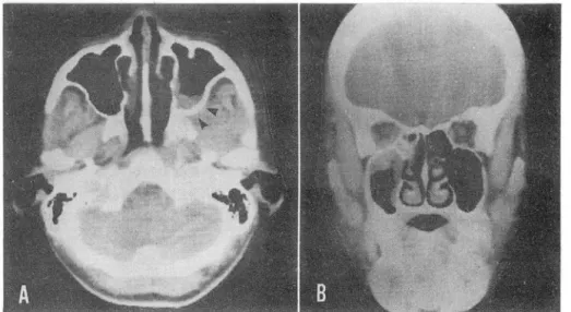
단순두개 및 부비동촬영은 시행된 전예에서 일측성 부 비동의 정 막바후나 증가음영을 보였으며 2 예에서
La- mina papyracea
와 상얀구열의 부분적 골파괴 를 관찰 할 수 있었마 (Fig_1) _
CT 소견은 변연이 비교겨 펑탄한 결칠성 종괴양상의 부비동벽 비후를 전예에서 판찰할 수 있었다. 부비동과 111강의 침윤범위는 일측성무로 (Fig_
1-3
)사골동, 접 형골동, 상악동 순이었으며 비인두 침벙이 1 예 있었다.부비동 뱅소의 얀와침융은 4예로
lamina papyracea
의 부분적 결손이냐 골경화와 동반된 종괴 양상으로 주 로 내직근과 하직근의 팽대를 동반하였으며 l 예(case
ill) 에선 안와첨부의 시신경비후를 보였다 (Fig.2-B, 5J;
두개강내 침윤은 3예에서 해연동 (Cavernous
sin.
us)
칩벙으로 인한 팽대와 2 예의 측두엽 농양으로 융Fig.
1.Case II Aspergillosis. Caldwell view demonstratss opacities in the left maxillary and ethmoid sinues with loss of anterior l amina papyracea ("').
Table
1.Surnrnary of cases with rhinocerebral as pergillosis
&mucorrn ycosis.
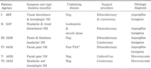
Patients: Syrnptorn and signl U nderlying Surgical Pthologic
Age/sex
duration (rnonths) disease procedure diagnosis
1: 28/F
Visual disturbance Neg Ethmoidectorny Aspergillus
&
hemiplegi al 3M
&craniectorny frrnigatu s
II:
52/FHeadache
&visual Leukopenia
disturbancel 6M
&Ethrnoidectorny Aspergillus
steroid abuse furnigatus
III:
331MPtosis & blindness Neg Ethrnoidectorny Aspergillus
headachel 3M
Craniectorny furnigatus
IV: 64/M
Facial painl 5M Post-TSA* Ethrnoidectorny Aspergillus
furnigatus
V:
40/MFacial painl 3M Neg. Caldwell-Luc Mucorrnycosis
V
I: 341MHeadache and Neg. Craniectorny Mucorrnycosis
herniplegial 2M
*TSA: Trans-sphenoidal approach for pituitay adenorna
깅동익 외 :
Rhinocerebral type
진균중의CT
소견 -Fig.2. Case II
Asper밍Ilosis. Axial CT(A) shows polypoid antrallesion with sclerosis of posteriormaxilIary wall. In spite of sclerosis , Invasion to infratemporal fossa can be seen
(~).Cor- onal CT(B) shows the involvement of medial
&inferior retus muscles in orbital apex.
Fig. 3. Case V Mucormycosis. Intra-antral soft tissue mass with sclerotic change. Extension to nasal cavity and infratemporal fossa
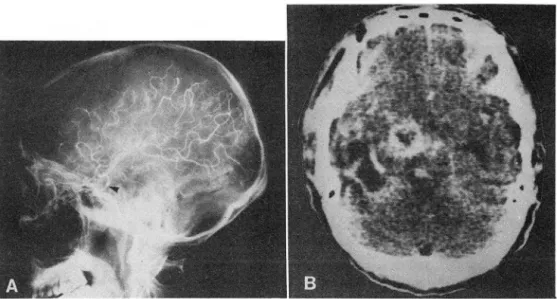
(~).상의 조영증강을 보이는 뱅소가 관찰되었으며 그 중 1 예(Case VI) 는 뇌경색의 소견을 보였다 (Fig.
4 , 6 B).
부바옹 주위 골조직은 전예에서 다발성 국소적 파손 이 얼어났고 알부 비갑캐들의 파손도 관찰되었다. 특。l 한 소견으로서는 i녕변주위 골벽
(bon
y wall) 이 정상 측보다 모든 환자에서 골경화를 초래한 것을 들 수 있£며 2 예에선 골파괴없이 경화된 골의 인접 부우l로의 침율을 보였다 (Fig.
2
-AB,
3).Fig. 4. Case III Aspergillosis. Coronal contrast CT shows m
i1d bulging of Lt cavemous sinus
(~)and soft tissue mass in sphenoid sinus & choana
안와첨부 (orbital apex)플 침윤한 경우 대부분 상 안와열의 확조L을 보였고 이를 통하여 전체 혹은 부분적 인 해변동무로 확산된 소견을 보였다 (Fig.5)
(Table
2 ). 이러한 경우 시력감소 및 시력소실, 동안근마비,동
통등의 임성적 증상을 호소하였고 뇌농양이 있었던 환 자는 뇌압상승 증상인 우통, 구토등을 수반하였 다.
그외 세균성 부비동엽의 소견인
fluid level
은 없- 大짧放射緣뽑學會誌 : 第 22卷 第
6
號1986-
었고 안연근육이냐 주위 연조직 (soft tissue)으로 파 에도 뚜렷한 호선은 판창되지 않았다.
급된 예도 거의 보이지 않았다. 또한 뇌동액조영검사를 시행한 3 예중 뇌놓양을 초래한 2 예를 포함한 전예에 서 경동액의 부분적 협착올 그리고 l 예에선 중뇌동액 분지의 다발성 동액류를 통반하였다 (Fig.6-A).
Mucorrnycosis 2
예와Aspergillosis 4
예 사이의 상이한 땅사선학적 소견을 판찰할 수 없었으며 추적C
T 를 시행한 4 예중 l 예를 제외하고 항진균제의 투여Fig. 5. Case 1 Aspergillosis. Coronal contrast CT shows infiltrating rnass in
superior orbital fissure.N.
고 찰부비동 혹은
ll]
강내 발발한 진균증은Mucorrnycos.
is
와Aspergillosis
가 대 표적 인 질환이 다 3)Mucor-
mycosis 는 우격균사 (non-septated
hyphae) 를 강 는Phycornycete
에 의해 발영되는 질환이 며 이 곰팡 이는 원래 정상인에첸 발영치 않는 부쟁진균이냐 당뇨 병이냐 연역기능이 현저히 저하펀 환자에서 싱각한 질 환을 야기시킨다.Phycornyce
te 의 종 (genus)중에서Rhizopus
,Mucor
,Absidia
등이 주된 t영원체이며 이 질환을 Phycornycosis 라 칭하기도 한다 4)이 질뱅은 1884 년 Faultary 에 의해 처음 발표되었 고 현재까지 다음
4
가지 유형으호 대옐된다. 첫째가cephalic
forrn_으-로 비강, 부비동 혹은 얀와에서 발영 하여 두개강내로 확산되는 가장 많은 유형이며 사oJ-율 이 매우 높다. 둘째로pulmonary
forrn으로 암환자에 서 항암제등을 투여한 군,종양없이도 심한 당뇨빵이 있 는 환자에게서 잘 발생하는 형태,세째 심한 영%냄측증 이 있는 환자의 회장이냐 대장에 발발한Intestinal fo-
rrn
, 네째 전신에 확산된Di sserninated
forrn 이다.특히
cephalic
forrn 에서는 영원체가 비점막, 인후부,군개에서 영변을 일으킨 후 직접척으로 국소칭윤을 하
Fig. 6. Case VI Mucorrnycosis. A , Focal narrowing of the supra
cJinoid segrnent of internaL carotid artery.
B , Ring-like enhancing lesion at the rnedical ternporallobe and infarction in the ternporallobe.
- 950-
깅 동익 외 :
Rhinocerebral type
진균중의CT
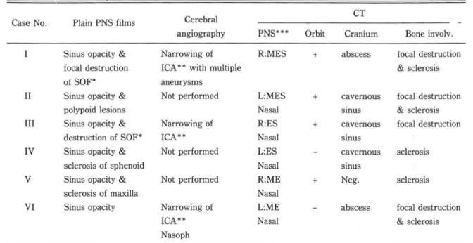
소견 -Table 2. Radiologic data of cases with rhinocerebral aspergillosis
&mucormycosis
Cerebral CT
Case No
‘Plain PNS films
angiography PNS*** Orbit Cranium Bone involv.
Sinus opacity
&Narrowing of R:MES
+abscess focal destruction
focal destruction ICA * * with multiple
&sclerosis
of SOF* aneurysms
II
Sinus opacity
&Not performed
L:MES
+cavernous focal destruction
polypoid lesions Nasal smus
&sclerosis
III
Sinus opacity
&Narrowing of R:ES
+cavernous focal destruction
destruction of SOF* ICA*' Nasal smus
IV Sinus opacity
&Not performed
L:ES cavernous sclerosis
sclerosis of sphenoid Nasal smus
V Sinus opacity
&Not performed R:ME
+Neg. sclerosis
sclerosis of maxilla Nasal
VI Sinus opacity Narrowing of
L:ME abscess focal destruction
ICA** Nasal
&sclerosis
Nasoph
* Superior otbital fissure * * Intern
a!carotid artery *
** R: right
L:left M:
maxill값y sinus E: ethmoid sinus S: sphenoid sinus거냐 혈판내로 파급되어 부바동, 안와, 사판(
cribrifo-
rm plate)
등을 칩엄하며 냐아가 안와 혈관조직,내경동액, 뇌악, 해연동, 뇌실질조직까지 파급된다. 혈판내에 서는 혈판벽의 탄력층을 따라 벤식하고 내악의 손상을 얼£쳐 동액염, 괴사, 화놓을 초래하여 。|후에 정맥파 임 파판도 동일한 명변을 일으컨다5) 근육층, 골조직,피부 등에서도 같은 결과를 유발시킬 수 있다.
부비동 침윤빈도는 사골동, 접헝골동. 상악동순이며 천 두동은 거의 칭융되지 않는다고 보고되었고 6) 저자들의 경우에도 같은 결고}흘 얻었다(사골동 6 예, 정형골동 및 상악동 각각 4 예).
CT 소견으로는 다발성 부비동 병변을 판찰할 수있 고 발영은 사골동이냐 접형골동에서 시작되어 일측정 영 변을 보이는 경우가 많다. 부III 옹벽의 비후는 결절성 종괴양 또는 펑탄한 형태등 모두 올 수는 있£냐, 주로 결절성 양상이 주된 소견이 라 발표되 었고 저자들도 모 두 이러한 형태를 경험하였다. 골손상은 다발성 점상
(spotty)
파손을 보이는 것이 대부분。 l 고, 원발성 부 비동암보다 골손상 뱀위가 작은 것우로 발표되었다 7)Green 등 8) 은 골손상 범위 및 형태와 다발성 부비동 침윤양상으로 악성종양파 강멸할 수 있다고 보고하기도 하였다.
부비동벽의 골경화현상에 대해 Addlestone 과
Bay-
lin등은 만성적으로 진행된 예에 대해 이를 기술한 적 이 있고 악성종양중 종양세포 번식이 느런 경우나 。l 차 세균감염에 의한 골수염의 일환으호 생킬 수 있마고도 발표하였다6) 그러냐 저자들의 경우처럼 모든예에서 확 연한 골경화를 볼 수 있는 점은 득이한 소견이라하겠 다.Mucormycosis 외에도 Aspergillosis 도 유사한 유 형의 영변을 유발시키냐 천자에 III 해 발생반도는 적고
Rhinocerebral type
이 아닌 비 강이 나 부비동에 국한 된 경우에만 빈도가 높다고 되어 있다. 또한 마흔 원인 질환없이 정상인에게서도 발뱅될 수 있다는 점, 부비동 중상악동침범이 제일 않마는 점이 상이한소견。 l 라하 겠다Jo) 보펀적으]로 CT 상 두 질환의 감벌은 불가능한 것으로 되어 있으냐 Kopp 11l의 보고에 의하면Asper-
gillosis
환자의56
%에서 부비동내 괴사된 영역에te- rtiary calcium
phosphate 와calcium
s 비fate , 중 금속영등이 침착된 2~IOmm 크기의 흡수계수가 높은 구조물을 판찰할 수 있마고도 하였 냐. 저 자들은 증례 가 석은 이유로 이를 발견하지 옷하였다.부 III동 빛 바강내 명변이 앞에서 기술했띤 경로 등을 통하여 우개 강내로 파급되기 해문에
Eiji
Yumoto응은- 大韓放射線뽑學會誌 : 第22卷 第
6
廳1986-
Invasive
Aspergillosis 에 의한 사 PJ-율을 감소시키기 위해 조기발견 및 그에 따른 척결한 치료블 강조하였다12) 뇌조직 변호}는 혈관손상, 혈괴형성, 점"'J-출혈, 01 차 적 뇌경색에 딱릎 CT 소견과 농양의 형성 등을 볼 수 있고, 경동백 촬영시 동액엽에 의한 협착, 동액류 등을 관찰할 수 있다. Courey 등 14) 은해변동에서 직접 파급 되거나 안동액
(oph thalmic artery)
으로부터 역행되 어 내경동액이 침윤됨을 기술하였고 이로 인한 동액염, 동액류, 혈천증, 뇌경색 등을 보고하였다. 저자들도 이 와 유사한 소견들 즉, 내경동맥 협착, 뇌농양, 해면동침 율등을 경험할 수 있었으며, Aspergillosis 에 의한 뇌 놓양이 있었던 1 예에서 뇌동액의 다발성 동맥류를 보 여, 뇌혈판조영 검사가Rhinocerebral
type 의 진균증 이 의심될 경우 펄수적인 검사염을 알 수 있었다.Rhinocerebral type
진균증은 부비동의 원발성 악 성종양이 주위 안와, 두개강내 로 파급된 양상과 유사하 고 강옐이 어려울 혜가 않으냐 과거력상 항암제냐 연역 억제제 사용, 심한 당뇨t영등이 있을 경우 반드시 진균 감염질환을 고려하여야 하겠다.V.
결 론최 근
3
년간 경 험 한Rhinocerebral type
의 진균증 6 예의CT
소견을 종합하여 마음 결과를 얻었다.1) Aspergillosis 4
예 ,Mucormycosis 2
예로서 남녀비4
: 2, 연령분표는 28~64 세이었다.2)
과거력상 l 예의 Aspergillosis 환자에서만 원인 불명의 백혈구 감소증과 장기간 부신피질호르온쩌|를 사 용한 뱅력이 있었고 냐머지 환자는’ 특기할만한 뱅력이 없었다.3)
CT 상 모두 결절성 종괴양상의 부비동백 비후,다 발성 국소적 골손상 및 주위 골조직의 골경화를 보였고fluid level
은 없었다. 또한 다흔 악성종양。 1 냐 영증 성질환에 111 하여 골파괴의 범위냐 정도가 심하지 않을 뿐 아니 라 골경화를 동반함。l 판찰되었다.4)
두개캉내 침윤은 5 예의 안와첨부, 해연동, 2 예의 뇌농양이 있었다.5)
부비동 발명빈도는 사골동。 l 제일 않았고 접형골 동 및 상악동의 순이었으며 전예가 일측성 뱅변을 보였다.
6)
CT 상 Mucormycosis 와Aspergillosis
와의 감별은불가능하였다.REFERENCES
1. Prockop LD , Hunter M5:
Cephalic mucormycosis. Arch NeuroI17:373-386. 1967.2
‘Abramson E
,Wìlson D
,Arky RA:
Rhinocerebral phycomycosis in association with diabetic ketoacidosis.Ann Inter Med
66:735
,1967
3. Harnsberger HR , Osborn AG , 5moker WR:
CT in the evaluation of the normal and diseased paranasal sinuses Semin ultrasound CT MR 7(긴:68-90, 19864. Anderson D
,Matìck H
,Naheedy MH
,5teìn K
Rhinocerebral mucormycosis with CT scan findings: A case report. Comput Radiol 8(.긴:113-117,.1984
5. Manìglìa A
J,Mìntz DH , Novak 5:
Cephalic phycompycosis:A case report of eight cases. Laryngoscope 92:755-760
, 1982
6. 8ergeron RT:
Head and neck imaging.
1st Ed:65-67
The CV ι10sby Co, St. Louis, 19847. Centeno
R5 , 8entson JR , Mancuso A
A: CT scanning in rhinocerebral mucormycosis and aspergillosis. Radiology 140:383-389,
1981.8. Green WH , Goldberg HI , Wohl GT:
Mucormycosis infec- tion of craniofacial structure. AjR 101:802-806, 1967.9. Addlestone R8
,8aylìn GJ:
Rhinocerebral mucormycosis.Radiology
111:113-111 , 1975
10. Lewìs RMJ:
Aspergillosis of the nose and paranasal sinuses.Laryngoscope