大합 Ií'-<[j‘U,f;l 영홉 !}~ 싹 갚、 껴\ 21 씬 까 6 싸 pp.876-882 , 1985 ] ournal of Korean Radiological Society, Vo1.21, N 0.6, 1985
淚臨좀훨 睡傷의 電算化斷層擬影術
서울大쩔校 짧科大學 放용t綠科쩔敎室
朴 贊 燮·金 榮 九·張 基 賢 - Abstract -
Computed Tomography of Lacrimal Fossa Tumors
Chan Sup Park, M.D., Young Goo Kim, M.D., Kee Hyun Chang, M.D.
Department of Radiologκ College of Medicine, Seoul National University
The lacrimal lossa can be involved by a wide spectrum 01 orbital pathology
The correct diagnosis is important to avoid unnecessary procecdure and to do appropriate management 14 patients with mass lesions in the lacrimal lossa were evaluated with computed tomography (CT) and clinical lindings
The results were as lollows
1. Final diagnosis 01 14 cases with lacrimal lossa tumors was pleomorphic adenoma in 3 cases, adenoid cystic carcinoll1a in 1 case, pseudotumor in S cases, lympholl1a in 2 cases, neurolibroll1a in 1 case, chloroma in 1 case and metastatic adenocarcinoma in 1 case
2. The duration 01 symptoms of pleomorphic adenoma was 1l10re than 1 year and characteristic CT lindings were globular masses with pressure erosion 01 the adjacent bone. Patient with adenoid cystic carcinoll1a had a short history 01 symptorm. CT showed a lusilorm ll1ass but intracranial extension with Irank destruc.
tion 01 sphenoid bone
3. Patients with pseudotumor and Iymphollla had sylllptollls lor less than 1 year. The CT lindings were ill delined inliltrative patterns 、vith scleral thickening and the dillerential diagnosis 01 thell1 was difficult 4. The Illargins 01 neurolibroma and chloroma were well delined while that 01 the Illetastatic adenocarcinoll1a
was ill-delined
5. The degree and the extent 01 the contrast enhancement gave no benelit in the differential diagnosis 01 each disease entities and even 01 the benign and Illalignant lesioi1
1.
序 論淚 H없 및 淚H없;찌 病變O양,1lN:球 外上方애 삐塊가 만져 지 따 안구룹출과 함께 안구를 內下方으효 밀고 視神혐 침 -운은 드볼어 디→은 U북정 1잉 띤보다 시 럭 간소가 덜 나타냐
이 논운은 1985 년 10월 23 일에 접수하。1 1985년 11 원 14 일에 채넥되었음.
므로 임상적으로 득이힌 증상을 갖는다 1)
그-더냐 이 」!L위늠 001러 가지 짙환이 올 수 있고 또한 각각의 治擔方法이 다르므로 正 M한 등?斷이 매우 중요
하다2)
펴f.\:f l::.斷}현밟彩pj!j (이하 CT로 약함)은 안와침환을 낀단하는데 널펴 이용되어 왔다 1,12,13,20) 淚服점 l잉변에 서도 임상증상무로 위치는 알 수 있지만 f申展程度,주위 付의 變化등을 운석 하여 組般의 특성 을 .!L디 더 正確히
- 朴贊燮 外: 淚服짧 睡훌의 電算化斷層짧影術 -
把握하는데 상당한 도움을 준다.
著者들은 淚服홈에 위치하는 여러 질환틀에서 각각특 징적인
CT
소견이 있는지를 알기 위하여 1979 년 12 월 부터 1985 년 7 월까지 본원에서 시행한 眼짧 CT 중淚眼 홈에 병변을 갖는 14 例의 임상증상 및CT
소견을 분석 하여 文敵考察과 함께 보고하는 바이다ll.
對象 및 方法1979 년 12월부터
1985
년 7월까지 시행한 眼홈 CT 중 안와의 外上方에 병소를 갖는 14 例를 대상ξ로 하 였다.著者들이 분석한
14
例중 多形性眼睡Cpleomorphic ad- enoma)
, 願樣훌뼈獨Cadenoid cystic carcinoma)
, 神經 織維睡 및 轉移性 H훌提의 6 ØlJ와 假性睡題의 1 例는 수 술 및 조직생검에 의하여 病理學的£로 確談되였으며,假性睡最
4
ØlJ는 임 상증상과CT
소견으로 假性睡題으 로 진단하여 스테로이드로 治廳하여 증상의 호전이 있었 던 예 이 며,
썩*밍睡2
f에는 각각 賴部빼밍節 및 骨體生檢 에서 惡性~巴睡즈ζ로 진단된 환자이었고, 緣色睡 I 例는 急性骨體性白血病으로 화학요법중 안구돌출을 호소하여CT 를 시행하였다.
CT
題影은GE CT /T 7
,800
또는 8 , 800 을 사용하 였으며, 走훌方向은 OM 선과 -10 도로 上下 각각4
내지 5 절편씩
5mm
두께 및 간격으로 橫斷爆影을 하였 으며, 이상소견이 발견된 경우5mm
간격으로 冠狀擺影 을하였다.造影齊IJ$入後走흉를 원칙£로 하였으며 120ml 의
Te- lebrix-30@을 bolus 로 주입 하여 쳤옮影하였 다.
m.
結 果 1. 임상소견多形性限陣과 H없樣쩔훌g힘짧은 睡塊와 안구돌출이 주증상 이었으며, 多形性線鍾은 병력이 1 년이상3로 길었으나 眼樣훌뼈擔은 4 개월로 짧았다.
假性睡앓과 빼러睡은 종창 및 동통의 염증성 증상이 특정적이었고 병력도 6 개월내로 짧았으며, 假性睡場은
1O ~24 세의 젊은 연령층이었다.
神經織維睡, 緣色睡 및 轉移性眼1옳은 전부 안구돌출이 주증상이 었 a며, 神經織維睡 환자는 피부에 색 소 침 착이 있어 임상적으로 神經織維睡효 Cneurofibromatosis) 의 환자이고, 녹색종 환자는 급성골수성백혈병으로 치료를 받던중이었£며, 전이성션암은 병력이 3 년무로 길었으 며 그 原發부위는 찾지 못하였다
CTable 1).
2.
CT 소견多形性眼睡은 3 例 모두 일측성이었요며 球狀이었으나
Table
1.Clinical Features
Diagnosis No. of Patients Age/Sex Chief complaints
Pleomorphic adenoma 3
35/FProptosis 10YA
59/F
Eye discomfort
,mass 5Y A 511M Proptosis
,1 Y A
Adenoid cystic carcinoma 1 511M Mass
,proptosis 4MA
Pseudotumor 5
10/FSwelling
,proptosis 2MA
131M
Proptosis
,swelling
,reddness 3MA
191MVisualloss
,pain lMA
20/F
Mass
,swelling
,pain 6MA
24/FMass
,swelling 6MA
Lymphoma 2
67/MSwelling
241M
Proptosis 2MA
N eurofibroma 1
17/FProptosis 1 Y A
Cutaneous pigmentation
Chloroma 1
131MProptosis
Metastasis 1
381MProptosis 3Y A
- 877-
- 大韓放射線훌훌學會誌 : 第 21卷 第6號
1985 -
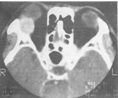
2 例는 경계가 분병하였고 1 例는 불분명하였다. 2 例에 서 주위 骨의 압박성 미 란
(pressure erosion)
이 보였으나(Fig.
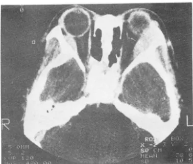
l), n용휠훌훌6힘}옳은 結鍾狀이었고 경계가 분명하였 으나 媒形骨破壞와 함께 腦內로 파급되 어 있었 다 CFig.2).
假性睡錫과 빼巴睡은 모두 경계가 불분명하였으며 침 윤적 인 양상을 보였고 假性睡場 5 (7IJ중 4 例 및 빼밍睡
Fig.
1.Pleomorphic adenoma, post-contrast scan, 51/M Well defined homogeneously e nhancing mass in right lacrimal fossa with pressure erosion of lateral orbital wall
A
Fig. 3.
PseudotumorA.
A. 20/F2 例중 1 例에서 養鷹비후가 觀察되었으며
CFig.3 , 4) ,
훌體비후가 없었던 假性睡場 1 例와 빼巴睡 1 例는 眼 홈전체에 병변이 있었던 경우였다.
神經鐵維睡과 緣色睡은 모두 경계가 분영하였£며 방 추상이었고 녹색종은 양측에 영변이 있었다 (Fig.6).
轉移性 A없病은 경계 는 불분명 하였으나 骨破壞는 없었다
(Fig.7) (Table ID.
Fig. 2. Adenoid cystic carcinoma, post-contrast scan Homogeneously enhancing well-defined mass in left lacrimal fossa and extension into middle cranial fossa with frank destruction of sphenoid bone.
Pathologically confirmed pseudotumo
r. Ill-defined enhancing mass in right lacrimal fossa with scleral thickening.
B.
191MIll
-defined homogeneously
enhancing mass in left lacrimal fossa with scleral thickening- 朴n燮外: 淚없짧 R훌훌의 훌算化斷層짧影術 -
Fig. 6.
Chloroma , bilateral
Fig. 4. Lymphoma , coronal scan , 67/M
Well-defined homogeneously enhancingmass in
Ill-defined mass in left lacrimal fossa with scleral both orbit.
thickening.
Fig.
5.Neurogenic tumo
r.Well-defined
mass in left lacrimal fossa.
조영제주업후 주위 節肉보다 조영증강이 잘된 경우를 양성으로 하였는데 多形性線睡의 2
f7
U. 假性睡짧의 3f7
U.緣色睡 그리 고 轉移性眼}찮은 均一한 조영증강을 보였으 며, 眼樣훌뼈廳과 假性睡錫의 1 例에서 不혐-한 조영증 강을 보였다 CTable I1
D.
N.
考 按淚線홈는 해부학적으로 眼홉의 外上方에 위치하며 前
Fig. 7. Metastatic adenocarcinoma
Ill
-defined mass with homogeneous enhancement
in right lacrimal fossa頭骨의 賴骨突起C
Zygomatic process)
안쪽에 있고, 상 직근 및 측직근의 인대부근에 위치한다 3’‘’')淚線 및 淚8몽홉병변시 다른 안와병변과는 달리 특이한 증상을 보이나, 여러 가지 질환이 올 수 있고 각 治擾方 法이 다르다. 그 예로 多形性願睡時 조직생겸으로 被體 을 다치거나 수술로 부분만 제거할 경우 被願파열로 주 위 骨과 軟組織에 파급되 며 재 발이. 잘되 어 惡性과 같이 되므로 6) , 각 영변의 조직적 특성을 아는 것이 중요하
다 2)
淚線의 병변중 약 半은 1:皮細뼈에서 생기며 多形性
- 879-
- 大韓放射線홈學會짧‘ : 第21卷 第6廳
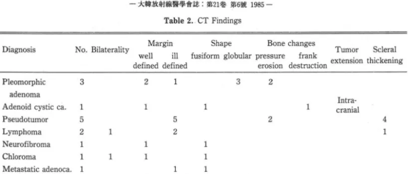
1985 Table 2. CT Findings
Margin Shape Bone changes
Tumor Scleral well
illfusiform globular pressure frank
extension thickening defined defined erosion destruction
No. Bilaterality Diagnosis
4 1
Intra-
cranial 2
2 3
1
--14
1i
1
5
2
21
1 1 1
3
1i
Fa
nι
1i
1i
1i
Pleomorphic adenoma Adenoid cystic ca.
Pseudotumor Lymphoma Neurofibroma Chloroma
Metastatic adenoca.
1
라고 淚服홈밖으로의 {申展이 있을 경우 惡性의 특이한 소견이 라고 보고하였다 6) 著者들의 경 우에 도 眼樣훌뼈 행이었는데 4 개월의 짧은 병력이었고
CT
상 방추형종 괴이었으며 경계가 분명하였으나 종괴가 娘形骨을 破壞 하여 腦內로 파급되어 惡性임을 쉽게 알 수있었으냐 주 위 骨의 硬化나 석회화는 없었다假性睡陽은 병리학적으로 다양한 찢효性所見을 보이 며 械밍球침윤이 자주 보여 빼밍睡과 비슷하냐 睡場性 t홉꺼훌이 아닌 反應性增쩌흉이며 9,10) 아직 그 원인은 밝혀 지지 않았다. 임상적으로 急性이며 동통, 종창등의 찢 tlË性효狀을 호소하나 熱이 없고 백혈구과다증이 없다 2, 10>.
일측성이 대부분이며 양측성모 있고 이 경우
Grave
흙 愚과의 강별이 어렵다 11) CT 소견은 경계가 불분명한 침윤성병변이며Bernardino
등은 外陽이나 手術의 병 력이 없을 경우 훌鷹 111 후가 特徵的所見이라고 보고하였다 1)9)
빼밍睡도 假性睡짧과 비슷한 소견을 보여 감별이 어려 우냐 U) 빼밍睡은 좀 더 나이 가 많고 骨破壞가 많으며 全身증상이 있고 15) 假性睡覆은 스테로이드로 治標한 후 증상의 호전등으로 강별할 수 있다고 하였다. 著者들의 경 우 假性睡傷과 ?하밍睡 모두 짧은 병 력 과 동통, 종창등 의 ~tlË性tlË狀을 호소하였으며.
CT
소견상 모두 경계가 불분명한 침윤성 병변이었고 꿇體비후도 동시에 보여 감 별이 힘들었다.神經織維睡은 경계가 분명하고 안와내 어느 부위라도 생길 수 있a며 서서히 자라므로 骨의 압박성미란이 흔 히 보이고 神經鐵維睡효과 동반이 잘되는 것으로 보고 되고 있다 12
’“’
16) 著者들의 경우 이학적검사상 피부색 소침착이 있어 神經鷹維鍾tlË으로 생각되는 환자。l며 .CT 1Table 3. Patterns of Contrast Enhancement
Diagnosis Heterogeneous
l
Homogeneous
3 2
1
No.
2/3
1/1
4/5
0/20/1 111 Pleomorphic
adenoma Adenoid
cystic ca Pseudotumor Lymphoma Neurofibroma Chloroma Metastatic
adenoca.
1H혔睡과 ?품睡이 같은 比率이 다. 나머 지 半은 빼밍睡 및 假性睡覆이 며 그外 肉흉睡
Cgranuloma)
, 類皮睡Cderm-oid)
및 轉移性램도 생길 수 있다고 보고되고 있다 3,까 多形性線睡은 임상적으로 1 년이상의 병력과 함께 통 증이 없요며 20 대 후반에서 60 대까지 넓은 연령분포 를 보인다까 CT소견은 경계가 뚜렷하고 骨破壞는 없 이 압박성마란이 觀察되며, 초영증강시 여러 形態의 조 영증강이 보인다고 보고되고 있다 3,8) • 著者들의 경우 3 例 모두 1 년이상의 긴 병력을 가졌으며 球狀이었고 2 例는 경제가 분명하였으나 1 例는 불분명하였£며 2 例에서 주위 骨의 압박성 미란이 觀察되었다.1품睡은 病理學的으로 R용樣컬훌§힘?끓, 惡性混合細R힘癡
Cm
a-lignant mixed cell
carcinαna) 및 R흉癡C adenocarc ino- ma)
이 있으며 그중 線樣훌뼈穩이 가장 많으며 또한 가 장 惡性이다 2) 임상적으로는 짧은 병력과 함께 동통이 주증상이며. Lloyd 등은 주위 骨破壞와 硬化,석회화 그111
- 朴贊燮 外: i핏腦’샘 8뀔傷의 끽J: rMr.없1iR줌뻐影術
소견상 종괴는 경계가 분명하였고 방추상이었으며 부수 적인 소견으로 우측 췄、突口蓋홈 (pterygopalat i ne fossa) 가 넓어져 있었다.
緣色睡은 granulocytic sarcoma, myeloid sarcoma 或 은 myeloblastoma 라고도 불리 워 지 는데 준로 어 린 이 그 중 男兒에게 많ξ 며, 병리학적으로 顆拉球의 전구물질이 침 착한 것 이 며 , 종양의 골수과산화효소 (myeloperoxida-
se) 에 의하여 녹색으로 보여 緣色睡이라고 불리운다
임 상증상은 다양하며 특히 안와를 잘 침 범하고 안구돌출 이 있을 경우 淚眼의 병떤일 경우가 많으며 백혈병의 혈 액 학적소견 前, 後 或은 동시에 다 출현이 可能하다 17, 18~
著者들의 경우 환자는 急性賴拉球性白血病으로 治擔도중 양측 팔의 통증과 안구돌출을 호소하여 방사선 학적 검 사상 양측 上統骨의 안쪽에 불규칙 한 골연화와 眼찜양측 에 경제가 좋은 종괴가 발견되어 緣色睡~로 생각되는 환자이다-
眼쩔의 轉移{生}파은 유방암이 가장 많으며 폐, 신장,대 장의 순이고 각각의 종양과 안와내의 병변의 위치와는 무관하다 CT 소견은 경제가 불분영한 종괴이며 약간 의 조영증강을 보일 수 있고 안와내 어느 곳도 병연을 일으컬 수 있으며 읍’破壞를 보인다 16,19) 著者들의 경우 경제가 불분명한 종괴이었지만 벙력도 길고 협·破짧도 길 어 惡{生睡傷으로 진단하기가 어려웠다
조영제주엽후 多形性線睡의 2 例, 假{生睡傷의 3 例,服 樣鍵뻔찮, 緣色鍾 그리고 轉移性秘은 均-한 조영증강 을 보였으냐 假性陣陽의 1 例는 不均-한 조영증강을 보 여 조영증강의 여부와 그 정도로 각 질환별 내지는 惡{生 및 良性의 감별은 어려웠다.
V. 結 응ð. a쩌
著者들은 1979 년 12월부터 1985 년 7월까지 서울 대학교병원 진단방사선과에서 시행한 眼홈CT 중 淚線 衝병변의 14 例를 분석하여 다음과 같은 結論을 얻었다
1. 淚眼점;병변 14 例증 多形性 H없睡은 3 例, 뼈樣삶 8힘經은 1 찌, 假性睡揚은 5 例, 없밍R꿇은 2 例이 었무며 神經鐵維睡, 緣色睡과 轉移{生經은 각 1 ØU씩이었다
2. 多形{生服握은 병력이 1년이상으로 겁었고. CT 소견은 球狀의 종괴와 함께 주위 삽의 압박성미린이 特 徵的이었으며 8명樣鍵뼈r:휩은 병력이 짧고 CT 상 방추상 의 종괴가 媒形骨破壞플 보이연서 腦內로 파급되어 惡 性임을 쉽게 진단할 수 있었다
3. 假性睡傷과 쩌*믿睡은 짧은 병 략의 ~llE性효狀을 보였으며 CT 소견은 경계가 불분영한 침윤적양상이었 으여 경훌願비후가 特徵的이었고 둘의 감별은 CT 소견만 으로는 어려웠다
4. 神經鐵維睡과 緣色睡은 각각 경계가 분명한 방추 상 종괴로 보였으며, 轉移性 R없랩은 경계가 불분명한 종 괴로보였다
5 • 조영증강여부와 그 정도로 各 질환별 내지는 惡 性 및 良性의 감별에 큰 도움을 주지 못했다.
REFERENCES
1. Forbes G5, 5heedy PF, Waller RR ’ Orbital tumors evaluated by computed tomograph
y.
Radiology 136:101-111, 19802. Wright jE, 5tewart WB, Krohel GB : Clinical presentation and management of lacrimal gland tumors. 8r. }.
Ophthalmol. 63:600-606, 1979
3. Hesselink jR, Davis KR, Dallow RL, et al : Computed tomography of masses in the lacrimal gland region.
Radiology 131:143-147, 1979
4. Dresner 5C, Rothfus WE, 51amovits TL, et al : Computed tomography of orbital metastasis. AjNR 5:351-354, 1984 5. Goss CM : Cray's anatomy. 28th ed.: 1065-1074, Lea &
Febigeι Philadelphia, 1966
6. Lioyd GA5 : Lacrimal gland tumors, the role of CT and Conventional radiolog
y.
8r. }. Radiology 54:1034-1038, 19817. Balchunas WR, Quencer RM, Byrne 5F : Lacrimal gland and fossa masses evaluation by computed tomography and A-mode echograph
y.
Radiology 149:751-758, 1981 8. Latchaw RE : Computed tomography of the head, neckand spine. 1st ed.: 370-412, Year book medical publishers, Chicagα 1985
9. Bernardino ME, Zimmerman RD, Citrin CM, et al : Sc/eral thickening, a CT sign of orbital pseudotumor. AjR 129.703-706, 1977
10. Nugent RA, Rootman j, Robertson WD, et al : Acute or- bital pseudotumors, c/assification ând CT features. AjR 137:957-962, 1981
11. Enzmann D, Donaldson 55, Marshall WH, et al Com puted tomography in orbital pseudotumor (Idiopathic or- bital inflammation). Radiology 120:597-601, 1976 12. Wende 5, Aulich A, Nover A, et al Computed
大韓IiX M綠醫웰會誌 : 第 21卷 第6號 1985-
tomography of orbitallesions. Neuroradiology 13:723-134, of granulocytic sarcoma. Am. j. Ophthalmol. 80‘ 975-99α
1977. 1975
13 홍성 언 , 서 수지 , 김 호균 등 : 안와병 소에 대 한 전 산 18. Pomeranz SF, Hawkins HH, Towbin R, et al ‘ Canulocytic 화단층촬영 . 대 한방사선의 학회 지 16:75-82,1980. sarcoma (chloroma), CT manifestations. Radiology 14. Rothfus WE, Curtin HD: Extraocular muscle enlargement 155:167-170, 1985
a CT review. Radiology 151:677-681, 7984. 19. Hesselink jR, Davis KR, Weber AL, et al : Radiological 15. Forbes GS, Earnest IV F, Waller RR : Computed evaluation of orbital metastasis, with emphasis on com
tomography of orbital tumors including late-generation puted tomography. Radiology 137:363-366
,
1980.scanning techniques. Radiology 142:-387-394, 1982. 20. Wright jE, Lloyd GAS, Ambrose j, et al : Computerized 16. Leeds NE : Symposium on neuroradiology. RCNA Vol. axial tomography in the detection of orbital space-
20, No. 1, 37-50, 1982. occupying lesions. Am. J. Ophthalmol. 80:78-84, 1975 17. Zimmerman LE, Font RL : Ophthalmologic manifestations