ORIGINAL ARTICLE
식도열공탈장이 없는 환자에서 내시경을 통한 산역류의 예측
김준영, 신인섭, 민양원, 김경아1, 이혁, 민병훈, 이준행, 김재준, 이풍렬
성균관대학교 의과대학 삼성서울병원 내과, 의생명정보센터1
Endoscopic Prediction for Acid Reflux in Patients without Hiatus Hernia
Jun Young Kim, In Seub Shin, Yang Won Min, Kyunga Kim1, Hyuk Lee, Byung-Hoon Min, Jun Haeng Lee, Jae J. Kim and Poong-Lyul Rhee
Department of Medicine; Biostatistics and Clinical Epidemiology Center, Research Institute for Future Medicine1, Samsung Medical Center, Sungkyunkwan University School of Medicine, Seoul, Korea
Background/Aims: A diagnosis of gastroesophageal reflux disease is challenging in patients who have reflux symptoms but do not respond to proton pump inhibitors nor have reflux esophagitis and hiatal hernia (HH) on endoscopy. This study examined the pre- dictive role of the endoscopic findings, including the flap valve grade for pathologic acid exposure (PAE) to establish an endo- scopic prediction model in patients with neither reflux esophagitis nor HH.
Methods: Five hundred seventy-eight patients who underwent upper endoscopy and 24 hours pH monitoring for reflux esoph- ageal symptoms without evidence of reflux esophagitis and HH were analyzed. The gastroesophageal flap valve (GEFV), esoph- ageal metaplasia, and chronic atrophic gastritis were assessed. The association between the endoscopic parameters and PAE was evaluated.
Results: Four hundred ninety-four patients were enrolled. The most common complaint was chest discomfort (42.3%) followed by globus (31.8%), dysphagia (7.9%), and heartburn (7.7%). PAE was present in 43 patients (8.7%). Multivariable analysis revealed PAE to be associated with the GEFV grade (p<0.001) and inversely associated with the chronic atrophic gastritis grade (p=0.005).
Using these features, a predictive model was established and showed an area under the receiver operating characteristic curve of 0.705 (95% CI 0.619-0.790). The cutoff value of 12.0 had a sensitivity and specificity of 44.0% and 84.0%, respectively.
Conclusions: A loosened GEFV is associated with a risk of PAE in patients with neither reflux esophagitis nor HH, while atrophic gastritis is preventive. On the other hand, the endoscopic predictive model revealed a low sensitivity for detecting PAE. Thus, re- flux testing needs to be performed further when gastroesophageal reflux disease is suspected, even without endoscopic evidence. (Korean J Gastroenterol 2020;76:134-141)
Key Words: Gastritis, atrophy; Gastroesophageal reflux; Risk factors
Received April 22, 2020. Revised May 25, 2020. Accepted June 4, 2020.
CC This is an open access article distributed under the terms of the Creative Commons Attribution Non-Commercial License (http://creativecommons.org/licenses/
by-nc/4.0) which permits unrestricted non-commercial use, distribution, and reproduction in any medium, provided the original work is properly cited.
Copyright © 2020. Korean Society of Gastroenterology.
교신저자: 민양원, 06351, 서울시 강남구 일원로 81, 성균관대학교 의과대학 삼성서울병원 내과
Correspondence to: Yang Won Min, Department of Medicine, Samsung Medical Center, Sungkyunkwan University School of Medicine, 81 Irwon-ro, Gangnam-gu, Seoul 06351, Korea. Tel: +82-2-3410-3409, Fax: +82-2-3410-6983, E-mail: yangwonee@gmail.com, ORCID: https://orcid.org/0000-0001-7471-1305
Financial support: None. Conflict of interest: None.
INTRODUCTION
In clinical practice, a diagnosis of gastroesophageal reflux disease (GERD) is often challenging. Ambulatory reflux mon- itoring is indispensable for a diagnosis in patients who do
not respond to proton pump inhibitors (PPI) therapy or have atypical reflux symptoms.1,2 On the other hand, the limited accessibility and discomfort associated with reflux monitoring limit its feasibility. In contrast to reflux monitoring, upper en- doscopy is simple to perform and provides objective evidence
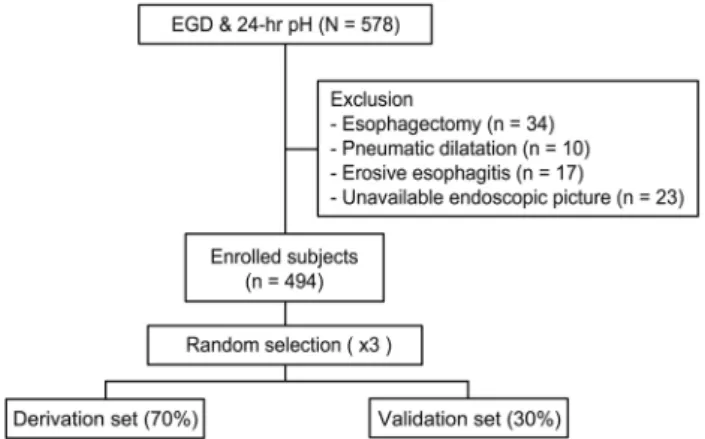
Fig. 1. Flow chart of patient selection. Among the 578 patients who underwent esophagogastroduodenoscopy (EGD) and 24 hours pH monitoring (24 hours pH) within a 1-month interval, patients who underwent an esophagectomy or pneumatic dilatation were excluded. Patients with erosive esophagitis on EGD were also excluded. After excluding 84 subjects, 494 subjects were enrolled.
To validate the prediction modeling procedure, the subjects were divided randomly into two groups in a ratio of 7:3 (derivation set:
validation set) three separate times.
of GERD, such as reflux esophagitis or Barrett’s esophagus (BE). In addition, endoscopy can assess the anatomic changes in the antireflux barrier.3
Although the pathogenesis of GERD is multifactorial, overt hiatal hernia (HH) is considered a key player, which is asso- ciated with most mechanisms underlying GERD.2,4 Hence, the major mechanism of GERD differs between patients with and without HH.5 Transient lower esophageal sphincter relaxations (TLESRs) are commonly associated with reflux episodes in normal subjects and mild GERD patients with HH.5,6 On the other hand, pathologic acid exposure (PAE) in GERD patients with HH compared to those without is caused by a dysfunction of the antireflux barrier.5
The LES, the crural diaphragm, and the anatomical flap valve make up the esophagogastric junction (EGJ). This com- plex functions as the antireflux barrier.7-12 Upper endoscopy can assess the flap valve and predict the reflux activity.13 In addition, several other endoscopic findings, such as HH, chronic atrophic gastritis (CAG), and endoscopically suspected esophageal metaplasia (ESEM) or BE, have been identified as the risk factors for GERD.4,10,14,15
This study evaluated the predictive role of the endoscopic findings for PAE and established an endoscopic prediction model for acid reflux in patients with neither reflux esophagitis nor HH.
SUBJECTS AND METHODS
1. Subjects
Five hundred seventy-eight patients over 18 years of age, who visited Samsung Medical Center for esophageal reflux symptoms, including chest pain, heartburn, regurgitation, dys- phagia, globus sensation, and chronic cough between June 2011 and February 2015, and who underwent 24 hours pH monitoring and upper endoscopy within a one-month interval were analyzed. The subjects had neither reflux esophagitis nor HH at endoscopy. Patients with a prior history of pneu- matic dilatation (n=10) or upper gastrointestinal surgery (n=34) were excluded. Finally, 494 patients were included in this study (Fig. 1). This study protocol was conducted in ac- cordance with the Declaration of Helsinki and approved by the Institutional Review Board of Samsung Medical Center on March 3, 2016 (No. 2016-03-001). The Institutional Review Board waived the requirement for informed consent
because de-identified data was used.
2. Data collection
The medical records, including demographic factors, such as age, sex, and chief complaint, were reviewed retrospectively.
All endoscopic images were reviewed by the consensus of two experienced endoscopists (Min YW and Shin IS). The status of ESEM, gastroesophageal flap valve (GEFV), and CAG were reviewed. The majority of images were clear enough to assess the EGJ because the routine endoscopy protocol includes care- ful observations of the EGJ in the authors’ institution.
This study used a single-use pH probe (Sandhill Scientific, Highlands Ranch, CO, USA) consisting of a 2.1 mm polyur- ethane catheter and a built-in pH probe positioned 5 cm above the upper margin of the LES. All patients were off medi- cations, including PPI, H2 receptor antagonists, and antacids, for seven days before reflux monitoring. After the measure- ments, the probes were withdrawn, and data were stored via a user interface on an IBM-compatible computer. Data analy- sis was performed using BioView MII software (Sandhill Scientific), and the data were also manually reviewed. The percentage acid exposure time for 24 hours was investigated.
3. Grades and definitions
The grade of CAG was diagnosed by evaluating the atrophic border in the EGD images. The atrophic pattern system de-
A B C D
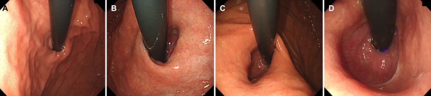
Fig. 2. Representative images of the gastroesophageal flap valve (GEFV) grade. (A) Grade 0. GEFV opening width ≤1 cm. (B) Grade 1. GEFV opening width less than 1.5 cm (1-1.5cm). (C) Grade 2. GEFV opening width less than 2 cm (1.5-2 cm). (D) Grade 3. GEFV opening width greater than 2 cm.
A B C
Fig. 3. Multivariable logistic regression with a stepwise selection. The patients were divided randomly into two groups at a ratio of 7:3 : a derivation group and a validation group. (A-C) show the results of the first, second, and third trials, respectively. In each trial, the derivation group was analyzed by multivariable logistic regression with stepwise selection. The endoscopic grades of the gastroesophageal flap valve and chronic atrophic gastritis showed consistent correlations with the pathologic acid exposure.
scribed by Kimura et al.16 was used. The subjects were div- ided into three groups. The first group consisted of patients without atrophic gastritis (no CAG). The second group included patients with mild atrophic gastritis, consisting of closed types 1 or 2 in the Kimura system (mild CAG). The third group was comprised of patients with significant atrophic gastritis, who had closed type 3 or above or open type gastritis (severe CAG).
The grade of GEFV was assessed by the width of the flap valve opening. The grade was divided into 0.5 cm intervals.
Grade 0 was defined as a GEFV opening width less than 1.0 cm; grade 1 was defined as a GEFV opening of 1.0 cm<
width<1.5 cm; grade 2 was defined as a GEFV opening of 1.5 cm<width<2.0 cm, and grade 3 is defined as a GEFV opening width greater than 2.0 cm (Fig. 2).
ESEM was assessed from the endoscopic findings without a biopsy. The presence of ESEM was defined as the maximal
(M) involvement of more than 5 mm based on the Prague system.17 PAE was defined when the intraesophageal pH was
<4 for more than 4.2% of the recording time.18
4. Statistical analyses
Univariate logistic regression analysis was used to explore the possible risk factors for PAE, including GEFV, ESEM, and CAG. A multivariate logistic regression model with a stepwise selection method was then used to predict the existence of PAE. The original data were divided randomly into derivation data and validation data at a ratio of 7:3, and used for pre- diction modeling and internal validation, respectively. The per- formance of the prediction model was evaluated from the area under the receiver operating characteristic curve, which was calculated using the test data. After validating the pre- diction model, the final prediction model for PAE was con- structed using the entire original data set. Statistical sig-
Fig. 4. Receiver operating characteristics curve of the established endoscopic prediction model for PAE. The area under the receiver operating characteristics curve of the final mathematical prediction model for PAE was 0.705 (95% CI 0.619-0.790). AUC, area under the receiver operating characteristic curve; CI, confidence interval; PAE, pathologic acid exposure.
Table 1. Baseline Characteristics of the Study Subjects
Variables Total patients (n=494)
Age (years) 53.1±12.2
Female 320 (64.8)
Chief complaint
Chest discomfort 209 (42.3)
Globus 157 (31.8)
Dysphagia 39 (7.9)
Heartburn 38 (7.7)
Chronic cough 25 (5.1)
Regurgitation 9 (1.8)
Rumination 7 (1.4)
Belching 4 (0.8)
Others 6 (1.2)
Pathologic acid exposure 43 (8.7)
Values are presented as mean±standard deviation or n (%).
Table 2. Endoscopic Findings of the Study Subjects
Variables Total patients (n=494)
Gastroesophageal flap valve
Grade 0 (≤1 cm) 321 (65.0)
Grade 1 (>1, <1.5 cm) 131 (26.5)
Grade 2 (1.5-2 cm) 32 (6.5)
Grade 3 (>2 cm) 11 (2.2)
ESEM
Absence 409 (82.8)
Presence 86 (17.4)
Chronic atrophic gastritis
None 194 (39.3)
Mild 214 (43.3)
Severe 87 (17.6)
Values are presented as n (%).
ESEM, endoscopically suspected esophageal metaplasia.
nificance was set to a p-value<0.05. Statistical analyses were conducted using SAS version 9.4 (SAS Institute, Cary, NC, USA) and R version 3.2.2 (Vienna, Austria; http://www.R-proj- ect.org).
Results
1. Characteristics of the subjects
Table 1 lists the baseline characteristics of the study population. The average age was 53±12.2 years old, and more than half of the subjects were female. The patients un- derwent upper endoscopy to examine the various esophageal reflux symptoms, the most common being chest discomfort (42.3%). The other common symptoms included globus (31.8%), dysphagia (7.9%), and heartburn (7.7%). Of the 494 subjects, PAE was found in 43 (8.7%).
2. Endoscopic findings
Table 2 lists the endoscopic findings. GEFV was tight (grade 0) in 64.8% of patients, and the majority of subjects (82.6%) did not have ESEM. CAG was present in 301 subjects (60.8%).
3. Association of endoscopic findings and PAE In univariate analysis, the grades of GEFV and CAG were associated significantly with PAE (GEFV, p=0.001; CAG, p=0.001) (Table 3). The CAG grade was inversely associated with PAE. ESEM showed a marginal association with PAE (p=0.061). Multivariate analysis showed that grades of GEFV and CAG were independent predictive factors for PAE (GEFV, p<0.001 and CAG, p=0.005) (Table 4).
Table 3. Univariate Analysis of the Associations between the Endoscopic Findings and Pathologic Acid Exposure
Variables OR (95% CI) p-value
Gastroesophageal flap valve 0.001
Grade 0 (≤1 cm) -
Grade 1 (>1, <1.5 cm) 1.53 (0.73-3.23)
Grade 2 (1.5-2 cm) 3.47 (1.28-9.41)
Grade 3 (>2 cm) 12.55 (3.52-44.69)
ESEM 0.061
Absence -
Presence 1.97 (0.97-4.02)
Chronic atrophic gastritis 0.001
No CAG -
Mild CAG 0.40 (0.20-0.80)
Severe CAG 0.22 (0.07-0.75)
OR, odds ratio; CI, confidence interval; ESEM, endoscopically suspected esophageal metaplasia; CAG, chronic atrophic gastritis.
Table 4. Multivariate Analysis of the Association between the Endoscopic Findings and Pathologic Acid Exposure
Parameter Estimate OR (95% CI) p-value
Gastroesophageal flap valve <0.001
Grade 0 (≤1 cm) 0 -
Grade 1 (>1, <1.5 cm) 0.4330 1.54 (0.72-3.29)
Grade 2 (1.5-2 cm) 1.1374 3.12 (1.13-8.61)
Grade 3 (>2 cm) 2.6849 14.66 (3.88-55.34)
Chronic atrophic gastritis 0.005
None 0 -
Mild -0.9668 0.38 (0.19-0.78)
Severe -1.5240 0.22 (0.06-0.77)
OR, odds ratio; CI, confidence interval.
4. Establishment of an endoscopic prediction model for PAE
After three trials of multivariable logistic regression with a stepwise selection, the grades of GEFV and CAG showed consistent relationships with PAE. Each trial was conducted on randomly selected patients followed by multiple logistic regression analysis, the establishment of a predictive model, and internal validation, which revealed good predictive power (AUROC 1st 0.783, 2nd 0.656, and 3rd 0.783) (Fig. 3). The following mathematical prediction model for PAE was estab- lished using two endoscopic factors:
Endoscopic reflux score=eQ/[1+eQ]×100, where Q=[-2.1631+
2.6859]×[GEFV grade 3+1.1374]×[GEFV grade 2+0.433]×[GEFV grade 1+(-1.524)×CAG grade 2+(-0.9668)×CAG grade 1 with GEFV grade 3=0 (no), =1 (yes); GEFV grade 2=0 (no), =1 (yes); GEFV grade 1=0 (no), =1 (yes); CAG grade 2=0 (no),
=1 (yes); CAG grade 1=0 (no), =1 (yes).
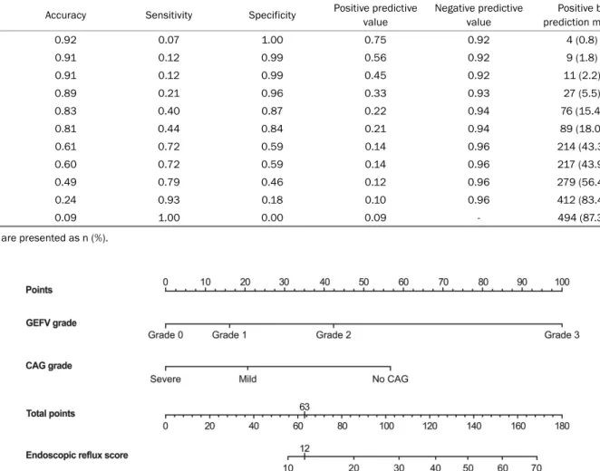
In this prediction model, an endoscopic reflux score of 12.0 showed the best performance for prediction. The cutoff value of 12.0 revealed an accuracy, sensitivity, specificity, positive predictive value, and negative predictive value of 81.0%, 44.0%, 84.0%, 21.0%, and 94.0%, respectively (Table 5). The final prediction model for PAE was established using the data from all subjects and showed an AUROC of 0.705 (95% CI, 0.619-0.790) (Fig. 4).
5. Nomogram for predicting PAE
A nomogram was developed using the prediction model (Fig. 5). In the nomogram, the assigned risk points of each endoscopic characteristic were expressed in the upper straightedge. The points for each predictor were added to ob- tain the total points. The straightedge was aligned with the
Table 5. Diagnostic Values of the Endoscopic Reflux Scores for Predicting Pathologic Acid Reflux in Patients with Neither Reflux Esophagitis nor Hiatus Hernia
Cutoff Accuracy Sensitivity Specificity Positive predictive value
Negative predictive value
Positive by prediction model
63.8 0.92 0.07 1.00 0.75 0.92 4 (0.8)
39.1 0.91 0.12 0.99 0.56 0.92 9 (1.8)
26.9 0.91 0.12 0.99 0.45 0.92 11 (2.2)
26.4 0.89 0.21 0.96 0.33 0.93 27 (5.5)
15.1 0.83 0.40 0.87 0.22 0.94 76 (15.4)
12.0 0.81 0.44 0.84 0.21 0.94 89 (18.0)
10.3 0.61 0.72 0.59 0.14 0.96 214 (43.3)
7.2 0.60 0.72 0.59 0.14 0.96 217 (43.9)
6.3 0.49 0.79 0.46 0.12 0.96 279 (56.4)
4.2 0.24 0.93 0.18 0.10 0.96 412 (83.4)
2.4 0.09 1.00 0.00 0.09 - 494 (87.3)
Values are presented as n (%).
Fig. 5. Nomogram for the endoscopic prediction of pathologic acid exposure in patients with reflux symptoms. Each point of the endoscopic parameter was calculated. The total points were added, and a vertical line was drawn from the total points’ row to obtain the endoscopic reflux score, which is associated with the PAE risk. The endoscopic reflux score cutoff value of 12, which corresponded to the total point cutoff value of 63, showed the best performance for predicting PAE with an accuracy, sensitivity, and specificity of 0.81, 0.44, and 0.84, respectively. GEFV gastroesophageal flap valve; CAG, chronic atrophic gastritis; PAE, pathologic acid exposure.
total points to determine the endoscopic reflux score at the bottom of the nomogram. An endoscopic reflux score over 12, which corresponded to total points of more than 63, sug- gested that the patients will have PAE in reflux monitoring.
DISCUSSION
Upper endoscopy is now the standard and basic tool for assessing gastrointestinal symptoms, including reflux symptoms.
Endoscopy can provide evidence of GERD and evaluate the anatomic changes in EGJ. Although the role of the mechanical antireflux barrier is lower in patients without HH than those with HH, an anatomical flap valve could be used to predict
reflux. If patients with reflux symptoms have neither reflux esophagitis nor HH on endoscopy, reflux monitoring could be necessary to confirm the acid reflux. This study evaluated the predictive role of the endoscopic findings and established a prediction model for PAE. A loosened GEFV and lower degree of atrophic gastritis were independent risk factors of PAE in patients with neither reflux esophagitis nor HH.
Previous studies have demonstrated the clinical sig- nificance of GEFV in GERD. The endoscopic GEFV grade was correlated with an increased incidence of regurgitation events, lower LES pressure, increased esophagitis grade, higher sur- gery rate, higher incidence of acid reflux, and more difficulty achieving symptomatic relief with PPI.10,19-21 Although the Hill
grade is the standard grading system for the flap valve,21 the GEFV grade was defined using measurements of the GEFV opening size with reference to the known diameter of an endo- scope to improve the intra-, inter-observer variations in this retrospective study. The results suggest that the impaired flap valve could contribute to acid reflux, even in patients without HH.
In the present study, the endoscopic severity of CAG was negatively correlated with the incidence of PAE. These results are compatible with those of previous studies.14 Several stud- ies have reported a significant inverse association between atrophic gastritis and reflux esophagitis and attributed the association to decreased gastric acidity.15,22-25 These findings suggest that PAE may be low when a patient with neither definite endoscopic evidence of GERD nor HH has chronic atrophic gastritis in a dose-dependent manner.
The study subjects did not have HH. Endoscopically HH was defined when the squamo-columnar junction above the visible stomach folds is displaced upwardly by more than 2 cm. On the other hand, a barium study is the only accurate means of measuring the HH size,26 but a diagnosis of HH could be inaccurate. In a previous study, the presence of HH was un- derestimated by endoscopy compared to a barium study.27 Therefore, some patients with HH could be included in this study. No patients showed Hill classification IV, but the possi- bility of misclassification may be quite low.
A prediction model for PAE with good discriminatory power was established. On the other hand, its positive predictive value was low because of the low incidence of PAE in this study population. Furthermore, because multifactorial factors can induce PAE, accurately predicting PAE just from the endo- scopic findings was necessarily unsatisfactory. The majority of study subjects may have symptoms not associated with acid reflux or symptoms associated with weakly acid or non-acid reflux.28-32 Therefore, the positive predictive value of the model can be improved by segregating patients with genuine reflux symptoms. In addition, 24 hours impedance-pH monitoring needs to be performed further when GERD is still suspected, even without endoscopic evidence, considering the low sensitivity of the endoscopic prediction model in pa- tients with neither reflux esophagitis nor HH.
This study had some limitations. First, its retrospective de- sign introduced inherent limitations, including endoscopic evaluations. In the present study, two experienced endo-
scopists reviewed the records and excluded those with endo- scopic images that were not clear enough to measure the necessary parameters. In addition, the routine upper endos- copy protocol of the authors’ institution includes a thorough examination of the morphology of GEFV, the presence of ESEM, and the degree of CAG sufficient to assess the endo- scopic parameter with accuracy. On the other hand, the varia- bles in the endoscopic findings were mostly subjective, and inter and intra-observer variations were unavoidable. Second, the study participants were all Koreans with a range of esoph- ageal symptoms. Thus, the findings may not be generalizable to other populations. Nevertheless, the endoscopic grading of GEFV and CAG have been proved to be independent pre- dictive factors for PAE in a validated prediction model. Third, all study participants with ESEM had an ultrashort-segment ESEM. This may have interfered with the evaluation of a rela- tionship between ESEM and PAE or may have been a barrier to generalization. Lastly, unknown previous antacid medi- cation could affect the presence of reflux esophagitis.
In conclusion, a loosened GEFV is associated with a risk of PAE in patients with neither reflux esophagitis nor HH, while atrophic gastritis is preventive. Given the low sensitivity of the endoscopic prediction, however, 24 hours impedance-pH monitoring is needed when GERD is still suspected, even with- out endoscopic evidence.
REFERENCES
1. Hershcovici T, Fass R. Step-by-step management of refractory gastresophageal reflux disease. Dis Esophagus 2013;26:27-36.
2. Boeckxstaens G, El-Serag HB, Smout AJ, Kahrilas PJ.
Symptomatic reflux disease: the present, the past and the future.
Gut 2014;63:1185-1193.
3. Kahrilas PJ, Boeckxstaens G, Smout AJ. Management of the pa- tient with incomplete response to PPI therapy. Best Pract Res Clin Gastroenterol 2013;27:401-414.
4. Herregods TV, Bredenoord AJ, Smout AJ. Pathophysiology of gas- troesophageal reflux disease: new understanding in a new era.
Neurogastroenterol Motil 2015;27:1202-1213.
5. van Herwaarden MA, Samsom M, Smout AJ. Excess gastro- esophageal reflux in patients with hiatus hernia is caused by mechanisms other than transient LES relaxations. Gastroenter- ology 2000;119:1439-1446.
6. Dodds WJ, Dent J, Hogan WJ, et al. Mechanisms of gastro- esophageal reflux in patients with reflux esophagitis. N Engl J Med 1982;307:1547-1552.
7. Bredenoord AJ, Pandolfino JE, Smout AJ. Gastro-oesophageal re- flux disease. Lancet 2013;381:1933-1942.
8. Curcic J, Roy S, Schwizer A, et al. Abnormal structure and function of the esophagogastric junction and proximal stomach in gastro- esophageal reflux disease. Am J Gastroenterol 2014;109:658-667.
9. Park CH, Kim KO, Baek IH, et al. Differences in the risk factors of reflux esophagitis according to age in Korea. Dis Esophagus 2014;27:116-121.
10. Kim GH, Song GA, Kim TO, et al. Endoscopic grading of gastro- esophageal flap valve and atrophic gastritis is helpful to pre- dict gastroesophageal reflux. J Gastroenterol Hepatol 2008;
23:208-214.
11. Cheong JH, Kim GH, Lee BE, et al. Endoscopic grading of gastro- esophageal flap valve helps predict proton pump inhibitor re- sponse in patients with gastroesophageal reflux disease. Scand J Gastroenterol 2011;46:789-796.
12. Mittal RK, Balaban DH. The esophagogastric junction. N Engl J Med 1997;336:924-932.
13. Koch OO, Spaun G, Antoniou SA, et al. Endoscopic grading of the gastroesophageal flap valve is correlated with reflux activ- ity and can predict the size of the esophageal hiatus in pa- tients with gastroesophageal reflux disease. Surg Endosc 2013;27:4590-4595.
14. Patti MG, Goldberg HI, Arcerito M, Bortolasi L, Tong J, Way LW.
Hiatal hernia size affects lower esophageal sphincter function, esophageal acid exposure, and the degree of mucosal injury. Am J Surg 1996;171:182-186.
15. Kim DH, Kim GH, Kim JY, et al. Endoscopic grading of atrophic gastritis is inversely associated with gastroesophageal reflux and gastropharyngeal reflux. Korean J Intern Med 2007;22:
231-236.
16. Kimura K, Satoh K, Ido K, Taniguchi Y, Takimoto T, Takemoto T.
Gastritis in the Japanese stomach. Scand J Gastroenterol Suppl 1996;214:17-23.
17. Sharma P, Dent J, Armstrong D, et al. The development and validation of an endoscopic grading system for Barrett's esophagus: the Prague C & M criteria. Gastroenterology 2006;
131:1392-1399.
18. Shay S, Tutuian R, Sifrim D, et al. Twenty-four hour ambulatory si- multaneous impedance and pH monitoring: a multicenter report of normal values from 60 healthy volunteers. Am J Gastroenterol 2004;99:1037-1043.
19. Kayaoglu HA. Correlation of the gastroesophageal flap valve grade with the surgery rate in patients with gastroesophageal re- flux disease. Surg Endosc 2013;27:801-807.
20. Lin BR, Wong JM, Chang MC, et al. Abnormal gastroesophageal flap valve is highly associated with gastroesophageal reflux dis- ease among subjects undergoing routine endoscopy in Taiwan.
J Gastroenterol Hepatol 2006;21:556-562.
21. Hill LD, Kozarek RA, Kraemer SJ, et al. The gastroesophageal flap valve: in vitro and in vivo observations. Gastrointest Endosc 1996;44:541-547.
22. Xia HH, Talley NJ. Helicobacter pylori infection, reflux esophagitis, and atrophic gastritis: an unexplored triangle. Am J Gastroenterol 1998;93:394-400.
23. Koike T, Ohara S, Sekine H, et al. Helicobacter pylori infection in- hibits reflux esophagitis by inducing atrophic gastritis. Am J Gastroenterol 1999;94:3468-3472.
24. Koike T, Ohara S, Sekine H, et al. Helicobacter pylori infection pre- vents erosive reflux oesophagitis by decreasing gastric acid secretion. Gut 2001;49:330-334.
25. Fujiwara Y, Higuchi K, Shiba M, et al. Association between gastro- esophageal flap valve, reflux esophagitis, Barrett's epithelium, and atrophic gastritis assessed by endoscopy in Japanese patients. J Gastroenterol 2003;38:533-539.
26. Ott DJ, Gelfand DW, Wu WC, Castell DO. Esophagogastric region and its rings. AJR Am J Roentgenol 1984;142:281-287.
27. Sloan S, Rademaker AW, Kahrilas PJ. Determinants of gastro- esophageal junction incompetence: hiatal hernia, lower esoph- ageal sphincter, or both?. Ann Intern Med 1992;117:977-982.
28. Savarino E, Zentilin P, Savarino V. NERD: an umbrella term includ- ing heterogeneous subpopulations. Nat Rev Gastroenterol Hepatol 2013;10:371-380.
29. Masiak W, Wallner G, Wallner J, Pedowski T, Solecki M. Combined esophageal multichannel intraluminal impedance and pH mon- itoring (MII -pH) in the diagnostics and treatment of gastro- esophageal reflux disease and its complications. Pol Przegl Chir 2011;83:488-496.
30. Khan MQ, Alaraj A, Alsohaibani F, Al-Kahtani K, Jbarah S, Al-Ashgar H. Diagnostic utility of impedance-pH monitoring in re- fractory non-erosive reflux disease. J Neurogastroenterol Motil 2014;20:497-505.
31. Hershcovici T, Fass R. Nonerosive reflux disease (NERD) - an update. J Neurogastroenterol Motil 2010;16:8-21.
32. Shi Y, Tan N, Zhang N, et al. Predictors of proton pump inhibitor failure in non-erosive reflux disease: a study with impedance-pH monitoring and high-resolution manometry. Neurogastroenterol Motil 2016;28:674-679.