Effects of Losartan on Catecholamine Release in the Isolated Rat Adrenal Gland
9
0
0
전체 글
(2) 328. HJ Noh, et al. blockade (losartan) of the renin-angiotensin-aldosterone system (RAS) in rats may decrease the excess sympathetic responses to stress in cardiovascular diseases as well as prevent the likely development of Type II diabetes mellitus (Uresin et al., 2004). In spontaneously hypertensive rats (SHRs), oral administration of AT1 antagonist (candesartan) can effectively block central actions of Ang II, regulating blood pressure and reaction to stress, and selectively and differentially modulating sympathoadrenal responses (Seltzer et al., 2004). Critchley and his colleagues (2004) have found that AT1 receptor antagonist candesartan, and the ACE inhibitor ramipril, increased basal CA release from the anaesthetized dog's adrenal gland along with decreases in blood pressure. However, it has been shown that AT2 stimulation induces CA secretion in cultured porcine chromaffin cells (Takekoshi et al., 2001). This suggests that AT2 receptors play a role in mediating CA secretion from the adrenal medulla of anesthetized dogs in response to Ang II receptor agonist administration in vivo. Furthermore, both PD 123319 and CGP 42112 inhibited the increase in adrenal CA secretion induced by local administration of Ang II (Martineau et al., 1999). Worck and his colleagues (1998) have speculated that Ang II through binding to both receptor subtypes (both AT1 and AT2) facilitates the sympathoadrenal reflex response by actions at several anatomical levels of the neural pathways involved in the sympathoadrenal reflex response elicited during insulin-induced hypoglycemia in conscious chronically instrumented rats. Thus, there seems to be some controversy about the effect of AT1 receptor blockade on the CA secretion in the adrenal gland. The aim of this study therefore was to determine whether losartan, a seletive antagonist of AT1 receptor, could influence the CA release in the isolated perfused model of the rat adrenal medulla.. METHODS Experimental procedure Male Sprague-Dawley rats, weighing 200 to 300 grams, were anesthetized with thiopental sodium (50 mg/kg) intraperitoneally. The adrenal gland was isolated by the methods described previously (Wakade, 1981). The abdomen was opened by a midline incision, and the left adrenal gland and surrounding area were exposed with the placement of three-hook retractors. The stomach, intestine and portion of the liver were not removed, but pushed over to the right side and covered with saline-soaked gauge pads and urine in the bladder was removed in order to obtain enough working space for tying blood vessels and cannulations. A cannula, used for perfusion of the adrenal gland, was inserted into the distal end of the renal vein after all branches of adrenal vein (if any), vena cava and aorta were ligated. Heparin (400 IU/ml) was injected into vena cava to prevent blood coagulation before ligating vessels and cannulations. A small slit was made into the adrenal cortex just opposite the entrance of the adrenal vein. Perfusion of the gland was started, making sure that no leakage was present, and the perfusion fluid escaped only from the slit made in the adrenal cortex. Then the adrenal gland, along with the ligated blood vessels and the cannula, was carefully removed from the animal and placed on a platform. of a leucite chamber. The chamber was continuously circuo lated with water heated at 37±1 C. Perfusion of adrenal gland The adrenal glands were perfused by means of a periⓇ staltic pump (ISCO pump, WIZ Co. U.S.A.) at a rate of 0.32 ml/min. The perfusion was carried out with Krebs-bicarbonate solution of following composition (mM): NaCl, 118.4; KCl, 4.7; CaCl2, 2.5; MgCl2, 1.18; NaHCO3, 25; KH2PO4, 1.2; glucose, 11.7. The solution was constantly bubbled with 95% O2+5% CO2 and the final pH of the solution was maintained at 7.4∼7.5. The solution contained disodium EDTA (10 μg/ml) and ascorbic acid (100 μg/ml) to prevent oxidation of catecholamines. Drug administration The perfusions of DMPP (100 μM) and Ang II for 1 or 2 minutes and/or a single injection of ACh (5.32 mM) and KCl (56 mM) in a volume of 0.05 ml were made into the perfusion stream via a three-way stopcock, respectively. McN-A-343 (100 μM), veratridine (100 μM), Ang II (100 nM), Bay-K-8644 (10 μM) and cyclopiazonic acid (10 μM) were also perfused for 4 min, respectively. In the preliminary experiments, it was found that upon administration of the above drugs, the secretory responses to ACh, KCl, McN-A-343, veratridine, Ang II, Bay-K-8644 and cyclopiazonic acid returned to preinjection level in about 4 min, but the responses to DMPP in 8 min. Collection of perfusate As a rule, prior to stimulation with the various secretagogues, the perfusate was collected for 4 min to determine the spontaneous secretion of CA (background sample). Immediately after the collection of the background sample, collection of the perfusates was continued in another tube as soon as the perfusion medium containing the stimulatory agent reached the adrenal gland. Stimulated sample's perfusate was collected for 4 to 8 min. The amounts secreted in the background sample have been subtracted from that secreted from the stimulated sample to obtain the net secretion value of CA, which is shown in all of the figures. To study the effect of losartan on the spontaneous and evoked secretion, the adrenal gland was perfused with Krebs solution containing losartan for 60 min, and then the perfusate was collected for a certain period (background sample). Then the medium was changed to the one containing the stimulating agent or along with losartan, and the perfusates were collected for the same period as that for the background sample. The adrenal gland's perfusate was collected in chilled tubes. Measurement of catecholamines The CA content of the perfusate was measured directly by the fluorometric method of Anton and Sayre (1962) without the intermediate purification alumina for the reasons described earlier (Wakade, 1981) using a fluorospectrophotometer (Kontron Co., Milano, Italy). A volume of 0.2 ml of the perfusate was used for the reaction. The CA content in the perfusate of the stimulated glands by secretagogues used in the present work was high enough to obtain readings several folds greater than the.
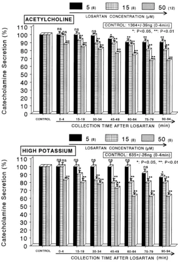
(3) Effects of Losartan on Catecholamine Release. reading of the control samples (unstimulated). The sample blanks were also lowest for perfusates of the stimulated and non-stimulated samples. The content of CA in the perfusate was expressed in terms of norepinephrine (base) equivalents. Statistical analysis The statistical difference between the control and pretreated groups was determined by the Student's t and ANOVA tests. A p value of less than 0.05 was considered to represent statistically significant changes unless specifically noted in the text. Values given in the text refer to means and the standard errors of the mean (SEM). The statistical analysis of the experimental results was made using a computer program described by Tallarida and Murray (1987).. 329. presence of losartan, CA releasing responses were inhibited to 68% of the corresponding control release. Also, the depolarizing agent, high potassium, markedly stimulated the CA secretion (635±26 ng for 0∼4 min). However, following + the pretreatment with losartan (5∼50 μM), high K (56 mM)-stimulated CA secretion was significantly inhibited to 63% of the control at last period (90∼94 min) as shown in Fig. 1 (lower). DMPP (100 μM), which is a selective nicotinic (NN) receptor agonist in autonomic sympathetic ganglia, evoked a sharp and rapid increase in CA secretion (1,317±33 ng for 0∼8 min). However, as shown in Fig. 2 (upper), DMPP-evoked CA secretion after pretreatment with losartan was greatly reduced to 71% of the control release (100%). McN-A-343 (100 μM), which is a selective. Drugs and their sources The following drugs were used: losartan, cyclopiazonic acid, acetylcholine chloride, 1.1-dimethyl-4-phenyl piperazinium iodide (DMPP), norepinephrine bitartrate, methyl-1, 4-dihydro-2,6-dimethyl-3-nitro-4-(2-trifluoro-methyl-phenyl)pyridine-5-carboxylate (BAY-K-8644), veratridine hydrochloride, angiotensin II (Sigma Chemical Co., U.S.A.), and (3-(m-chloro-phenyl-carbamoyl-oxy)-2-butynyltrimethyl ammonium chloride [McN-A-343]) (RBI, U.S.A.). Drugs were dissolved in distilled water (stock) and added to the normal Krebs solution as required except Bay-K-8644, which was dissolved in 99.5% ethanol and diluted appropriately with Krebs-bicarbonate solution (final concentration of alcohol was less than 0.1%). Concentrations of all drugs are expressed in terms of their molar base.. RESULTS Effects of losartan on CA secretion evoked by ACh, + high K , DMPP and McN-A-343 from the perfused rat adrenal glands After the perfusion with oxygenated Krebs-bicarbonate solution for 1 hr, basal CA release from the isolated perfused rat adrenal glands amounted to 22±3 ng for 2 min (n=12). Since a number of previous studies have indicated that the selective blockade of AT1 receptors failed to abolish the increase in adrenal CA secretion induced by Ang II (Bunn and Marley, 1989; Wong et al., 1990; Powis and O’Brien, 1991; Martineau, et al., 1995), it was attempted initially to examine the effects of losartan itself on CA secretion from the perfused model of the rat adrenal glands. However, in the present study, losartan (5∼50 μM) itself did not produce any effect on basal CA output from perfused rat adrenal glands (data not shown). Therefore, it was decided to investigate the effects of losartan on cholinergic receptor stimulation- as well as membrane depolarization-mediated CA secretion. Secretagogues were given at 15 min-intervals. Losartan was present for 90 minutes after the establishment of the control release. When ACh (5.32 mM) in a volume of 0.05 ml was injected into the perfusion stream, the amount of CA secreted was 1,364±39 ng for 4 min. However, in the presence of losartan in the range of 5∼50 μM for 90 min, ACh-stimulated CA secretion was inhibited in both a concentration- and time-dependent fashion. As shown in Fig. 1 (upper), in the. Fig. 1. Dose-dependent effects of losartan on the secretory responses of catecholamines (CA) evoked by acetylcholine (ACh, upper) and high potassium (lower) from the perfused rat adrenal medullas. The CA secretion by a single injection of ACh (5.32 mM) and K+ (56 mM) in a volume of 0.05 ml was evoked at 15 min intervals after preloading with 5, 15 and 50 μM of losartan for 90 min as indicated at an arrow mark, respectively. Numbers in the parenthesis indicate number of rat adrenal glands. Vertical bars on the columns represent the standard error of the mean (S.E.M.). Ordinate: the amounts of CA secreted from the adrenal gland (% of control). Abscissa: collection time of perfusate (min). Statistical difference was obtained by comparing the corresponding control (CONTROL) with each concentration-pretreated group of losartan. ACh- and high K+-induced perfusates were collected for 4 minutes, respectively. *p<0.05, **p<0.01. ns, statistically not significant..
(4) 330. HJ Noh, et al. Fig. 2. Dose-dependent effects of losartan on the CA secretory responses evoked by DMPP (upper) and McN-A-343 (lower) from the perfused rat adrenal medullas. The CA secretion by perfusion of DMPP (100 μM) and McN-A-343 (100 μM) for 2 min was induced at 15 and 20 min intervals after preloading with 5, 15 and 50 μM of losartan for 90 min, respectively. Statistical difference was obtained by comparing the corresponding control (CONTROL) with each concentration-pretreated group of losartan. DMPP- and McN-A-343-induced perfusates were collected for 8 and 4 minutes, respectively. Other legends are the same as in Fig. 1. *p<0.05, **p<0.01. ns, statistically not significant.. muscarinic M1-receptor agonist (Hammer and Giachetti, 1982), perfused into an adrenal gland for 4 min also caused an increased CA secretion (524±25 ng for 0∼4 min). However, in the presence of losartan, McN-A-343-evoked CA secretion was markedly depressed to 67% of the corresponding control secretion (100%) as depicted in Fig. 2 (lower). Effect of losartan on CA secretion evoked by Bay-K8644, cyclopiazonic acid, veratridine and Ang II from the perfused rat adrenal glands Since Bay-K-8644 is known to be a calcium channel activator, which enhances basal Ca2+ uptake (Garcia et al., 1984) and CA release (Lim et al., 1992), it was of interest to determine the effect of losartan on Bay-K-8644-evoked CA secretion from the isolated perfused rat adrenal glands. Bay-K-8644 (10 μM)-evoked CA secretion in the presence of losartan (15 μM) was greatly blocked to 75% of the con-. Fig. 3. Time-course effects of losartan on the CA release evoked by Bay-K-8644 (upper) and cyclopiazonic acid (lower) from the perfused rat adrenal medullas. Bay-K-8644 (10 μM) and cyclopiazonic acid (10 μM) were perfused into an adrenal vein for 4 min at 15 min intervals after preloading with losartan (15 μM) for 90 min, respectively. Other legends are the same as in Fig. 1. *p<0.05, **p<0.01. ns, statistically not significant.. trol at 75∼94 min period as compared to the corresponding control release (512±28 ng for 0∼4 min) from 7 adrenal glands as shown in Fig. 3 (upper). Cyclopiazonic acid, a mycotoxin from Aspergillus and Penicillium, has been described as a highly selective in2+ hibitor of Ca -ATPase in the skeletal muscle sarcoplasmic reticulum (Goeger and Riley, 1989; Seidler et al., 1989). The inhibitory action of losartan on cyclopiazonic acid-evoked CA secretory response was observed as shown in Fig. 3 (lower). In the presence of losartan (15 μM) from 8 adrenal glands, cyclopiazonic acid (10 μM)-evoked CA secretion was also inhibited to 72% of the control response (464±23 ng for 0∼4 min). + The voltage-dependent Na channels consist of the principal α-subunit, which is associated with noncovalently attached β1-subunits, and a disulfide-linked β2-subunit (Catterall, + 2000). It has also been known that veratridine-induced Na + 2+ influx mediated through Na channels increased Ca influx via activation of voltage-dependent Ca2+ channels and produced the exocytotic secretion of CA in cultured bovine adrenal medullary cells (Wada et al., 1985). To characterize the pharmacological action of losartan on voltage-depend-.
(5) 331. Effects of Losartan on Catecholamine Release. Fig. 5. High dose-effects of losartan on the ACh-evoked CA secretory responses from the perfused rat adrenal medullas. The CA secretion by a single injection of ACh (5.32 mM) in a volume of 0.05 ml was evoked at 15 min intervals after preloading with 150 and 300 μM of losartan for 90 min as indicated at an arrow mark. ACh-induced perfusate was collected for 4 minutes. Other legends are the same as in Fig. 1. *p<0.05, **p<0.01. ns, statistically not significant.. Fig. 4. Time-course effects of losartan on the CA release evoked by veratridine (upper) and angiotensin II (lower) from the perfused rat adrenal medullas. Veratridine (100 μM) and angiotensin II (100 nM) was perfused into an adrenal vein for 4 min and 1 min at 15 min intervals after preloading with losartan (15 μM) for 90 min, respectively. Other legends are the same as in Fig. 1. **p<0.01.. the CA secretion, in the presence of high doses (150 and 300 μM) of losartan, the CA secretory responses evoked by ACh-stimulation were examined. In the presence of losartan (150 μM) for 90 min, ACh-evoked CA release was not affected at initial periods (0∼49 min), but since then significantly enhanced to 106% of the corresponding control release as illustrated in Fig. 5. Moreover, after treatment with higher concentration (300 μM) for 90 min, ACh-evoked CA release was greatly enhanced to 123% of the corresponding control release during all periods (Fig. 5).. DISCUSSION +. ent Na channels, the effect of losartan on the CA secretion induced by veratridine was examined here. As shown in Fig. 4 (upper), veratridine greatly produced CA secretion (1,259±27 ng for 0∼4 min). However, in the presence of losartan (15 μM), veratridine (100 μM)-evoked CA secretion was greatly inhibited to 68% of the corresponding control release. Since Hano and his colleagues (1994) have suggested that Ang II increase epinephrine release from the adrenal medulla via the AT1 receptors, it was likely interesting to examine the effect of Ang II on the CA rease. Ang II (100 nM) significantly evoked the CA secretory response (469±54 ng for 0∼4 min) whereas, in the presence of losartan (15 μM), Ang II (100 nM)-evoked CA secretion was greatly inhibited to 46% of the corresponding control release (Fig. 4-lower). High dose effects of losartan on CA release evoked by ACh from the perfused rat adrenal glands As shown in Fig. 1∼4, it has also been shown that losartan inhibits the CA secretory response evoked by several secretagogues in the perfused rat adrenal glands. Therefore, in order to study the high dose effects of losartan on. These results obtained from the present study suggest that losartan can inhibit the CA secretion evoked by cholinergic stimulation (both nicotininc and muscarinic receptors) and membrane depolarization from the rat adrenal medulla. This inhibitory effect of losartan seems to be medi+ 2+ ated by blocking the influx of Na and Ca ions through their channels as well as by inhibiting the release of Ca2+ from cytoplasmic store through the blockade of Ang II AT1 receptors located on the presynatic membrane of the rat adrenomedullary chromaffin cells, which are relevant to adrenal nicotinic receptor blockade. In support of the present results, previously immobilisation stress has been shown to cause increase in plasma norepinephrine (NE) and epinephrine (E) levels (Saiki et al., 1997; Kubo et al., 2001). Intracerebroventricular application of ARBs inhibits the increases in plasma NE and E levels during stress exposure, indicating that the central Ang II system has an excitatory role in sympathetic responses to stress (Saiki et al., 1997). Armando and his colleagues (2001) found that pre-treatment with candesartan, an ARB, eliminated the increase in adrenal NE and E concentrations induced by isolation stress. On the other hand, it has been shown that acute and chronic stress stimulates.
(6) 332. HJ Noh, et al. the RAS to increase the levels of Ang II, both in the plasma and brain (Yang et al., 1993). It was also found that isolation stress enhanced Ang II receptor expression to a similar extent as occurs during repeated immobilisation stress (Castrén et al., 1988; Saavedra, 1992; Aguilera et al., 1995). Uresin and his colleagues (2004) have speculated that chronic blockade (losartan) of RAS in rats may decrease the excess sympathetic responses to stress in cardiovascular diseases and prevent the likely development of Type II diabetes mellitus. The AT1 antagonist, losartan blocked both inhibition and facilitation of secretion by AngII in cultured bovine chromaffin cells (Teschemacher and Seward, 2000). The results of this study showed that activation of multiple types of G-proteins and transduction pathways by single neuromodulator acting through one receptor type can produce concentration-dependent, bi-directional regulation of exocytosis (Teschemacher and Seward, 2000). Based on previous findings, the present results that losartan dose- and time-dependently reduced the CA secretory responses evoked by ACh, high potassium, DMPP and McN-A-343 from the perfused rat adrenal medulla might be due to the blockade of AT1 receptors located presynaptically on rat adrenomedullary chromaffin cells. Moreover, it has been shown that, in spontaneously hypertensive rats (SHRs), oral administration of AT1 antagonist (candesartan) can effectively block central actions of Ang II, regulating blood pressure and reaction to stress, and selectively and differentially modulating sympathoadrenal response and the hypothalamic-pituitary-adrenal stimulation produced by brain Ang II-effects of potential therapeutic importance (Seltzer et al., 2004). Barber and his co-workers (1999) have also suggested that, in SHR, AT2 receptor activation can facilitate the initial depressor response caused by an AT1 receptor antagonist. In the present study, as shown in Fig. 4 (lower), losartan also greatly inhibited Ang II-evoked CA release from the rat adrenal medulla. This finding indicates that losartan can inhibit the CA release evoked by cholinergic stimulation as well as by membrane depolarization. On the other hand, it has been demonstrated that, in cultured porcine chromaffin cells, AT2 stimulation induces CA 2+ through voltage-dependent secretion by mobilizing Ca 2+ channels without affecting intracellular pools and Ca that these effects could be mediated by a decrease in cGMP production (Takekoshi et al., 2001). Worck and his colleagues (1998) have also speculated that angiotensin II through binding to both receptor subtypes (both AT1 and AT2) facilitates the sympathoadrenal reflex response by actions at several anatomical levels of the neural pathways involved in the sympathoadrenal reflex response elicited during insulin-induced hypoglycemia in conscious chronically instrumented rats. In contrast, Takekoshi and his co-workers (2001) have demonstrated that CGP 42112 (AT2-R agonist) reduces both TH-enzyme activity and TH-synthesis biosynthesis in cultured porcine adrenal medullary cells and that these inhibitory effects could be mediated by decrease of cGMP production. Moreover, Martineau and his co-workers (1999) have suggested that AT2 receptors play a role in mediating CA secretion by the adrenal medulla of anesthetized dogs in response to AngII receptor agonist administration in vivo. PD 123319 and CGP 42112 were devoid of any agonist actions with respect to CA output by the adrenal gland in vivo. Furthermore, both PD 123319 and CGP 42112 inhibited the increase in adrenal CA secretion induced by local administration of Ang II. In light of these results, the present. findings seem to be disagreement with those results that adrenal CA secretion is mediated through AT2 receptors. Armando and his colleagues (2004) have demonstrated that both adrenomedullary AT1 and AT2 receptor types maintain and promote the adrenomedullary CA synthesis and the transcriptional regulation of TH in rats. Instead of opposing effects, however, these results indicate a complex synergistic regulation between the AT1 and AT2 receptor types. The nicotinic receptor is a neurotransmitter-gated cationconducting ion channel that is opened by binding of agonists such as ACh and DMPP (McGehee and Role, 1995). The opening of this channel triggers Ca2+ uptake and secretion of CA from chromaffin cells (Wada et al., 1985). To determine if the inhibition of DMPP-stimulated secretion by AT1 antagonist was due to an effect on the activity of the nicotinic receptor, the effect of losartan, an AT1-selective agonist, on DMPP-stimulated CA secretion was examined. As shown in Fig. 2, treatment with losartan greatly inhibited DMPP-evoked CA secretion, reducing by 71% of the control release. The present data are similar to the result that chronic immobilization stress increased plasma glucose, NE, E and corticosterone levels in the rats, and that the ARB losartan significantly prevented these increments induced by chronic stress when given before the stress regimen (Uresin et al., 2004). It is likely plausible that losartan can activate a signal transduction pathway that is altering the activity of both + nicotinic receptors and voltage-sensitive Na channels. It has been shown that most of AngII’s physiological effects, such as those exerted on the cardiovascular system and fluid volume homeostasis, are mediated by AT1; these effects are linked to 1,4,5-inositol triphosphate (IP3) production after phospholipase C activation, resulting in mobilization of 2+ intracellular Ca (Timmermans et al., 1993). Activation 2+ of such a pathway could result in elevated levels of Ca , diacylgIycerol, and inositol trisphosphate in the cells. Con2+ sequently Ca -dependent and protein kinase C (PKC)dependent pathways may be activated. PKC has been reported to attenuate the activity of both nicotinic receptors + (Swope et al., 1992) and voltage-sensitive Na channels (Catterall, 1992). Thus, these previous findings are in accordance with the present results that losartan inhibited the CA secretion evoked by ACh, DMPP and veratridine. In the present study, losartan, an AT1-selective antagonist inhibited the CA secretory responses by high potassium, a direct membrane depolarizer, as well as by Bay-K-8644, 2+ an activator of L-type Ca channels, which facilitates the influx of Ca2+ into the cells. The observation that AT1selective antagonist inhibited the CA secretion evoked by Bay-K-8644 was surprising, as Takekoshi et al. (2001) have reported that removal of external Ca2+ significantly suppressed either AngII plus CV-11974 (AT1 antagonist, 100 nM; which simulates specific AT2 stimulation) or CGP 42112 (AT2 agonist)-induced CA secretion in cultured porcine adrenomedullary chromaffin cells. It is unclear how the blockade of AT1 receptors results in the inhibition of secretion seen in these cells. The simplest interpretation 2+ is that the decrease in Ca uptake by losartan is responsible for the observed inhibition of the CA secretion. However, such an interpretation is complicated by the complexity of the relationship between the CA secretion and 2+ intraceIlular free Ca levels. Both the intracellular loca2+ tion of the Ca level increase (Cheek, 1989; Ghosh and 2+ Greenberg, 1995) and the magnitude of the Ca level in-.
(7) Effects of Losartan on Catecholamine Release. crease (Holz et al., 1982) can affect the relationship be2+ tween intracellular free Ca levels and secretion. Holz et 2+ al. (1982) have reported that when Ca uptake is large, 2+ changes in Ca uptake resulted in less than proportional changes in CA secretion. Consequently, although the de2+ crease in Ca uptake (influx) into the adrenal chromaffin cells may explain the decrease by losartan in CA secretion, it is still unclear whether this is only or even most important factor contributing to the inhibition of CA secretion by the AT1 antagonist. However, in view of the results so far obtained from the present study, it is felt that the volt2+ channels located on chromaffin cell age-sensitive Ca membrane of the rat adrenal medulla could be the target site for losartan-mediated inhibition of CA secretion. In the present study, losartan also inhibited the CA secretory responses evoked by cyclopiazonic acid, which is 2+ known to be a highly selective inhibitor of Ca -ATPase in skeletal muscle sarcoplasmic reticulum (Goeger and Riley, 1989; Seidler et al., 1989). Therefore, it is felt that the inhibitory effect of losartan on the CA secretion evoked by cholinergic stimulation as well as by membrane-depolarization may be associated with the mobilization of intra2+ cellular Ca in the chromaffin cells. This indicates that the blockade of AT1 receptors causes an inhibitory effect on the release of Ca2+ from the intracellular pools induced by stimulation of muscarinic ACh receptors, which is weakly responsible for the secretion of CA. In the present work, losartan time- and concentration-dependently produced the inhibition of CA secretion evoked by McN-A-343, a selective muscarinic M1-agonist. This fact suggests new other concept that losartan can modulate the CA secretory process induced by activation of muscarinic M1-receptors as well as neuronal nicotinic receptors in the rat adrenal medulla. In supporting this finding, it has been shown that cyclopiazonic acid easily penetrates into the cytoplasm through 2+ the plasma membrane and reduces Ca -ATPase activity in sarcoplasmic/endoplasmic reticulum, resulting in in2+ crease in the subsequent Ca release from those storage 2+ + sites and thereby increase of Ca -dependent K -current (Suzuki et al., 1992). Moreover, in bovine adrenal chromaffin cells, stimulation of muscarinic ACh receptors is also proposed to cause activation of phosphoinositide metabolism, resulting in the formation of inositol 1,4,5-trisphosphate, 2+ which induces the mobilization of Ca from the intracellular pools (Cheek et al., 1989; Challiss et al., 1991). However, in the present study, it is uncertain whether the 2+ inhibitory effect of the losartan on Ca movement from intracellular pools is due to their direct effect on the PI response or the indirect effect as a result of AT1 receptor blockade by losartan. Based on these previous results, this finding of the present work suggests that AT1 receptor blockade-induced inhibition may be involved in regulating CA secretion evoked by muscarinic M1-receptor stimulation in the rat adrenal medullary chromaffin cells. Furthermore, Ang II is a secretogogue for CA release that is believed to be mediated through IP3 production by AT1 (Wong et al., 1990; Dendorfer et al., 1998). Indeed, Wong and his colleagues (1990) demonstrated that AngII-induced CA release is mediated by AT1 in the rat adrenal medulla. AT1-mediated phospholipase C activation and subsequent IP3 formation may increase cytosolic Ca2+ levels by releasing Ca2+ from intracellular storage, with subsequent activation of CA release (Israel et al., 1995). Indeed, it has been shown that addition of IP3 to permeabilized bovine chromaffin cells re2+ leases intracellular Ca (Stoehr et al., 1986). Furthermore,. 333. addition of Ca2+ to permeabilized bovine chromaffin cells was reported to cause CA secretion (Dunn and Holz, 1983). On the other hand, in the present work, high concentrations of losartan (150 and 300 μM) significantly enhanced ACh-evoked CA secretory responses. As this result alone, there seems to be difficult for interpretation of the enhancement of ACh-evoked CA secretion by high dose of losartan. In support of this idea, the research results of Vijayapandi and Nagappa (2005) showed biphasic effects of losartan potassium on immobility in mice: reducd immobility at lower dose (0.1 and 5 mg/kg, i.p.) and enhanced immobility in higher dose (100 mg/kg, i.p.). These biphasic effects were further confirmed by interaction of losartan potassium with reserpine and antidepressant drugs, nortriptylline and fluoxetine (Vijayapandi and Nagappa, 2005). Nahmod and his colleagues (1978) found Ang II to cause 5-HT release and accelerate its synthesis in biphasic manner, stimulating at high doses and inhibiting at lower doses. Vijayapandi and Nagappa (2005) have obtained that the biphasic effect of losartan potassium on immobility in mice might be due to inhibitory effect on AT1 receptor at lower dose and pronounced effect on AT2 receptor at higher dose (large concentrations of losartan potassium can displace Ang II from its AT1 receptor to AT2 receptor). In chronic studies with losartan potassium even at lower dose (3 mg/kg, P.O.) potentiated immobility in mice, which might be due to continuous blockade of AT1 receptor resulting in unopposed AT2 receptor stimulation (Vijayapandi and Nagappa, 2005). It has also been previously reported that the treatment of Ang II for 4 h has a biphasic effect on Na+ transport in the primary cultured rabbit renal proximal tubule cells (PTCs) ; a pico molar range of Ang II stim+ ulates Na transport, whereas a micro molar range of Ang II inhibits it (Han et al., 2000). Based on these previous results, in the present study, it seems that biphasic effects of losartan on the CA secretion in the perfused rat adrenal medulla are due to inhibitory effect on AT1 receptor at lower dose (5∼50 μM) and pronounced effect on AT2 receptor at higher dose (150 and 300 μM), indicating that large concentrations of losartan can displace Ang II from its AT1 receptor to AT2 receptor. However, the detailed relationship between AT1 and AT2 receptors in adrenomedullary CA secretion should be confirmed in the future study. Taken together, these experimental results suggest that losartan at low concentrations inhibits the CA secretion evoked by cholinergic stimulation (both nicotininc and muscarinic receptors) as well as by membrane depolarization from the rat adrenal medulla, but at high concentration it rather inhibits Ach-evoked CA secretion. It seems that losartan has dual action acting as both agonist and antagonist at nicotinic receptors of the rat adrenal medulla, which might be dependent on the concentration. It is also thought that this inhibitory effect of losartan may be mediated by + 2+ blocking the influx of both Na and Ca through their channels into the rat adrenomedullary chromaffin cells as 2+ well as by inhibiting the Ca release from its cytoplasmic calcium store, which is thought to be relevant to AT1 receptor blockade, in addition to its unknown enhancement effect on the CA release.. ACKNOWLEDGEMENTS This study was supported partly by the fund of Chosun University (2008)..
(8) 334. HJ Noh, et al. REFERENCES Aguilera G, Kiss A, Luo X. Increased expression of type 1 angiotensin II receptors in the hypothalamic paraventricular nucleus following stress and glucocorticoid administration. J Neuroendocrinol 7: 775−783, 1995. Anton AH, Sayre DF. A study of the factors affecting the aluminum oxidetrihydroxy indole procedure for the analysis of catecholamines. J Pharmacol Exp Ther 138: 360−375, 1962. Armando I, Carranza A, Nishimura Y, Hoe KL, Barontini M, Terron JA, Falcon-Neri A, Ito T, Jourio AV, Saavedra JM. Peripheral administration of and angiotensin II AT1 receptor antagonist decreases the hypothalamic-pituitary-adrenal response to isolation stress. Endocrinology 142: 3880−3889, 2001. Armando I, Jezova M, Bregonzio C, Baiardi G, Saavedra JM. Angiotensin II AT1 and AT2 receptor types regulate basal and stress-induced adrenomedullary catecholamine production through transcriptional regulation of tyrosine hydroxylase. Ann NY Acad Sci 1018: 302−309, 2004. Barber MN, Sampey DB, Widdop RE. AT(2) receptor stimulation enhances antihypertensive effect of AT(1) receptor antagonist in hypertensive rats. Hypertension 34: 1112−1116, 1999. Bunn SJ, Marley PD. Effects of angiotensin II on cultured, bovine adrenal medullary cells. Neuropeptides 13: 121−132, 1989. Castrén E, Saavedra JM. Repeated stress increases the density of angiotensin II binding sites in the rat paraventricular nucleus and subfornical organ. Endocrinology 122: 370−372, 1988. Catterall WA. Cellular and molecular biology of voltage-gated sodium channels. Physiol Rev 72: 15−48, 1992. Catterall WA. From ionic currents to molecular mechanisms: the structure and function of voltage-gated sodium channels. Neuron 26: 13−25, 2000. Challiss RA, Jones JA, Owen PJ, Boarder MR. Changes in inositol 1,4,5-trisphosphate and inositol 1,3,4,5-tetrakisphosphate mass accumulations in cultured adrenal chromaffin cells in response to bradykinin and histamine. J Neurochem 56: 1083−1086, 1991. Cheek TR, O'Sullivan AJ, Moreton RB, Berridge MJ, Burgoyne RD. Spatial localization of the stimulus-induced rise in cyrosolic Ca2+ in bovine adrenal chromaffin cells: Distinct nicotinic and muscarinic patterns. FEBS Lett 247: 429−434, 1989. Critchley L, Ding B, Fok B, Wang D, Tomlinson B, James A, Thomas GN, Critchley J. The effects of candesartan and ramipril on adrenal catecholamine release in anaesthetized dogs. Eur J Pharmacol 489: 67−75, 2004. Dendorfer A, Raasch W, Tempel K, Dominiak P. Interactions between the renin-angiotensin system (RAS) and the sympathetic system. Basic Res Cardiol 93: 24−29, 1998. Dunn LA, Holz RW. Catecholamine secretion from digitonin-treated adrenal medullary chromaffin cells. J Biol Chem 258: 4989− 4993, 1983. Garcia AG, Sala F, Reig JA, Viniegra S, Frias J, Fonteriz R, Gandia L. Dihydropyridine Bay-K-8644 activates chromaffin cell calcium channels. Nature 309: 69−71, 1984. Ghosh A, Greenberg ME. Calcium signaling in neurons: molecular mechanisms and cellular consequences. Science 268: 239−247, 1995. Goeger DE, Riley RT. Interaction of cyclopiazonic acid with rat skeletal muscle sarcoplasmic reticulum vesicles. Effect on Ca2+ binding and Ca2+ permeability. Biochem Pharmacol 38: 3995− 4003, 1989. Hammer R, Giachetti A. Muscarinic receptor subtypes: M1 and M2 biochemical and functional characterization. Life Sci 31: 2992− 2998, 1982. Han HJ, Park SH, Koh HJ, Taub M. Mechanism of regulation of Na+ transport by angiotensin II in primary renal cells. Kidney Int 57: 2457−2467, 2000. Hano T, Mizukoshi M, Baba A, Nakamura N, Nishio I. Angiotensin II subtype 1 receptor modulates epinephrine release from isolated rat adrenal gland. Blood Press 5: 105−108, 1994. Holz RW, Senter RA, Frye RA. Relationship between Ca2+ uptake and catecholamine secretion in primary dissociated cultures of. adrenal modulla. J Neurochem 39: 635−640, 1982. Israel A, Strömberg C, Tsutsumi K, Garrido MR, Torres M, Saavedra JM. Angiotensin II receptor subtypes and phosphoinositide hydrolysis in rat adrenal medulla. Brain Res Bull 38: 441−446, 1995. Kubo T, Numakura H, Endo S, Hagiwara Y, Fukumori R. Angiotensin receptor blockade in the anterior hypothalamic area inhibits stress-induced pressor responses in rats. Brain Res Bull 56: 569−574, 2001. Lim DY, Kim CD, Ahn KW. Influence of TMB-8 on secretion of catecholamines from the perfused rat adrenal glands. Arch Pharm Res 15: 115−125, 1992. Livett BG, Marley PD. Non cholinergic control of adrenal catecholamine secretion. J Anat 183: 277−289, 1993. Martineau D, Lamouche S, Briand R, Yamaguchi N. Functional involvement of angiotensin AT2 receptor in adrenal catecholamine secretion in vivo. Can J Physiol Pharmacol 77: 367−374, 1999. Martineau D, Yamaguchi N, Briand R. Inhibition by BMS 186295, a selective nonpeptide AT1 antagonist, of adrenal catecholamine release induced by angiotensin II in the dog in vivo. Can J Physiol Pharmacol 73: 459−464, 1995. McGehee DS, Role LW. Physiological diversity of nicotinic acetylcholine receptors expressed by vertebrate neurons. Annu Rev Physiol 57: 521−546, 1995. Nahmod VE, Finkielman S, Benarroch EE, Pirola CJ. Angiotensin regulates release and synthesis of serotonin in brain. Science 202: 1091−1093, 1978. Phillips MI, Speakman EA, Kimura B. Levels of angiotensin and molecular biology of the tissue rennin angiotensin systems. Regul Pept 43: 1−20, 1993. Plunkett LM, Correa FM, Saavedra JM. Quantitative autoradiographic determination of angiotensin-converting enzyme binding in rat pituitary and adrenal glands with 124I−351A, a specific inhibitor. Regul Pept 28: 263−272, 1985. Powis DA, O'Brien KJ. Angiotensin II increases catecholamine release from bovine adrenal medulla but does not enhance that evoked by K+ depolarization or by carbachol. J Neurochem 57: 1461−1469, 1991. Saavedra JM. Brain and pituitary angiotensin. Endocr Rev 13: 329− 380, 1992. Saiki Y, Watanabe T, Tan N, Matsuzaki M, Nakamura S. Role of central ANG-II receptors in stress-induced cardiovascular and hyperthermic responses in rats. Am J Physiol 272: 26−33, 1997. Seidler NW, Jona I, Vegh N, Martonosi A. Cyclopiazonic acid is a specific inhibitor of the Ca2+-ATPase of sarcoplasimc reticulum. J Biol Chem 264: 17816−17823, 1989. Seltzer A, Bregonzio C, Armando I, Baiardi G, Saavedra JM. Oral administration of an AT1 receptor antagonist prevents the central effects of angiotensin II in spontaneously hypertensive rats. Brain Res 1028: 9−18, 2004. Stoehr SJ, Smolen JE, Holz RW, Agranoff BW. Inositol trisphosphate mobilizes intracellular calcium in permeabilized adrenal chromaffin cells. J Neurochem 46: 637−640, 1986. Suzuki M, Muraki K, Imaizumi Y, Watanabe M. Cyclopiazonic acid, an inhibitor of the sarcoplasmic reticulum Ca2+-pump, reduces Ca2+-dependent K+ currents in guinea-pig smooth muscle cells. Br J Pharmacol 107: 134−140, 1992. Swope SL, Moss SJ, Blackstone CD, Huganir RL. Phosphorylation of ligand-gated ion channels: a possible mode of synaptic plasticity. FASEB J 6: 2514−2523, 1992. Takekoshi K, Ishii K, Kawakami Y, Isobe K, Nakai T. Activation of angiotensin II subtype 2 receptor induces catecholamine release in an extracellular Ca2+-dependent manner through a decrease of cyclic guanosine 3′,5′-monophosphate production in cultured porcine adrenal medullary chromaffin cells. Endocrinol 142: 3075−3086, 2001. Tallarida RJ, Murray RB. Manual of pharmacologic calculation with computer programs. 2nd ed. Speringer-Verlag, New York, p 132, 1987. Teschemacher AG, Seward EP. Bidirectional modulation of exocytosis by angiotensin II involves multiple G-protein-regulated.
(9) Effects of Losartan on Catecholamine Release. transduction pathways in chromaffin cells. The J Neurosci 20: 4776−4785, 2000. Timmermans PB, Wong PC, Chiu AT, Herblin WF, Smith RD. New perspectives in angiotensin system control. J Hum Hypertens 7: 19−31, 1993. Uresin Y, Erbas B, Ozek M, Ozkök E, Gürol AO. Losartan may prevent the elevation of plasma glucose, corticosterone and catecholamine levels induced by chronic stress. J Renin Angiotensin Aldosterone Syst 5: 93−96, 2004. Vijayapandi P, Nagappa AN. Biphasic effects of losartan potassium on immobility in mice. Yakugaku Zasshi 125: 653−657, 2005. Wada A, Takara H, Izumi F, Kobayashi H, Yanagihara N. Influx of 22Na through acetylcholine receptor-associated Na channels: relationship between 22Na influx, 45Ca influx and secretion of catecholamines in cultured bovine adrenal medulla cells. Neuroscience 15: 283−292, 1985.. 335. Wakade AR. Studies on secretion of catecholamines evoked by acetylcholine or transmural stimulation of the rat adrenal gland. J Physiol 313: 463−480, 1981. Wong PC, Hart SD, Zaspel AM, Chiu AT, Ardecky RJ, Smith RD, Timmermans PB. Functional studies of nonpeptide angiotensin II receptor subtype-specific ligands: DuP 753 (AII-1) and PD123177 (AII-2). J Pharmacol Exp Ther 255: 584−592, 1990. Worck RH, Frandsen E, Ibsen H, Petersen JS. AT1 and AT2 receptor blockade and epinephrine release during insulin-induced hypoglycemia. Hyperten 31: 384−390, 1998. Yang G, Xi Z, Wan Y, Wang H, Bi G. Changes in circulating and tissue angiotensin II during acute and chronic stress. Biol Signals 2: 166−172, 1993. Zaman MA, Oparil S, Calhoun DA. Drugs targeting the reninangiotensin-aldosterone system. Nat Rev Drug Discov 1: 621− 636, 2002..
(10)
수치


관련 문서