임상적용을 위한 전자선의 선량분포 특성에 대한 고찰
차동수
영남대학교병원 방사선종양학과
11)
A Consideration on the Characteristics of
Electron Beam Dose Distributions for Clinical Applications
Dong Soo Cha
Department of Radiation Oncology, Yeungnam University Hospital Abstract
High energy electron beams were to concentrically dose inside a tumor and more energy is a shape decreased of dose. Therefore, it is useful to radiation therapy of a tumor. Also high energy electron beams ionized into collision with a atom in structure material of tissue and it has big changes to dose distribution by multiple scattering. The study had to establish characteristic of electron beams from interaction of electron beams and materials. Experiment method was to measure dependence of electron beam central axis for depth dose curve, field flatness and symmetry and field size dependence. The results were able to evaluate data for a datum pint of electron beam. Also radiotherapy has to be considered for not only energy pencil of lines but characteristic, electron guide and isodose curves distribution.
Key Words : Electron beams, Multiple scattering, Depth dose, Field flatness, Field symmetry
Ⅰ. 서 론
고 에너지 전자선은 물 또는 연부조직에서 1cm를 진 행하는데 약 2 MeV의 에너지를 소실하고 어느 깊이까 지는 주어진 선량을 부여하고 이상에서 선량이 급격히 떨어지는 물리적 특성을 가지기 때문에 임상적으로 유 용한 고 에너지 전자선의 범위는 6~20 MeV이다.
1의료
Received April 12, 2010, 1st Revised May 3, 2010, Accepted May 25, 2010.
Corresponding Author: 차동수
(705-717) 대구광역시 남구 대명5동 317-1번지 영남대학교의료원 방사선종양학과
Tel: 053) 620-3050 Fax: 053) 620-3057 E-mail: [email protected]
용 선형가속기에서 방출되는 전자선(electron beam)의 물리적 특성은 산란박(scattering foil), 조준기(colli- mator)의 개방(opening), 조사통(electron cone)의 구 조, 조사면의 크기 및 형태 그리고 차폐물 또는 조직 구성물질의 밀도 및 두께에 따라 결정 된다.
2~4이러한 고 에너지 전자선의 특성을 물질 내에서 전자선의 비정 (range)과 비적(track)을 규명하고 종양 치료에 적합한 에너지(probable energy)와 조사통, 인체를 구성하는 조직의 구성 등에 따른 전자선의 선량분포의 특성을 정 확하게 측정하여 임상에 적용해야만 한다.
일반적으로 전자선 치료는 1문 조사(one portal)를
시행하며 상대적으로 편평하고 균질적인 조직의 경우
선량분포는 적절한 등선량분포를 이용할 수 있으나 실
제 인체에 대한 전자선 치료는 이러한 단순성은 거의
없으며 표면은 거의 편평하지 않고 많은 경우 뼈 (bone), 폐(lung), 공기(air) 같은 비 균질성이 존재하 기 때문에 선량분포 특성은 매우 복잡한 관계를 갖게 된다. 현재 임상적으로 복잡한 인체에 대한 전자선의 선량분포 특성을 측정할 수 없으며 가상적인 모형으로 Pencil beam 또는 Monte Carlo simulation을 이용하 고 있다.
5~6본 연구는 중심축 심부선량 곡선(central axis depth dose curve), 조사면의 평탄도(field flatness)와 대칭 도(symmetry), 조사면 의존성(field size dependence) 등에 대한 전자선의 선량분포를 측정하고 이론적 배경 을 고찰함으로서 방사선치료계획 수립을 위한 기본적인 자료를 제공하자 하였다.
Ⅱ. 재료 및 방법
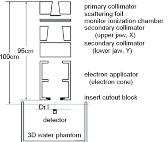
실험을 위하여 선형가속기(Clinac 2100 C/D, Varian, USA)에서 출력되는 전자선의 명목에너지(given energy) 6, 9, 12, 15, 18 MeV를 대상으로 하였다. Fig. 1은 물 팬텀(water phantom)을 이용하여 중심축 심부선량 곡 선(central axis depth dose curve)과 조사면의 평탄도 (field flatness)와 대칭도(symmetry)를 측정하는 기하 학적 구조를 보여준다. 심부선량 측정은 선원과 물 표 면까지의 거리를 100 cm로 고정시키고 심부선량은 전 리함을 물 표면에서 10 cm 깊이까지 이동시키면서 측정
Fig. 1. Geometry of the Clinac 2100C/D used for central axis depth dose curve, field flatness and
symmetry of measurement
하였으며 평탄도와 대칭도는 최대선량 지점에서 좌우측 방향으로 측정하였다. 중심선속 최대선량깊이(maximum dose depth)를 결정하기 위하여 전리함을 1 mm/sec의 속도로 심부로 이동시켜 측정하였으며 신호 변화가 심 한 영역에서는 1 mm 깊이마다 선량변화를 기록하고 그 외 영역에서는 2 mm 깊이마다 기록하였다. 조사면의 평탄도와 대칭도 측정은 심부선량 측정에 얻어진 최대 선량깊이에서 중심선속에서 좌측에서 우측으로, 갠트리 방향에서 테이블 방향으로 각각 측정하였다.
심부선량 및 조사면의 평탄도와 대칭도 측정에 사용 되어진 물 팬텀(MP3-M, PTW, Germany)은 규격이 636×634×523 mm
3이고 팬텀 내에서 검출기의 3차원적 인 이동을 위하여 소형 제어기(local control unit)가 부착되어 있으며 개인용 컴퓨터에 설치된 소프트웨어 (MEPHYST, PTW, Germany)를 이용하여 검출기의 제 어가 가능하다. 검출기와 전위계는 0.125 cm
3원통형 이온함(Semiflex, PTW, Germany)과 수집전압이 400 V인 전위계(UNIDOS, PTW, Germany)를 이용하였으며 국제적 교정규약(calibration protocol)
7~9에 따라 표준 기술연구소(National Institute of Standards and Technology, NIST)에서 불확정도가 ±1% 미만으로 교 정되었다.
Fig. 2는 조사면 의존성(field size dependence) 측 정을 보여준다. 조사면 의존성 측정은 조사통에 삽입되 는 차폐물(insert shielding block)의 크기를 2×2 cm
2에서 25×25 cm
2까지 1×1 cm
2간격으로 제작하였다. 선
Fig. 2. Geometry of the Clinac 2100C/D used for
field size dependence of measurement
원에서 고체 팬텀(solid dry phantom)의 표면까지의 거리를 100 cm으로 하여 심부선량 측정에서 얻어진 각 전자선의 명목에너지에 대한 최대선량깊이에서 조사선 량 100모니터 단위(monitor unit)을 조사하여 측정하였 다. 조사면 의존성 측정을 위하여 사용되어진 고체 팬 텀(RW3 slab, PTW, Germany)의 규격은 30×30 mm
2의 면적에 1 mm, 2 mm, 5 mm, 10 mm 두께를 가진 판 으로 임의의 측정 깊이를 재현할 수 있도록 고안되었으 며 물과 등가인 물질로 구성되어 있다.
Ⅲ. 결과 및 고찰
전자선의 명목에너지 6, 9, 12, 15, 1 8MeV를 대상으로 중심축 심부선량 곡선(central axis depth dose curve), 조사면의 평탄도(field flatness)와 대칭도(symmetry), 조사면 의존성(field size dependence)을 측정하여 비 교한 결과는 다음과 같다.
1. 중심축 심부선량 곡선
(Central Axis Depth Dose Curve)
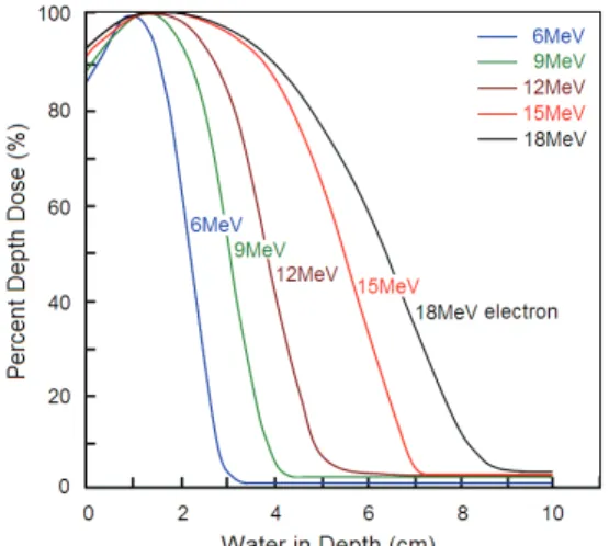
Fig. 3은 조사면 10×10 cm
2크기에서 각 전자선 명 목에너지에 대한 중심축 심부선량 곡선을 보여준다. 결 과에서 최대선량에 대하여 약 80%와 90%의 선량 깊이 는 보편적으로 MeV 단위의 전자선 명목에너지의 1/3과 1/4에 비슷한 경향을 보였으며 이 값은 전자선 에너지 와 직선관계를 따르지는 않지만 Khan 등
10의 연구에서 Ep.0를 전자선의 최대에너지(probable energy)라 할
Fig. 3. Comparison of central axis depth dose distributions of the electron energies(10×10 cm
2)
때 Ep.0( MeV)/2.8 cm와 Ep.0( MeV)/3.2 cm의 근사적 값과 일치하였다. 임상적으로 유용한 전자선 에너지 영 역에 있는 경우 심부선량 곡선은 종양이 존재하는 유용 한 깊이까지는 충분한 선량 전달을 하고 그 깊이 이상 에서는 급격하게 떨어져 정상조직 보호효과가 있음을 알 수 있었다.
또한 Table 1에서 최대선량에 대한 표면선량의 비로서 정의되는 표면선량백분율(percent surface dose)은 낮은 에너지의 전자일수록 산란각이 증가되고 전자선량이 짧 은 거리에 걸쳐 빠르게 부여되기 때문에 에너지의 증가 함에 따라 증가하는 것으로 보인다. 이러한 전자의 산란 에 의한 심부선량의 변화는 산란박(scattering foil)의 두께와 재질, 조리개의 개방(collimator opening) 크기, 조사면의 크기 및 형태, 조사통 끝에서 피부 표면까지 거리 등에 의존한다고 보고하였다.
11~16따라서 임상에서 선속 에너지만을 고려하는 것은 충분하지 못하며 개별 적인 선형가속기의 특성 및 조사통, 조사면에서의 등선 량곡선 분포들이 요구될 것으로 사료된다.
2. 조사면의 평탄도 및 대칭도 (Field Flatness and Symmetry)
미국의학물리학회(AAPM)
9에서 조사면 평탄도는 균일 지수(uniformity index)를 이용하여 중심선속 팬텀 표 면에서 기하학적 조사면에 대하여 기준점 깊이에서 중 심축 선량의 90%를 넘는 면적의 비로 정의하였으며 균 일지수가 0.8을 초과하는 결과를 기준으로 하였다. 선 속 내에 저 에너지 전자들이 존재하기 때문에 조사면의 평탄도는 깊이에 따라 의존성을 갖는다는 것을 알 수 있었으며 중심축 선량에 대한 선량의 변화는 10×10 cm
2와 같거나 그 이상인 조사면의 기하학적 가장자리 2 cm 안의 선들로 국한된 면적에 걸쳐 ±3% 이내에 들 어옴을 알 수 있었다.
조사면의 대칭도는 중심축의 한 면과 반대 면의 선량 측면도를 비교하였다. 측정한 결과 기준면에서 교차하 는 선속 측면도가 중심축의 대칭되는 양면에 위치된 어 느 한 쌍의 점들에서 ±2% 이내에 들어옴을 알 수 있었 다.
3. 조사면 의존성(Field Size Dependence)
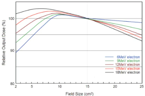
Fig. 4는 최대선량 깊이에서 조사면 15×15 cm
2를 기 준으로 다른 조사면에 대한 조사면 의존성을 보여준다.
최대선량 깊이에서 흡수선량의 비는 모든 에너지에서
Table 1. The defining of the parameters on according to the electron energies and field size for central axis depth dose curves for the electron beam of the Clinac 2100C/D
Parameters Field
(cm
2)
Electron energies(MeV)
Range Energy
R100[mm] Rp[mm] R50[mm] R80[mm] Ds[%] Ep.0[MeV] E0[MeV]
6×6
6 1.32 3.08 2.37 1.97 80.95 6.00 5.40
9 1.93 4.31 3.50 2.95 85.21 8.77 8.04
12 2.69 5.91 4.61 4.06 91.13 11.69 11.05
15 1.72 7.88 6.31 5.15 96.24 14.69 14.61
18 1.08 9.93 7.73 5.91 97.41 17.93 17.91
10×10
6 1.26 3.01 2.28 1.88 81.25 6.00 5.19
9 1.94 4.32 3.50 2.94 85.29 8.77 8.03
12 2.78 5.94 4.81 4.10 89.83 11.69 11.08
15 2.42 7.93 6.40 5.36 95.07 14.79 14.89
18 1.37 10.1 8.05 6.43 96.23 17.95 18.64
15×15
6 1.24 2.94 2.34 1.94 80.69 5.94 5.33
9 2.06 4.38 3.87 3.02 84.53 8.76 8.20
12 2.76 5.90 4.86 4.16 89.54 11.80 11.23
15 2.27 7.90 6.52 5.46 94.62 14.84 15.07
18 2.28 9.97 8.17 6.62 95.44 18.02 18.92
20×20
6 1.36 2.99 2.36 1.96 80.88 5.98 5.38
9 2.04 4.40 3.59 3.03 84.51 8.80 8.25
12 2.76 5.95 4.88 4.16 90.64 11.90 11.25
15 2.41 7.90 6.52 5.47 94.88 14.85 15.07
18 1.84 9.97 8.18 6.61 95.17 18.05 18.94
R100 : reference depth, Rp : practical range, R50,R80 : 50%, 80% depth dose, Ds : surface dose, Ep.0 : probable energy, E0 : mean energy
Fig. 4. Variation of relative dose at maximum dose through a 15×15 cm
2cone with change of field size
조사면이 클수록 증가하다가 어떤 조사면의 크기에서 최대치를 나타내었으며 조사면이 커질수록 감소되었다.
이것은 조리개(collimator)와 팬텀으로부터 증가되는 산
란 때문에 선량은 조사면에 따라 증가한다. 출력과 팬
톰에 의한 산란만으로 중심축 심부선량 곡선에 대한 조
사면의 효과는 측정점과 조사면의 가장자리 사이의 거
리가 측면으로 산란된 전자의 비정보다 짧은 한도 내에
서는 중요할 것으로 사료된다. 따라서 전자선의 측면
산란을 고려하여 선형가속기의 조리개를 조사면의 크기
에 맞게 조정한다면 작은 조사면의 경우 흡수선량은 넓
은 선속 분포와 비교할 때 상당히 감소할 것이다.
Ⅳ. 결 론
임상적으로 유용한 고 에너지 전자선은 전자 자신의 산란(scattering)이나 충돌(collision)에 의한 에너지 손 실보다는 조직 내 방사선의 선량분포와 종양에 대한 처 방선량을 우선적으로 고려하고 있기 때문에 의료용 선형 가속기의 명목에너지(given energy), 산란박(scattering foil), 조리개의 개방여부(collimator opening), 조사통 의 크기 및 모양, 선원 표면간 거리(source surface distance. SSD)에 따른 선량의 변화에 더 많은 비중을 두고 있다.
본 연구는 중심축 심부선량 곡선(central axis depth dose curve), 등선량곡선(isodose curve), 조사면의 평 탄도(field flatness)와 대칭도(symmetry), 조사면 의존 성(field size dependence)등에 대한 전자선의 선량분 포를 측정하고 전자선의 이론적 행동 연구를 통하여 이 론적 배경을 고찰함으로서 방사선치료계획 수립을 위한 정확한 자료를 제공하자 하였다.
참고문헌