https://doi.org/10.5468/ogs.2019.62.2.103 pISSN 2287-8572 · eISSN 2287-8580
Introduction
Uterine leiomyosarcoma (LMS) is a rare uterine cancer that accounts for approximately 1% of all uterine cancers [1,2].
However, because of its aggressive biology and resistance to chemotherapy, LMS causes approximately 70% of deaths caused by uterine malignant tumors [3,4]. The symptoms and presenting characteristics of uterine LMS are almost in- distinguishable from those of uterine leiomyoma [1]. Palpable mass and abnormal vaginal bleeding followed by weight loss and general weakness are the most common symptoms of uterine LMS [5,6]. Intraoperatively, it is difficult to differenti- ate between LMS and leiomyoma based on visible evidence
Prognostic factors for recurrence and survival in uterine leiomyosarcoma: Korean single center experience with 50 cases
E Sun Paik
*, Jae Hong Kang
*, Jihye Kim, Yeon-Joo Lee, Chel Hun Choi, Tae-Joong Kim, Byoung-Gie Kim, Duk-Soo Bae, Jeong-Won Lee
Department of Obstetrics and Gynecology, Samsung Medical Center, Sungkyunkwan University School of Medicine, Seoul, Korea
Objective
The aim of this study was to determine the possible prognostic factors in patients with uterine leiomyosarcoma (LMS).
Methods
This study retrospectively investigated 50 patients with uterine LMS treated at the Samsung Medical Center between 2001 and 2017. To analyze the prognostic significance of factors for recurrence-free survival (RFS), overall survival (OS), and survival after recurrence, the log-rank test and Cox proportional hazards model were used for univariate and multivariate analysis.
Results
Of the 50 patients, 30 (60.0%) experienced recurrence and 16 (32.0%) died within a median follow-up period of 21 (range, 3–99) months. Multivariate analysis revealed that older age, absence of residual tumor after surgery, lower mitotic count, and a history of adjuvant radiotherapy at first treatment were significantly associated with better RFS.
Presence of residual tumor after surgery and severe nuclear atypia were associated with poor OS. In the analysis of survival after recurrence, hematogenous recurrence, severe nuclear atypia, and presence of residual tumor at primary surgery were significantly associated with worse prognosis. Notably, residual tumor status at primary surgery was associated with RFS, OS, and survival after recurrence.
Conclusion
We demonstrated the possible prognostic factors for RFS, OS, and survival after recurrence for patients with LMS.
These results may provide useful information for patients with LMS.
Keywords: Uterine neoplasm; Leiomyosarcoma; Prognostic factor; Survival; Recurrence
Received: 2018.06.26. Revised: 2018.09.25. Accepted: 2018.10.23.
Corresponding author: Jeong-Won Lee
Department of Obstetrics and Gynecology, Samsung Medical Center, Sungkyunkwan University School of Medicine, 81 Irwon- ro, Gangnam-gu, Seoul 06351, Korea
E-mail: [email protected] https://orcid.org/0000-0002-6110-4909
*These authors contributed equally to this work.
Articles published in Obstet Gynecol Sci are open-access, distributed under the terms of the Creative Commons Attribution Non-Commercial License (http://creativecommons.
org/licenses/by-nc/3.0/) which permits unrestricted non-commercial use, distribution, and reproduction in any medium, provided the original work is properly cited.
Copyright © 2019 Korean Society of Obstetrics and Gynecology
alone [7], and uterine LMS is often diagnosed by histologic evaluation of tumor specimens.
Surgical removal of tumors, including hysterectomy and/
or bilateral salpingo-oophorectomy, is the standard manage- ment for uterine LMS. However, surgical staging is regarded as less important because uterine LMS is known to have early hematogenous metastasis and rare lymphatic spread [8].
Recently, there has been an increase in the frequency of ad- juvant chemotherapy and radiation therapy for the treatment of uterine LMS. However, adjuvant treatment has not shown a definite survival advantage [3].
Although in most cases, the tumor is limited to the uterus, it remains difficult to predict the disease course of uterine LMS. Stage is the strongest predictor of survival. Various other prognostic factors have previously been suggested for uterine LMS, but these remain controversial and the available data is limited [9,10]. We aimed to determine the possible prognostic factors for recurrence, overall survival (OS), and survival after recurrence in patients with uterine LMS.
Materials and methods
1. Patients and treatment
The medical records of 50 patients with uterine LMS, who were diagnosed and treated at Samsung Medical Center from 2001 to 2017, were retrospectively reviewed. Per the routine protocol in patients with uterine LMS, patients un- derwent total hysterectomy and salpingo-oophorectomy (ei- ther bilateral or unilateral). The decision to perform adjuvant therapy (chemotherapy/radiation therapy) was made on the basis of the physician’s opinion and patient’s situation. To treat recurrent disease, chemotherapy, radiotherapy, and/or surgical resection of tumor were considered, if feasible. Avail- able histological slides were also reviewed by a gynecologic pathologist. LMS was histologically defined by the presence of 2 of the following 3 criteria: 1) significant nuclear atypia, 2) >10 mitotic counts per 10 high-power field (HPF), and 3) coagulative tumor cell necrosis [11]. The modified 2009 In- ternational Federation of Gynecology and Obstetrics (FIGO) staging for LMS was used.
Clinicopathologic, surgical, and survival data were retro- spectively gathered from electronic medical records. Prog- nostic variables that were included in this study were age at primary diagnosis, tumor suspected as leiomyoma with clini-
cal characteristics before surgery, FIGO stage, residual disease status after primary surgery, tumor size, symptoms at diagno- sis, grade, nuclear atypia, mitotic count (per 10 HPF), history of adjuvant therapy after primary surgery (chemotherapy/ra- diotherapy), recurrence pattern (peritoneal/hematogenous), and treatment after recurrence. Characteristics were assigned to categories for descriptive purposes and statistical analysis.
Tumor size was recorded on the basis of the maximum dimension of the tumor at pathologic analysis. The tumor grade, nuclear atypia, and mitotic count were recorded on the basis of final pathologic reports. Symptoms at diagnosis were limited to those related to the uterine mass at the time of diagnosis. Recurrence patterns were categorized into peri- toneal and hematogenous metastases. Peritoneal recurrences were defined as the recurrence of metastatic lesions in the peritoneum of the pelvis and abdominal areas. Hematog- enous recurrences were defined as visceral metastases such as those in the liver and lung parenchyma.
Recurrence-free survival (RFS) was defined as the time of initial diagnosis to the date of recurrence, death, or loss to follow-up. OS was described as the time between diagnosis and the patient’s death or loss to follow-up. Survival after re- currence was described as the time between the diagnosis of recurrence and the patient’s death or loss to follow-up.
2. Statistical analysis
Summary statistics were used to describe the data. Median (range) or mean (standard deviation) was used to describe continuous variables. Categorical variables were presented as frequencies (percentages). After confirming normal dis- tributions using the Shapiro-Wilk test, the Mann-Whitney test was used to compare median values, and the Student’s t-test was used to compare mean values. Categorical vari- ables were presented as frequencies (percentages). Fisher’s exact test or χ
2test was used to analyze the distribution of characteristics. Survival curves were drawn by the Kaplan- Meier method and were compared using the log-rank test.
Cox proportional hazards model was used for univariate and
multivariate analyses to evaluate the prognostic significance
of clinicopathologic features for RFS, OS, and survival after
recurrence. For multivariate analysis, a stepwise backward
elimination method was used. Variables that were signifi-
cantly associated with RSF, OS, and survival after recurrence
with a significance level of P<0.10 in univariate analysis were
selected for possible inclusion in multivariate logistic regres-
sion models, as it was previously suggested that this value could be used as an appropriate threshold [12]. Multivari- ate P-values were used to represent the significance of each feature. A 95% confidence interval (CI) was used to quantify the correlation between survival time and each independent feature. All P-values were 2-sided, and P-values <0.05 were regarded as statistically significant. All statistical analyses were accomplished using IBM SPSS ver. 21.0 (IBM Corp., Ar- monk, NY, USA).
Results
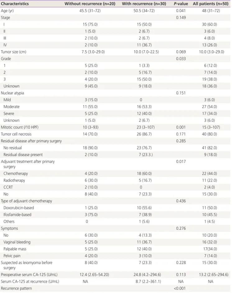
The clinicopathologic characteristics of 50 patients with uter- ine LMS are presented in Table 1. At the time of analysis, 30 patients (60.0%) experienced recurrence, and 16 patients (32.0%) died within a median follow-up period of 21 (range, 3–99) months. Median age was 48 (range, 31–72) years, and 60.0% of the patients had stage I disease. Median tumor size was 10 (range, 3.0–29.0) cm, and moderate atypia was the most common feature (54.0%). Peritoneal recurrence was more common than hematogenous recurrence (66.7%
vs. 33.3%) among patients with recurrence, and lymphatic recurrence was not observed in our data. Upon comparing characteristics between patients with and without recur- rence, age, grade, mitotic count (10 HPF), and type of adju- vant therapy after primary surgery showed significant differ- ences between the 2 groups (Table 1).
Univariate and multivariate analyses were performed to identify prognostic factors for RFS, OS, and survival after recurrence. In analysis for RFS (Table 2), age, stage, tumor suspected as leiomyoma before surgery, residual status after primary surgery, tumor size, grade, nuclear atypia, mitotic count, and history of adjuvant chemotherapy/radiotherapy were significant variables in univariate analysis. Among these factors, age (hazard ratio [HR], 1.091; 95% CI, 1.045–1.140;
P<0.001), residual disease (HR, 5.066; 95% CI, 1.880–
13.651; P<0.001), mitotic count (>10/10 HPF, HR, 3.976;
95% CI, 1.420–11.131; P=0.009), and history of radiother- apy (HR, 0.209; 95% CI, 0.076–0.578; P=0.003) were sig- nificant variables in multivariate analysis. Fig. 1A and B show Kaplan-Meier curves for the time influenced by significant prognostic factors, residual disease status, and mitotic count.
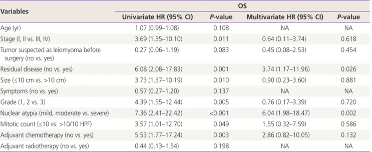
Significant prognostic factors for OS in univariate analysis were stage, residual disease after primary surgery, tumor
size, grade, nuclear atypia, mitotic count, and adjuvant che- motherapy (Table 3). In multivariate analysis, the presence of residual disease after primary surgery (HR, 3.740; 95%
CI, 1.170–11.956; P=0.026) and severe nuclear atypia (HR, 6.041; 95% CI, 1.977–18.465; P=0.002) were significant prognostic factors associated with OS. Fig. 1C and D show survival curves influenced by residual disease status and nuclear atypia.
For analysis of survival after recurrence, only patients with recurrence were assessed (n=30). In univariate analysis, nu- clear atypia was a significant factor (Table 4). In multivariate analysis, the presence of residual disease status (HR, 11.304;
95% CI, 2.009–65.598; P=0.006), severe nuclear atypia (HR, 17.237; 95% CI, 2.902–102.384; P=0.002), and he- matogenous recurrence (HR, 4.189; 95% CI, 1.032–17.000;
P=0.045) were significantly associated with worse prognosis in survival after recurrence. The survival curve for survival af- ter recurrence is shown in Fig. 1E and F.
Discussion
In our study, retrospective analysis of 50 patients with uterine LMS demonstrated possible prognostic factors for RFS, OS, and survival after recurrence. Age, residual disease status, mitotic count, and history of adjuvant radiotherapy were sig- nificant factors for RFS. Residual disease status and nuclear atypia were significant factors for OS, while residual disease status, nuclear atypia, and recurrence pattern were signifi- cant factors for survival after recurrence. Notably, complete resection of tumor at primary surgery was significantly asso- ciated with RFS, OS, and survival after recurrence.
A number of studies on the prognostic factors for uterine
LMS have previously been published. The most important
prognostic factor for survival in uterine LMS remains tumor
stage at diagnosis. In 2009, FIGO presented a new classifica-
tion for uterine LMS that includes tumor size, extrauterine
involvement, and invasion to abdominal tissue [6]. In a previ-
ous study of Surveillance, Epidemiology, and End Results data
of patients with uterine LMS from 2000 to 2012, almost
half of patients were stage I, 14% were stage II and III, and
31% were stage IV [13]. Survival outcomes of uterine LMS
are poor regardless of stage with 5-year disease-free survival
of 65.7% in total the cohort [14]. In addition to FIGO stage,
other reported prognostic factors include age, tumor size,
Table 1. Characteristics of study cohorts (n=50)
Characteristics Without recurrence (n=20) With recurrence (n=30) P-value All patients (n=50)
Age (yr) 45.5 (31–72) 50.5 (34–72) 0.041 48 (31–72)
Stage 0.149
I 15 (75.0) 15 (50.0) 30 (60.0)
II 1 (5.0) 2 (6.7) 3 (6.0)
III 2 (10.0) 2 (6.7) 4 (8.0)
IV 2 (10.0) 11 (36.7) 13 (26.0)
Tumor size (cm) 7.5 (3.0–29.0) 10.0 (7.0–22.5) 0.069 10.0 (3.0–29.0)
Grade 0.033
1 5 (25.0) 1 (3.3) 6 (12.0)
2 2 (10.0) 5 (16.7) 7 (14.0)
3 4 (20.0) 15 (50.0) 19 (38.0)
Unknown 9 (45.0) 9 (18.0) 18 (36.0)
Nuclear atypia 0.151
Mild 3 (15.0) 0 3 (6.0)
Moderate 11 (55.0) 16 (53.3) 27 (54.0)
Severe 5 (25.0) 12 (40.0) 17 (34.0)
Unknown 1 (5.0) 2 (6.7) 3 (6.0)
Mitotic count (/10 HPF) 10 (3–93) 23 (3–107) 0.001 15 (3–107)
Tumor cell necrosis 14 (70.0) 26 (86.7) 0.171 40 (80.0)
Residual disease after primary surgery 0.285
No residual 18 (90.0) 23 (76.7) 41 (82.0)
Residual disease present 2 (10.0) 7 (23.3.) 9 (18.0)
Adjuvant treatment after primary surgery
0.017
Chemotherapy 4 (20.0) 18 (60.0) 22 (44.0)
Radiotherapy 6 (30.0) 5 (16.7) 11 (22.0)
CCRT 2 (10.0) 0 2 (4.0)
No 8 (40.0) 7 (23.3) 15 (30.0)
Type of adjuvant chemotherapy 0.436
Doxorubicin-based 1 (25.0) 10 (55.6) 11 (50.0)
Ifosfamide-based 3 (75.0) 7 (38.9) 10 (45.5)
Others 0 1 (5.6) 1 (4.5)
Symptoms 0.276
No 6 (30.0) 4 (13.3) 10 (20.0)
Vaginal bleeding 5 (25.0) 11 (36.7) 16 (32.0)
Palpable mass 5 (25.0) 12 (40.0) 17(34.0)
Pelvic pain 4 (20.0) 3 (10.0) 7 (14.0)
Suspected as leiomyoma before
surgery 8 (40.0) 7 (23.3) 0.228 15 (30.0)
Preoperative serum CA-125 (U/mL) 12.4 (2.65–54.20) 24.8 (4.2–294.6) 0.113 13.2 (2.65–294.6)
Serum CA-125 at recurrence (U/mL) NA 8.7 (2.2–361.1) NA NA
Recurrence pattern <0.001
mitotic index, and lymphovascular invasion [10,15-18]. Not all of these factors are included in current staging, yet they are related to prognosis.
In a study analyzing prognostic factors using the National Cancer Database [19], surgical resection remained the best effective management for uterine LMS. In the current study, stage-defining variables and other factors were used to analyze prognostic factors, and residual tumor status at pri- mary surgery was also associated with RFS, OS, and survival after recurrence. With regard to surgical resection of uterine
LMS, total hysterectomy with/without bilateral salpingo- oophorectomy is the standard treatment for patients with uterine-confined disease. Tumor removal should be en bloc, with an effort to avoid intraoperative rupture, morcellation, or spillage in the peritoneal cavity. Despite having metastatic disease, patients with both intraperitoneal and extraperito- neal disease are considered to be appropriate candidates for surgical resection due to limited systemic treatment options.
In a previous retrospective study performed at the Memorial Sloan Kettering Cancer Center showed that optimal surgical Table 1. Continued
Characteristics Without recurrence (n=20) With recurrence (n=30) P-value All patients (n=50)
No 20 (100.0) 20 (40.0)
Peritoneal 20 (66.7) 20 (40.0)
Hematogenous 10 (33.3) 10 (20.0)
Treatment type at recurrence <0.001
No 20 (100.0) 20 (40.0)
Chemotherapy 9 (30.0) 9 (18.0)
Radiotherapy 15 (50.0) 1 (2.0)
CCRT 1 (3.3) 1 (2.0)
Target therapy 2 (6.7) 2 (4.0)
Surgical treatment 15 (50.0) 15 (30.0)
Conservative 2 (6.7) 2 (4.0)
Values are presented as median (interquartile range) or number (%).
HPF, high-power field; CCRT, concurrent chemo-radiation therapy; CA-125, cancer-antigen 125; NA, not available.
Table 2. Univariate and multivariate Cox proportional hazards analysis for recurrence-free survival to adjust risk associated with prognos- tic clinical features (n=50)
Variables RFS
Univariate HR (95% CI) P-value Multivariate HR (95% CI) P-value
Age (yr) 1.05 (1.02–1.09) 0.004 1.09 (1.05–1.14) <0.001
Stage (I, II vs. III, IV) 3.15 (1.49–6.68) 0.003 0.75 (0.19–2.92) 0.682
Tumor suspected as leiomyoma before
surgery (no vs. yes) 0.39 (0.16–0.96) 0.041 0.83 (0.28–2.46) 0.733
Residual disease (no vs. yes) 5.06 (2.09–12.26) <0.001 5.07 (1.88–13.65) <0.001
Size (≤10 cm vs. >10 cm) 2.90 (1.33–6.29) 0.007 1.25 (0.48–3.23) 0.652
Symptoms (no vs. yes) 2.36 (0.82–6.82) 0.112
Grade (1, 2 vs. 3) 2.97 (1.36–6.48) 0.006 0.49 (0.13–1.89) 0.298
Nuclear atypia (mild, moderate vs. severe) 2.32 (1.09–4.95) 0.029 2.09 (0.77–5.70) 0.150
Mitotic count (≤10 vs. >10/10 HPF) 3.19 (1.28–7.92) 0.013 3.98 (1.42–11.13) 0.009
Adjuvant chemotherapy (no vs. yes) 3.72 (1.69–8.20) 0.001 0.74 (0.17–3.20) 0.684
Adjuvant radiotherapy (no vs. yes) 0.36 (0.15–0.89) 0.028 0.21 (0.08–0.58) 0.003
RFS, recurrence-free survival; HR, hazard ratio; CI, confidential interval; HPF, high-power field.
resection was associated with improved progression-free sur- vival [20]. Patients with hematogenous metastasis can also be considered as candidates for surgical resection. Several studies showed improved survival outcomes after pulmonary metastasectomy, and pulmonary metastasectomy is an effec- tive option for selected patients with metastatic uterine LMS [21-24]. According to previous studies, surgical resection for
uterine LMS should be performed for better survival out- comes in the appropriate candidates.
For uterine LMS, the effect of adjuvant therapy is still un- clear due to its poor efficacy [25]. In our results, administra- tion of adjuvant radiotherapy was associated with improved RFS. However, this was not significant in long-term survival, and survival advantage may not persist. Patients who un- Fig. 1. Kaplan-Meier curve considering the influence of significant prognostic factors for recurrence-free survival (A, B), overall survival (C, D), and survival after recurrence (E, F).
Cumulative survival pr obability Cumulative survival pr obability Cumulative survival pr obability Cumulative survival pr obability Cumulative survival pr obability Cumulative survival pr obability
1.0 0.8 0.6 0.4 0.2 0.0
1.0 0.8 0.6 0.4 0.2 0.0
1.0 0.8 0.6 0.4 0.2 0.0
1.0 0.8 0.6 0.4 0.2 0.0 1.0 0.8 0.6 0.4 0.2 0.0 1.0 0.8 0.6 0.4 0.2 0.0
Residual disease No Yes
Residual disease No Yes
Nuclear atypia mild-mod severe
Recurrence type peritoneal hematogenous Nuclear atypia mild-mod severe Mitotic count <10 >10