Received: 2012.5.4. Revised: 2012.8.15. Accepted: 2012.9.17.
Corresponding author: Dong-Soo Suh
Department of Obstetrics and Gynecology, Pusan National University Hospital, Pusan National University College of Medicine, 179 Gudeok-ro, Seo-gu, Busan 602-739, Korea Tel: +82-51-240-7283 Fax: +82-51-248-2384
E-mail: dssuh@pusan.ac.kr
Articles published in Obstet Gynecol Sci are open-access, distributed under the terms of the Creative Commons Attribution Non-Commercial License (http://creativecommons.
org/licenses/by-nc/3.0/) which permits unrestricted non-commercial use, distribution, and reproduction in any medium, provided the original work is properly cited.
Copyright © 2013 Korean Society of Obstetrics and Gynecology
Introduction
Among malignant tumors in Korean women, cervical can- cers have been reported as the fifth most common form of cancer after breast, stomach, colorectal, and thyroid cancers [1]. Recently, however, the occurrence of cervical cancer has decreased as it takes considerable amount of time to develop invasive cancers through a progress of dysplasia and intraepi- thelial carcinoma, and early detection of precancerous lesions is readily available due to periodic screening and the develop- ment of cervical cancer examination methods utilizing colpos-
Clinicopathological significance of atypical glandular cells on Pap smear
Sun-Suk Kim 1 , Dong-Soo Suh 1 , Ki-Hyung Kim 1 , Man-Soo Yoon 1 , Kyung-Un Choi 2
Departments of
1Obstetrics and Gynecology and
2Pathology, Pusan National University College of Medicine, Busan, Korea
Objective
To investigate the clinical significance of atypical glandular cells (AGC) by analyzing the prevalence and histologic outcomes of patients with AGC according to Pap smear.
Methods
The medical records of 83 patients who were diagnosed AGC on Pap tests at the Pusan National University Hospital outpatient department and health care center from January 1998 to March 2006 were reviewed.
Results
The prevalence of AGC was 55 of 54,160 (0.10%) and 28 of 54,160 (0.05%) for AGC-not otherwise specified (NOS) and neoplastic associated AGC, respectively. The histopathologic results of the AGC-NOS group (n=55) were as follows: low-grade squamous intraepithelial lesion, 7 (12.7%); high-grade squamous intraepithelial lesion, 4 (7.2%);
adenocarcinoma of cervix, 3 (5.4%); endometrial carcinoma, 2 (3.6%); and other malignancies including 2 ovarian cancer cases and 1 breast cancer case, 3 (5.4%). The histopathologic results for the AGC-associated neoplastic group (n=28) were as follows: low-grade squamous intraepithelial lesion, 1 (3.5%); high-grade squamous intraepithelial lesion, 3 (10.7%); adenocarcinoma of cervix, 5 (17.8%); endometrial carcinoma, 4 (4.8%); and additional malignancies including 3 stomach cancer cases, 2 ovarian cancer cases, and 2 breast cancer cases; 7 (25%).
Conclusion
AGCs may represent a variety of benign and malignant lesions. AGC-associated neoplastic findings may be related to gynecological or extrauterine malignancies. Thus, when AGCs, especially neoplastic AGCs, are encountered, it is best to evaluate the cervix not only for typical maladies, but also for gynecological and non-gynecological malignancies.
Keywords: Atypical glandular cell; Pap smear http://dx.doi.org/10.5468/OGS.2013.56.2.76
pISSN 2287-8572 · eISSN 2287-8580
copy and human papillomavirus (HPV) tests [2].
Pap smears are the most frequently utilized method for cervical cancer screening. The Pap smear technique was first developed by Papanicolaou and Taut [3], in 1941, and al- though it is a convenient, inexpensive, and safe method for cervical cancer screening, the rate of false negative errors is between 6% and 55%. In order to improve upon this error rate, liquid-based Pap [4], colposcopy, and human papil- lomavirus tests have been suggested as alternative screen- ing methods [5]. Likewise, ‘The Bethesda System’ (TBS) was established by the National Cancer Institute meeting, held in Bethesda, USA in 1988, to address problems regarding diag- nostic classification of Pap smear results. Currently, a variety of classification methods are utilized around the world, es- pecially in the US. Specifically, atypical squamous cells of un- determined significance (ASCUS) and atypical glandular cells of undetermined significance (AGUS) are among the most important characteristics of TBS. According to TBS, ASCUS is defined as foremost by abnormal cells observed are worse than either reparative or reactive cells, and secondly as cells that do not satisfy the quantitative and qualitative criteria of squamous intraepithelial lesions [6], where AGUS are de- fined as those cells that satisfy the range of positive reactive changes but are not enough sufficient to be diagnosed as invasive adenocarcinomas [7]. In 2001, the Bethesda III clas- sification, the terminology of both ASCUS and AGUS, were updated as ASC and atypical glandular cell (AGC), respec- tively; where ASC is subdivided into ASC-US and ASC-H while AGUS is subdivided into AGC-not otherwise specified (NOS) and AGC-favor neoplastic, respectively, and are now utilized in clinical diagnoses [8].
ASCUS findings accounts for 3% to 5% of Pap smear re- sults, although this figure has been reported to vary between 10% to 20% and 3% to 5% of the diagnosed patients pos- sessed the risk of cervical intraepithelial neoplasia (CIN)1 and CIN2 or CIN3, respectively. A significant proportion of ASCUS
patients with CIN1 are accompanied by positive HPV infec- tion, the lesions of which have been reported to spontane- ously disappear in more than 60% of cases. Thus, follow-up analysis via an outpatient clinic after confirming whether le- sions of grade CIN2 or CIN3 are present in the patients who were diagnosed ASCUS appears to be an important research avenue [9-13]. Indeed, the prevalence of AGC accounts for 0.08% to 5.96% of known cases, and it has been shown that 8% of diagnosed patients are associated with malignant lesions, thereby requiring more attention and additional his- tological examinations and outpatient clinic follow-up for the patients clinically diagnosed with AGC [14].
Thus, the objective of this study was to investigate the clini- cal significance of AGC (AGC-NOS, AGC-favor neoplastic) by analyzing the final diagnosis results obtained from biopsies of female patients diagnosed with AGC based on the TBS classification method according to Pap smear.
Materials and Methods
1. Subjects
The medical records of 83 patients who were diagnosed AGC (AGC-NOS, 55 subjects; AGC-favor neoplastic, 28 subjects) were analyzed retrospectively among 54,160 subjects who underwent Pap smear analysis in our hospital between Janu- ary 1998 and March 2006 (Table 1). The average age of the patients was 49.2 ± 8.89 with a range of 33 to 74 years old.
The average gravidity and parity were 3.91± 2.3 times and 2.01±1.1, respectively (Table 2).
2. Methods
1) Pap smear and biopsy
Slides of patients with AGUS and AGC were reviewed by a pathology specialist based upon the 2001 revision of TBS III classification system (Fig. 1). Results of punch biopsy and
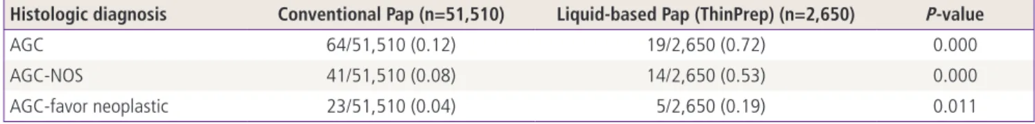
Table 1. Prevalence of AGC using conventional Pap and liquid-based Pap (ThinPrep)
Histologic diagnosis Conventional Pap (n=51,510) Liquid-based Pap (ThinPrep) (n=2,650) P-value
AGC 64/51,510 (0.12) 19/2,650 (0.72) 0.000
AGC-NOS 41/51,510 (0.08) 14/2,650 (0.53) 0.000
AGC-favor neoplastic 23/51,510 (0.04) 5/2,650 (0.19) 0.011
Pearson-chi square analysis.
Values are presented as number (%).
AGC, atypical glandular cell; NOS, not otherwise specified.
cervical curettage in AGC patients were analyzed retrospec- tively.
2) Correlation with clinicopathologic examination results Age, presence or absence of HPV infection, and CA-125 val- Table 2. Clinicopathological relevance of AGC-NOS and AGC-favor neoplastic
Parameters AGC-NOS
55 (66.3) AGC-favor neoplastic
28 (33.7) Total
83 (100)
Age (yr) 47.3 ± 7.8 51.9 ± 10.1 49.2 ± 8.89
Obstetrical history
Gravidity 3.35 ± 1.9 5.15 ± 2.4 3.91 ± 2.3
Parity 1.79 ± 1.0 2.51 ± 1.2 2.01 ± 1.1
HPV infection
Positive 4/16 (25.0) 5/10 (50.0) 9/26 (34.6)
Negative 11/16 (68.7) 5/10 (50.0) 16/26 (61.6)
CA-125 (U/mL) at diagnosis 24.4 ± 36.5 97.6 ± 155.4 50.8 ± 101.6
Values are presented as number (%) or mean ± standard deviation.
Data evaluated by unpaired t test.
AGC, atypical glandular cell; NOS, not otherwise specified; HPV, human papillomavirus (DNA-chip method results are presented and poly- merase chain reaction results are excluded).
Fig. 1. (A) Atypical endocervical cells, not otherwise specified (cervical smears by Pap stain, 34-year-old woman). Groups of cells show round to oval nuclei with nuclear enlargement, small nucleoli, and smooth nuclear membrane. Follow-up revealed chronic cervici- tis (×400). (B) Atypical endocervical cells, favor neoplastic (cervical smears by Pap stain, 67-year-old woman). Sheet of cells show en- larged nuclei with hyperchromasia, some variation in nuclear size, small nucleoli, and feathering. Follow-up revealed endocervical adenocarcinoma (×400). (C) Atypical endometrial cells, favor neo- plastic (cervical smears by Pap stain, 53-year-old woman). A small group of cells have mildly hyperchromatic nuclei, small nucleoli, and a vacuolated cytoplasm. Histologic diagnosis was endometrial adenocarcinoma (×400).
A B
C
ues during diagnosis were analyzed in patients with AGC-NOS and AGC-favor neoplastic AGC patients (Table 2).
3) Histologic correlation of AGC-NOS and AGC-favor neoplastic Punch biopsy and hysterotrachelectasia were performed in patients with AGC. Depending upon indications, several examinations including conization, gastrointestinal examina- tions, and laparotomy were performed. The cases were clas- sified into AGC-NOS and AGC-favor neoplastic cases, and the
histologic results of the patient groups were compared (Tables 3, 4).
3. Results and statistical analysis
Statistical analyses were performed using SPSS ver. 10.0 (SPSS Inc., Chicago, IL, USA). Statistical significance was determined by Pearson chi-square test. Differences of P<0.05 were con- sidered to be statistically significant different.
Table 3. Histological outcome of AGC-NOS and AGC-favor neoplastic on follow-up
Histologic diagnosis AGC-NOS
55 (66.3) AGC-favor neoplastic
28 (33.7) Total
83 (100) Cervix disease
Negative Benign pathology
a)34 (61.8) 7 (25) 41 (49.3)
LSIL Mild dysplasia
Flat condyloma 7 (12.7) 1 (3.6) 8 (9.6)
HSIL Moderate dysplasia 1 (1.8) 0 (0) 7 (8.4)
Severe dysplasia 1 (1.8) 0 (0)
Carcinoma in situ 2 (3.6) 3 (10.7)
Adenocarcinoma in situ 0 (0) 0 (0) 0 (0)
Squamous cell carcinoma 0 (0) 0 (0) 0 (0)
Adenocarcinoma 3 (5.4) 5 (17.8) 8 (9.6)
Other site disease
Endometrial hyperplasia 2 (3.6) 1 (3.6) 3 (3.6)
Endometrial cancer 2 (3.6) 4 (14.2) 6 (7.2)
Breast cancer 1 (1.8) 2/28 (7.1) 3 (3.6)
Ovarian cancer 2 (3.6) 2 (7.1) 4 (4.8)
Stomach cancer 0 (0) 3 (10.7) 3 (3.6)
Values are presented as number (%)
AGC, atypical glandular cell; NOS, not otherwise specified; LSIL, low grade squamous intraepithelial lesion; HSIL, high grade squamous in- traepithelial lesion.
a)