Pathologic and molecular characterization of subsp. infection in neonatal piglets JVS
5
0
0
전체 글
(2) 314 Sang-Ik Oh et al.. Fig. 2. Case 2. Farm floor and gross findings. (A) Concrete residue on the pen floor. (B) Note the blood-stained concrete. (C) Hip joint of piglet. Note the yellowish fragile materials and redness of synovial cavity with periarticular thickening (arrows).. Fig. 1. Case 1. Gross and histopathological findings. (A) Piglet. Note the enlarged tarsal joint (arrow). (B) Thoracic cavity. Yellowish materials (asterisks) were attached to the epicardium. (C) Cerebrum. Suppurative encephalitis with multifocal microabscesses (circle) was observed. Hematoxylin and eosin (H&E) stain. 400×. (D) Kidney. Note the multifocal microabscesses (circle) with intralesional bacteria (arrow). H&E stain. 400×. (E) Heart. Note the multifocal microabscesses with intralesional bacteria (arrow). H&E stain. 400×. (F) Heart. Gram-positive cocci were observed in the myocardium. Gram stain. 1,000×. Scale bars = 20 μm (C–F).. suppurative encephalitis with multiple microabscesses in three piglets. Those microabscesses consisted of abundant polymorphonuclear cells (panel C in Fig. 1). Multiple abscesses with intralesional bacteria were observed in the kidney (panel D in Fig. 1) and epicardium (panels E and F in Fig. 1) in one piglet. In case 2, no specific histopathologic lesions were observed, except for suppurative arthritis. Bacterial cultures from carpal, tarsal, and knee joint, meninges, and epicardium samples from case 1, as well as femoral joint samples from case 2, were performed. Samples were cultured on 5% sheep blood agar (Asan Pharm., Korea) under 5% CO2 at o 37 C for 24 h. The β-hemolytic colonies were obtained from the carpal, tarsal, and knee joints and meninges of four piglets, as well as from the epicardium of one piglet in case 1 and the right femoral joint of one piglet in case 2. To identify the isolates, 16S ribosomal RNA gene sequencing was performed by using an ABI Prism BigDye Terminator Cycler Sequencing Ready Reaction Kit (Applied Biosystems, USA). Sequences obtained. Journal of Veterinary Science. from cases 1 and 2 were compared with sequences in public sequence databases (National Center for Biotechnology Information, USA); all of the isolates presented 99.9% similarity to the SDSE reference strain (GenBank accession No. CP002215). To identify the Lancefield serological group, a Strep LA grouping kit (Denka Seiken, Japan) was used. Isolates from both cases were categorized into Lancefield group C; this result is consistent with that of a previous study reporting the classification of SDSE isolated from a pig with systemic suppurative inflammation into group C [5]. In both cases in the present study, other pathogenic bacteria causing systemic infection, such as Haemophilus parasuis or Streptococcus suis, were not isolated. The samples were additionally negative for all tested viruses, including classic swine fever virus, porcine circovirus type 2, and porcine reproductive and respiratory syndrome virus. The results suggest that veterinarians should consider SDSE as a potential causative agent in piglets presenting with severe arthritis or sudden death; particularly, when abrasion of feet, due to a coarse floor surface as found in case 2, is additionally observed. The observations in the present study highlight the importance of management of floor surfaces in farrowing pens because coarse flooring is a risk factor for SDSE infection. Antimicrobial therapy is widely used to treat streptococcosis in pigs and humans [3,9]. In this study, MICs were determined via a broth microdilution system (Sensititre System; TREK Diagnostic Systems, UK) with 96-well antimicrobial testing plates (BOPO6F; TREK Diagnostic Systems) in accordance with the manufacturer’s instructions and Clinical and Laboratory Standards Institute guidelines [2]. Streptococcus pneumoniae ATCC 49619 was used as the quality control strain. Two SDSE isolates from the tarsal joint sample in case 1 and femoral joint sample of case 2 were found to have the same MIC values for all antibiotics except gentamicin (Table 1). Notably, low MIC values were observed for the β-lactams tested; this is consistent with the results of previous studies in pigs and humans worldwide [5,6,9,15]. These results suggest the importance of β-lactam agents for the treatment of SDSE infection in pigs. Meanwhile, some human-derived SDSE strains that are resistant to β-lactams require treatment with cell-penetrating antimicrobials, preferably macrolides, or, in the case of.
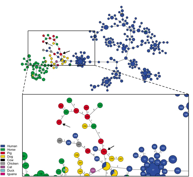
(3) Streptococcus dysgalactiae subsp. equisimilis infection in piglets 315. Table 1. Minimum inhibitory concentration (MIC) values, virulence genes, and multilocus sequence typing data for Streptococcus dysgalactiae subspecies equisimilis isolates from piglets Case 1 2. MIC (μg/mL) TIO. PEN. AMP. CTE OTE DAN ENR FFC GEN NEO. ≤ 0.25 ≤ 0.12 ≤ 0.25 > 8 > 8 > 1 ≤ 0.25 ≤ 0.12 ≤ 0.25 > 8 > 8 > 1. Case. Lancefield group. 1 2. C C. 1 1. 4 4. SDM. SXT. CLI. TYL. TIL. TUL. SPT. TIA. 8 > 32 > 256 ≤ 2 > 16 > 32 > 64 > 64 > 64 > 32 > 16 > 32 > 256 ≤ 2 > 16 > 32 > 64 > 64 > 64 > 32. Virulence genes slo. sagA. pSTKP8. − −. + +. + +. Allelic profile*. ST†. 32-13-28-30-39-48-29 30-13-6-6-12-56-33. 315 252. TIO, ceftiofur; PEN, penicillin; AMP, ampicillin; CTE, chlortetracycline; OTE, oxytetracycline; DAN, danofloxacin; ENR, enrofloxacin; FFC, florfenicol; GEN, gentamicin; NEO, neomycin; SDM, sulphadimethoxine; SXT, trimethoprim/sulfamethoxazole; CLI, clindamycin; TYL, tylosin; TIL, tilmicosin; TUL, † tulathromycin; SPT, spectinomycin; TIA, tiamulin. *Allelic profile, gki-gtr-murI-mutS-recP-xpt-atoB. ST, sequence type.. macrolide-resistant strain, tetracyclines, fluoroquinolones, or clindamycin [10]. However, the swine-derived isolates in the present work, as well as in previous studies, showed relatively high MIC values and resistance to macrolides, tetracyclines, and clindamycin [5,6]. Given that SDSE isolates from animals are known to have a potential zoonotic risk, the antimicrobial resistance observed in the present swine-derived strains warns of the possibility of similar treatment failure in human SDSE infection. Therefore, the current results, which are of potential public health significance, indicate that it is necessary to establish antimicrobial stewardship guidelines for SDSE infection, both in human and veterinary medicine. In the present study, three non-superantigenic virulence genes were screened by polymerase chain reaction targeting streptolysin O (slo), streptolysin S (sagA), and streptokinase (pSTKP8) in two isolates from the tarsal joint sample in case 1 and femoral sample of case 2. The primer sequences were as described in previous studies, and amplification was carried out o under the following conditions [1,4]: 30 cycles of 94 C for 1 o o min, 55 C for 1 min, and 72 C for 1 min. The virulence gene profiles of isolates from both cases was slo−/sagA+/pSTKP8+ (Table 1), which is consistent with the results in a previous report investigating SDSE strains from Yorkshire pigs in Japan [5]. However, the result was inconsistent with that of a previous + + + study of human-derived SDSE (slo /sagA /pSTKP8 ), implying that isolates of swine origin may possess different virulence gene profiles from those recovered from humans [5,14]. A recent study suggested that slo represents a cytotoxic factor that likely contributes to SDSE-mediated necrotizing soft tissue infections [12]. This may represent a plausible explanation for the observed discrepancy in virulence gene profiles between humans and pigs. To elucidate their molecular epidemiology, the two isolates. from the joint samples of each case were characterized by using the MLST scheme for SDSE provided by the public MLST databases (pubMLST, UK), a scheme that is based on internal fragments of seven housekeeping genes gki, gtr, murI, mutS, recP, xpt, and atoB. Unique sequences at each locus were assigned allele numbers, and a combination of seven allele numbers for each isolate was used to define its sequence type (ST). Isolates from cases 1 and 2 were identified as a new ST and as ST252, respectively. The novel ST of the case 1 isolate was submitted to the pubMLST and assigned as ST315 (Table 1). PHYLOViZ v2.0 [7] was used to establish a minimum spanning tree, which represents the relationships among STs from the pubMLST (Fig. 3). Based on the MLST database, the isolate from case 1 (ST315) was identified as a triple-locus variant (TLV) of a ST234 horse-derived strain from the United States. The ST of case 2 (ST252) had previously been identified in pigs with peritonitis in Germany. Interestingly, ST252 was a single-locus variant (SLV) of ST253 and ST255 derived from pigs in Sweden and humans in Portugal, respectively. Additionally, ST252 was a TLV of ST254 derived from dogs in the United Kingdom. Given that one of the SDSE isolate in this study was linked at the SLV level with that isolated from human, the result supports the hypothesis that this bacterium may have a zoonotic possibility [11,13]. Nevertheless, future studies should aim to perform molecular characterization of large numbers of SDSE isolates from pigs to gain a better understanding of the SDSE transmission dynamics, zoonotic possibilities, and microevolutionary events.. Acknowledgments This work was supported by the Animal and Plant Quarantine Agency (B-1543069-2017-18) and Korea Institute of Planning. www.vetsci.org.
(4) 316 Sang-Ik Oh et al.. Fig. 3. A goeBURST diagram for MLST data of Streptococcus dysgalactiae subspecies equisimilis isolates. Each circle in the diagram represents a sequence type (ST), with the size of the circle and its colored segments proportional to the number and origin of isolates, respectively. Numbers on branches represent the number of loci different from that of the founder ST. Isolates from case 1 (ST315) and case 2 (ST252) in this study are indicated by dashed arrow and arrow, respectively.. and Evaluation for Technology in Food, Agriculture and Forestry (114058-03) through the Agri-Bio Industry Technology Development Program, funded by Ministry of Agriculture, Food and Rural Affairs of Korea.. Conflict of Interest The authors declare no conflicts of interest.. Journal of Veterinary Science. References 1. Caballero AR, Lottenberg R, Johnston KH. Cloning, expression, sequence analysis, and characterization of streptokinases secreted by porcine and equine isolates of Streptococcus equisimilis. Infect Immun 1999, 67, 6478-6486. 2. Clinical and Laboratory Standards Institute (CLSI). Performance Standards for Antimicrobial Disk and Dilution Susceptibility Tests for Bacteria Isolated from Animals; Approved Standard–Fourth Edition. CLSI document VET01-A4. CLSI, Wayne, 2013..
(5) Streptococcus dysgalactiae subsp. equisimilis infection in piglets 317. 3. Gottschalk M. Streptococcosis. In: Straw BE, Zimmerman JJ, D’Allaire S, Taylor DJ (eds.). Diseases of Swine. 10th ed. pp. 841-855, Blackwell Publishing Professional, Ames, 2012. 4. Ikebe T, Murayama S, Saitoh K, Yamai S, Suzuki R, Isobe J, Tanaka D, Katsukawa C, Tamaru A, Katayama A, Fujinaga Y, Hoashi K, Watanabe H, The Working Group for Streptococci in Japan. Surveillance of severe invasive group-G streptococcal infections and molecular typing of the isolates in Japan. Epidemiol Infect 2004, 132, 145-149. 5. Kasuya K, Yoshida E, Harada R, Hasegawa M, Osaka H, Kato M, Shibahara T. Systemic Streptococcus dysgalactiae subspecies equisimilis infection in a Yorkshire pig with severe disseminated suppurative meningoencephalomyelitis. J Vet Med Sci 2014, 76, 715-718. 6. Moreno LZ, da Costa BL, Matajira CE, Gomes VT, Mesquita RE, Silva AP, Moreno AM. Molecular and antimicrobial susceptibility profiling of Streptococcus dysgalactiae isolated from swine. Diagn Microbiol Infect Dis 2016, 86, 178-180. 7. Nascimento M, Sousa A, Ramirez M, Francisco AP, Carriço JA, Vaz C. PHYLOViZ 2.0: providing scalable data integration and visualization for multiple phylogenetic inference methods. Bioinformatics 2017, 33, 128-129. 8. Pinho MD, Erol E, Ribeiro-Gonçalves B, Mendes CI, Carriço JA, Matos SC, Preziuso S, Luebke-Becker A, Wieler LH, Melo-Cristino J, Ramirez M. Beta-hemolytic Streptococcus dysgalactiae strains isolated from horses are a genetically distinct population within the Streptococcus dysgalactiae taxon. Sci Rep 2016, 6, 31736. 9. Rantala S. Streptococcus dysgalactiae subsp. equisimilis bacteremia: an emerging infection. Eur J Clin Microbiol Infect Dis 2014, 33, 1303-1310. 10. Savini V, Catavitello C, Talia M, Manna A, Pompetti F, Di Bonaventura G, Di Giuseppe N, Febbo F, Balbinot A, Di Zacomo S, Esattore F, D'Antonio D. Beta-lactam failure in. 11.. 12.. 13.. 14.. 15.. 16.. treatment of two group G Streptococcus dysgalactiae subsp. equisimilis pharyngitis patients. J Clin Microbiol 2008, 46, 814-816. Schrieber L, Towers R, Muscatello G, Speare R. Transmission of Streptococcus dysgalactiae subsp. equisimilis between child and dog in an Aboriginal Australian community. Zoonoses Public Health 2014, 61, 145-148. Siemens N, Kittang BR, Chakrakodi B, Oppegaard O, Johansson L, Bruun T, Mylvaganam H, INFECT Study Group, Svensson M, Skrede S, Norrby-Teglund A. Increased cytotoxicity and streptolysin O activity in group G streptococcal strains causing invasive tissue infections. Sci Rep 2015, 5, 16945. Silva LG, Genteluci GL, Corrêa de Mattos M, Glatthardt T, Sá Figueiredo AM, Ferreira-Carvalho BT. Group C Streptococcus dysgalactiae subsp. equisimilis in south-east Brazil: genetic diversity, resistance profile and the first report of human and equine isolates belonging to the same multilocus sequence typing lineage. J Med Microbiol 2015, 64, 551-558. Sunaoshi KS, Aburahashi H, Kobayashi R, Yamamoto Y, Okuzumi K, Yoshida A, Misawa Y, Adachi K, Ubukata K. [Emm typing by genetic identification of Streptococcus dysgalactiae subsp. equisimilis and susceptibility to oral antibiotics]. Kansenshogaku Zasshi 2006, 80, 488-495. Japanese. Wajima T, Morozumi M, Hanada S, Sunaoshi K, Chiba N, Iwata S, Ubukata K. Molecular characterization of invasive Streptococcus dysgalactiae subsp. equisimilis, Japan. Emerg Infect Dis 2016, 22, 247-254. Zoric M, Sjölund M, Persson M, Nilsson E, Lundeheim N, Wallgren P. Lameness in piglets. Abrasions in nursing piglets and transfer of protection towards infections with Streptococci from sow to offspring. J Vet Med B Infect Dis Vet Public Health 2004, 51, 278-284.. www.vetsci.org.
(6)
수치

관련 문서
