Adriamycin Induced Apoptosis of H9c2 Cardiomyocytes via a Caspase-Independent Pathway
Mi-Hee Jeon
1, Ho-Joong Youn, MD
2, Jung-Hee Lee
1and Jeong-Hwa Lee, MD
11
Department of Biochemistry, College of Medicine, The Catholic University of Korea, Seoul,
2
Division of Cardiology, Department of Internal Medicine, College of Medicine, The Catholic University of Korea, St. Mary’s Hospital, Seoul, Korea
ABSTRACT
Background : The cardiotoxicity of adriamycin limits its clinical usefulness as a powerful drug for solid tumors and malignant hematological disease. Although the exact mechanism by which it causes cardiac damage is not yet known, several reports have suggested apoptosis to be the principal process in adriamycin-induced cardio- myopathy, which exhibits DNA fragmentation, cytochrome C release, and caspase activation. However, no direct evidence has linked the critical involvement of caspase-3 in adriamycin-induced apoptosis. Methods : To deter- mine the requirements for the activation of caspase-3 in adriamycin-treated cardiac cells, we examined the effect of caspase inhibitor on the cell survival and apoptotic changes, using MTT assay, microscopy and Western blotting.
Results : Exposure of H9c2 cells to adriamycin resulted in time- and dose-dependent cell death and cleavage of pro-caspase-3 and the nuclear protein poly (ADP-ribose) polymerase (PARP). However, neither the reduction of cell viability nor the characteristic morphological changes induced by adriamycin was prevented by pretreatment with the general caspase inhibitor z-VAD.FMK. In contrast, caspase inhibition effectively blocked the apoptosis induced by H
2O
2in H9c2 cells, but was not essential for adriamycin-induced apoptosis in H9c2 cells. We also observed that p53 expression was increased by adriamycin, and this increase was not affected by the inhibition of caspase activity, suggesting a role for p53 in adriamycin-induced caspase-independent apoptosis in cardiac toxi- city. Conclusions : Our results demonstrate that adriamycin specifically activates an apoptotic pathway that is not dependent upon the activation of caspase. (Korean Circulation J 2004;34 (1):76-83)
KEY WORDS : Adriamycin; H9c2 cells; Apoptosis; Caspase; p53.
Introduction
Adriamycin is a powerful chemotherapeutic agent for solid tumors and malignant hematological disease. How- ever, cardiac toxicity, including the development of car- diomyopathy and, ultimately, congestive heart failure, limits the clinical usefulness of the drug.
1)Therefore, identifying the mechanisms that underlie adriamycin- induced cardiac damage is essential to developing strate-
gies to interrupt the cardiotoxic action of adriamycin.
Several hypotheses have been proposed to account for adriamycin cardiotoxicity, including the generation of free radicals, impaired adrenergic regulation, release of vasoactive amines, changed calcium handling, mitochon- drial dysfunction, suppression of muscle-specific genes, and ceramide generation.
2-9)The length of this list indi- cates that the molecular mechanisms leading to adriamy- cin-induced cardiomyopathy are not yet clearly understood.
Recent studies have shown that apoptosis is one of the major processes leading to the progressive deterioration of myocardial function induced by adriamycin. In isolated adult rat cardiac myocytes, treatment with adriamycin resulted in DNA fragmentation, chromatin condensation, and nuclear shrinkage- all of which are classical features
Received:September 18, 2003Accepted:October 4, 2003
Correspondence:Jeong-Hwa Lee, MD. Department of Bio
-
chemistry, College of Medicine, The Catholic University of Korea, 505 Banpo-Dong, Seocho-gu, Seoul 137-701, Korea Tel:82-2-590-1180, Fax:82-2-596-4435 E-mail:[email protected]of apoptosis.
10)Despite increasing evidence of the occu- rrence of apoptosis, the apoptotic signaling pathway acti- vated by adriamycin in cardiac cells still remains to be elucidated. A common and critical event in the execution of apoptosis is the activation of a group of cysteine pro- teases called caspases.
11)At least 14 distinct caspases have so far been cloned and divided into two groups, the initiator and effector caspases, according to their func- tions during the apoptotic process.
12)Of these, caspase-3 is the main effector caspase and cleaves the DEVD mo- tif of many cellular substrates, inducing the fundamental features of apoptotic cell death.
13)Adriamycin also in- creases cytochrome c efflux, which is a representative caspase activator, in rat heart mitochondria.
14)Moreover, caspase-3 activity increases when adriamycin is admini- stered to the H9c2 rat cardiac myocyte cell line, as deter- mined using a fluorogenic substrate.
10)Although these findings suggest that the activation of caspase might be involved in adriamycininduced apoptosis in cardiac myo- cytes, it is still possible that the processing of caspase-3 is an accompanying or subsequent event after apoptosis has been triggered, rather than a critical requirement for the induction of apoptosis.
Therefore, to determine whether caspase activation is necessary for adriamycin-induced apoptosis in H9c2 rat cardiac myocyte cells, we examined the effects of caspase inhibition, using the general caspase inhibitor z-VAD.FMK in effective concentrations, on the survival of and mor- phological changes in H9c2 cells exposed to adriamycin.
We describe here that caspase activation is not essential for the death of cardiac cells caused by adriamycin. A possible role for p53 in the caspase-independent apopto- sis induced by adriamycin is briefly discussed.
Methods
Chemicals and antibodies
Adriamycin (Doxorubicin) and MTT (2-(4,5-di- methyltriazol-2-yl)-2,5-diphenyl tetrazolium bromide) were purchased from Sigma Chemical Co (ST. Lous, MO, USA). Caspase-3 colorimetric assay kit (Cat. No.
K2027-1) was obtained from Clontech Laboratories, Inc.
( Palo Alto, USA) and general caspase-3 inhibitor (z- VAD.FMK) (Cat. No. 627610) was obtained from Cal- biochem Co. (Darmstadt, Germany). Anti-PARP anti- body was obtained from Pharmigen and anti-β-actin antibody was purchased from Sigma Chemical Co (ST.
Lous, MO, USA). Anti-p53 antibody and anti-caspase-3 antibody were obtained from Cell Signaling Technology, Inc. (Beverly, USA).
Cell cultures and adriamycin treatment
The cardiac muscle cell line H9c2 was obtained from American Type Culture Collection (ATCC). The cells were maintained at 37℃ in a 5% CO
2humidified atmo- sphere in Dulbecco’s modified Eagle’s medium supple- mented with 10% heat-inactivated fetal bovine serum and antibiotic solutions (100 Units/mL of penicillin and 100 μg/mL streptomycin) (Gibco-BRL, NY, USA).
Cells were subcultured at a 1:4 ratio every 3 days.
Cell viability analysis
Cell viability was monitored by the classical MTT assay. Briefly, 50 μL of the MTT reagent (final concen- tration: 500 μg/mL) was added to each well. Three hours later at 37℃, the cell supernatants were discarded, MTT crystals were dissolved with acid isopropanol, and absorbance was measured at 570 nm. All assays were performed in triplicate, and data were presented as ave- rage±SEM value. Percent of viability was defined as re- lative absorbance of treated versus untreated control cells.
Analysis of nuclear morphology
Cells were plated in 6 well chamber slides and were
allowed to adhere. Adriamycintreated and untreated cells
were fixed with methanol: acetic acid=3:1 (v/v) for
10 min after which staining was carried out with Hoechst
33342 (1 mg/mL). The slides were then washed in disti-
lled water and mounted in a mounting medium (contai-
ning citric acid and disodium orthophosphate dissolved
in distilled water and glycerol [1:1]) and examined by
an epifluorescence microscope (Micros, Austria). Apop-
totic cells were defined on the basis of nuclear changes, such as chromatin condensation and fragmentation.
Caspase-3 activation assay
Caspase-3 activity was measured with an ApoAlert
®Caspase-3 Colorimetric Assay kit (Clontech, Palo Alto, USA). Briefly, 1×10
5cells were collected and lyzed with 50 μL of chilled lysis buffer and incubated on ice for 10 min. Cell lysates were centrifuged at maximum speed for 5 min at 4℃, after which 50 μL of 2× reac- tion buffer/DTT mix and 5 μL of 1 mM caspase-3 substrate (DEVD-pNA) were added to each reaction and incubated at 37℃ for 1 h. Subsequently, 105 μL of supernatant was transferred to a 96-well plate and read at 405 nm in a microplate reader (Molecular Device).
Final caspase-3 activity was calculated by dividing the net OD at 405 nm with the slope of the calibration curve obtained with different concentrations of pNA.
Immunoblot analysis
Adriamycin-treated cells were harvested and lysed in ice-cold RIPA buffer (50 mM Tris/HCl, pH 7.4, 150 mM NaCl, 1% NP-40, 0.5% deoxycholate, 0.1% sodium dodecyl sulfate). Equivalent protein extracts from each sample were separated by 12.5% SDS-PAGE. Proteins were transferred onto PVDF membranes (Immobilon-P, Milipore, Bedford, USA). The membranes were blocked with PBS containing 5% nonfat milk and incubated for 1h at room temperature with primary antibodies (1/500).
The blots were washed four times for 15 min with 0.3%
Tween 20-containing PBS and incubated for 1 h with peroxidase-labeled anti-mouse or anti-rabbit immuno- globulin (1/5000). The membrane was washed again four times. The blots were developed using an enhanced chemiluminescence detection system (ECL, Amersham Corp., Cardiff, UK).
Results
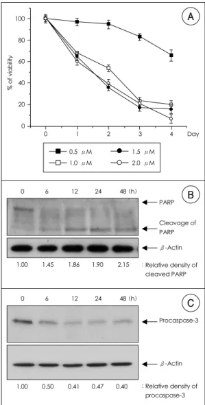
As previously described, exposure to adriamycin re- sulted in dose- and time-dependent toxicity in H9c2 cells (Figure 1A). The reduction in cell viability was
Figure 1. Adriamycin induces apoptosis in H9c2. A: H9c2 cells were treated with various concentrations of adria- mycin for the indicated times, and cell viability was measured by MTT assay. Relative cell viability was deter- mined as a percentage of viability at 0 h, which was adjusted to 100%. The displayed values are the means
±S.D of three independent experiments. B: proteolytic cleavage of PARP was shown during the time course of adriamycin-induced apoptosis. H9c2 cells were treated with 1 μM of adriamycin for various time periods and analyzed by Western blotting using anti-PARP antibody.
113 kD PARP protein was specifically cleaved into 89 kD fragment. C: adriamycin led decrease in procaspase-3 from each time point. H9c2 cells were treated with 1 μM of adriamycin. Cell lysates were analyzed for Western blotting using anti-caspase-3 antibody. β-Actin level was also examined as a loading control.
0 6 12 24 48 (h)
1.00 1.45 1.86 1.90 2.15
PARP
Cleavage of PARP β-Actin
: Relative density of : cleaved PARP
1.00 0.50 0.41 0.47 0.40 0 6 12 24 48 (h)
Procaspase-3
β-Actin
: Relative density of : procaspase-3 0.5 μM 1.5 μM 1.0 μM 2.0 μM
% of viability
100
080 060 040 020
000
0 1 2 3 4 Day
C
B
A
not noticeable until three days after treatment with 0.5 μ M of adriamycin, when determined by MTT assay. At a concentration of 1.0 μM of adriamycin, cell viability was reduced to about 68 % during the first 24 h, and to 54% two days after treatment. The time-course of the decline in cell viability was similar at doses above 1.0 μ M. Therefore, we continued to use a 1.0 μM con- centration of adriamycin in the subsequent experiments to analyze caspase-3 activity and the cleavage of poly ( ADP-ribose) polymerase (PARP), two well-established hallmarks of apoptosis. PARP is activated by breaks in DNA and, in extreme cases, results in ATP depletion and cell death. During apoptosis, Caspase 3 cleaves the 112kDa PARP protein to yield a 24kDa DNA-binding fragment and an 85kDa catalytic fragment. Western blot analysis showed that PARP cleavage was first evident at
about 6 h after treatment with adriamycin with a kinetic profile similar to that for the decrease in procaspase-3, which reflects the activation of caspase-3 (Figure 1B, C). These results suggest that caspase-3 activation is in- volved in the apoptotic pathway induced by adriamycin in H9c2 cells.
We then examined whether the observed increase in caspase activity was critical for adriamycin-induced cell death in H9c2 cells using the general caspase inhibitor z- VAD.FMK. Pretreatment with 100 μM of z-VAD.FMK for 1 h before the addition of 1 μM of adriamycin failed to protect H9c2 cells from adriamycin-induced cell death in MTT assay (Figure 2A). This lack of protection might have been due to a dose of z-VAD.FMK insufficient to inhibit caspase-3 or to an incomplete transfer of z- VAD.FMK across the plasma membrane. To exclude
Figure 2. Caspase inhibitor failed to block H9c2 cells from adriamycin-induced cell death. H9c2 cells were treated with or without 100 μM of z-VAD.FMK, the general caspase inhibitor. And the pre-treated cells were incubated with 1 μM of adriamycin for 24 h (A, B) or 250 μM of H2O2 for 4 h (C, D). A, C: cell viability was measured using MTT assay as described in materials and method. B, D: caspase-3 activity was measured using ApoAlert® Caspase-3 Colo- rimetric Assay. Data represent the means±S.D of the three independent experiments.
% Viability
100
080 060 040 020
000
Non-treated ADM z-VAD.FMK+ADM
% Viability
100
080 060 040 020
000
Non-treated H2O2 z-VAD.FMK+ H2O2
Caspase 3 Activity
0.20
0.15
0.10
0.05
0.00
Non-treated H2O2 z-VAD.FMK+ H2O2
Caspase 3 Activity
0.20
0.15
0.10
0.05
0.00
Non-treated ADM z-VAD.FMK+ADM
B A
D
C
these possibilities, we measured the caspase-3-like acti- vity in cell lysates after treatment with adriamycin, with or without z-VAD.FMK, by evaluating the cleavage of the substrate DEVD-pNA. Treatment with adriamycin caused a three-fold increase in caspase-like activity rela- tive to that of the control, and this was down-regulated to two-fold by pretreatment with 100 μM of z-VAD.FMK, indicating that z-VAD.FMK effectively functions as a caspase inhibitor in H9c2 cells (Figure 2B). Further- more, z-VAD.FMK blocked the death of H9c2 cells caused by H
2O
2, which is known to induce cell death through a caspase-dependent pathway
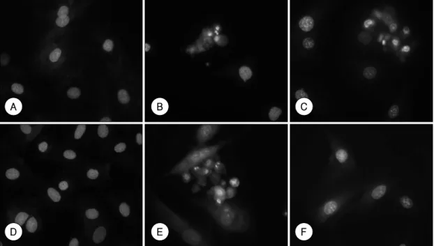
15)accompanied by a concomitant decrease in caspase-3 activity (Figure 2C, D). These findings imply that adriamycin induced apoptosis in H9c2 cells via a caspase-independent path- way. To confirm this, we observed the morphological changes in H9c2 cells after treatment with adriamycin in the presence and absence of the caspase inhibitor. Fl- uorescence microscopy with Hoechst 33342 staining
revealed that H9c2 cells treated with adriamycin exhi- bited chromatin condensation and nuclear fragmentation typical of apoptosis
10)(Figure 3B). These characteristics were also detected in cells pretreated with the caspase inhibitor (Figure 3C). However, in agreement with the cell-death assay, z-VAD.FMK prevented the morpholo- gical changes induced by H
2O
2(Figure 3E, F). Our results, taken together, strongly suggest that, although caspase activation occurs in H9c2 cells after adriamycin treatment, as observed in this study and by other inves- tigators, the activation of caspase is not an essential step in the promotion of apoptosis by adriamycin in these cells. Therefore, a caspase-independent pathway might be the main effector downstream from adriamycin that leads H9c2 cells into apoptotic cell death.
A recent report indicated p53 to regulate apoptosis without any involvement of the caspase family.
16)Fur- thermore, adriamycin was reported to induce p53 ex- pression in many cell types.
17)18)Therefore, to evaluate
Figure 3. Adriamycin-induced alterations in nuclear morphology are caspase-independent. Nuclear changes were observed under the fluorescence microscope at magnification ×400 after Hoechst 33342 staining. H9c2 cells were treated with 1 μM of adriamycin or 250 μM of H2O2. A: non-treated control cells. B: cells treated with 1 μM of adria- mycin, C: cells pretreated with 100 μM of z-VAD. FMK prior to the addition of 1 μM of adriamycin. D: non-treated control cells. E: cells treated with 250 μM of H2O2, F: cells pretreated with 100 μM of z-VAD.FMK prior to the addition of 250 μM of H2O2.
A B C
D E F
p53 as a candidate effector molecule in adriamycin- induced apoptosis, we examined whether adriamycin increases the expression of p53 in H9c2 cells as in other cells, and whether or not this expression is affected by caspase inhibitor. Levels of p53 protein were up-re- gulated with increasing concentrations of adriamycin ( Figure 4A). Time-course analysis revealed that the induction of p53 was detectable as early as 6 h after treatment with 1 μM of adriamycin. This increase was maintained for up to 48 h after treatment and was not prevented by pretreatment with the caspase inhibitor (Figure 4B). These findings raise the possibility that p53 participates as a key molecule in the adriamycin- induced apoptotic pathway, which is not dependent upon the activation of caspase.
Discussion
Apoptosis has been regarded as one of the mecha- nisms underlying the cardiotoxic effects of adriamycin.
This is supported by the fact that several typical apopto- tic features, including mitochondrial dysfunction, DNA
fragmentation, and caspase activation, are induced by adriamycin in the H9c2 rat cardiac myocyte cell line, which is used as a model for adriamycin-induced car- diac toxicity.
19)Recently, an insulin-like growth factor I (IGF-1) was shown to inhibit the apoptosis induced by adriamycin in H9c2 cells, concomitant with attenuating caspase 3 activation, suggesting caspase-3 activation as the cause of cell death in adriamycin-induced apopto- sis.
10)In this investigation, we also observed, by western blotting, that caspase-3 activation occurred during adria- mycin-induced apoptosis, as revealed by a decrease in procaspase-3 and the processing of the nuclear protein PARP, a well-known substrate of caspase-3-like activity, and by an increase in DEVD-cleaving activity. However, caspase inhibition had no effect on the survival of H9c2 cells exposed to adriamycin. Moreover, the morpholo- gical changes induced by adriamycin, such as chromatin condensation and nuclear fragmentation, were not pre- vented by pretreatment with the caspase inhibitor. The independence of adriamycin-induced apoptosis from caspase activation in H9c2 cells, described in the current study, seemed to be specific because z-VAD.FMK effi- ciently suppressed the DEVD-cleaving activity increased by adriamycin or H
2O
2. This inhibitor also blocked the cell death induced by H
2O
2in H9c2 cells almost com- pletely. It is likely, therefore, that the activation of cas- pase accompanies or occurs after cell death, rather than as an essential step central to the process of adriamycin- induced cell death in H9c2 cells.
Recently, it has been reported that several stimuli, such as chloroquine, camptothecin, low concentrations of ni- tric oxide, and potassium depletion, induce apoptosis without the involvement of caspase activation.
20)Bcl-2 family proteins or the p53 protein seem to regulate some caspase-independent apoptotic processes.
21)Our results show that p53 expression was induced by adriamycin in H9c2 cells, and that this was not affected by the caspase inhibitor, implying a role for p53 as a key molecule in caspase-independent apoptosis. Furthermore, exposure of H9c2 cells to adriamycin or hypoxia increases the ex- pression of Bax, a downstream effector of p53, and this
Figure 4. p53 is up-regulated by adriamycin in H9c2cells. A: H9c2 cells were exposed to various concentra- tions of adriamycin for 48 h and analyzed for p53 expre- ssion by Western blotting. B: H9c2 cells were treated with 1 μM of adriamycin for various time periods before assessing p53 expression. The 6th lane shows that p53 expression was not affected by the caspase inhibitor. As a loading control, the expression of β-Actin was also shown.
0 0.25 0.5 1.0 1.5 2.0 (μM)
1.00 1.07 2.38 2.87 3.78 3.19
p53
β-Actin : Relative density of : p53
0 6 12 24 48 48 (h)
- - - - - + z-VAD.FMK
1.00 1.36 2.52 3.23 4.42 3.81
p53 β-Actin : Relative density of : p53
A
B
induction is diminished by IGF-1 or PMA treatment, res- pectively, with restoration of cell viability.
22)23)Several causes of neuronal apoptosis, such as potassium deple- tion and amyloid β, are dependent on Bax, but not on caspase.
24)Therefore, it is possible that the adriamycin- induced p53 observed in this study had pro-apoptotic activity by transactivating downstream target genes such as PUMA, Noxa, and Bax.
25)26)These, in turn, regulate mitochondrial membrane permeability, leading to the release of potentially toxic mitochondrial proteins. Of these proteins, apoptosis-inducing factor (AIF) translo- cates to the nucleus, where it binds to DNA and induces caspase-independent chromatin condensation.
27)However, further studies using a system in which p53 expression is specifically regulated are required to define the exact role of p53 and its downstream pathway in the caspase- independent apoptosis induced by adriamycin.
The generation of reactive oxygen species (ROS) has been implicated as one mechanism involved in adria- mycin-induced cardiotoxicity.
28)Because H
2O
2, which generates ROS, induces apoptosis via a caspase-depen- dent pathway, as shown in our results, and because ROS mediate adriamycin-induced apoptosis in the absence of p53 in Saos-2 cells,
29)adriamycin probably induces ap- optosis via two different pathways. One is a caspase- independent but p53-dependent pathway, and the other a caspase-dependent but p53-independent pathway, which involves the generation of ROS. Of these, the former seems to predominate in H9c2 cells, based on the results presented in this study. Therefore, a more extensive in- vestigation of the molecular mechanisms of the caspase- independent pathway induced by adriamycin will pro- vide useful information for developing pharmacological agents, other than caspase inhibitors, that interfere with the progression of cardiotoxicity when adriamycin is used in anti-cancer therapy.
■
Acknoewledgments
This work was supported by a Korean Society of Circu- lation Grant (2002).
REFERENCES
1)
Mott MG. Anthracycline cardiotoxicity and its prevention.
Ann N Y Acad Sci 1997;824:221-8.
2)
Doroshow JH. Effect of anthracycline antibiotics on oxygen radical formation in rat heart. Cancer Res 1983;43:460-72.
3)
Olson RD, Mushlin PS. Doxorubicin cardiotoxicity: analy- sis of prevailing hypotheses. FASEB J 1990;4:3076-86.
4)
Tong J, Ganguly PK, Singal PK. Myocardial adrenergic ch- anges at two stages of heart failure due to adriamycin treatment in rats. Am J Physiol 1991;260:H909-16.
5)
Bristow MR, Sageman WS, Scott RH, Billingham ME, Bowden RE, Kernoff RS, et al. Acute and chronic cardio- vascular effects of doxorubicin in the dog: the cardiova- scular pharmacology of drug-induced histamine release. J Cardiovasc Pharmacol 1980;2:487-515.
6)
Singal PK, Panagia V. Direct effects of adriamycin on the rat heart sarcolemma. Res Commun Chem Pathol Pharma- col 1984;43:67-77.
7)
Doroshow JH, Davies KJ. Redox cycling of anthracyclines by cardiac mitochondria: II. formation of superoxide anion, hydrogen peroxide, and hydroxyl radical. J Biol Chem 1986;261:3068-74.
8)
Kurabayashi M, Jeyaseelan R, Kedes L. Doxorubicin repre- sses the function of the myogenic helix-loop-helix transcrip- tion factor MyoD: involvement of Id gene induction. J Biol Chem 1994;269:6031-9.
9)
Andrieu-Abadie N, Jaffrezou JP, Hatem S, Laurent G, Le- vade T, Mercaider JJ. L-carnitine prevents doxorubicin- induced apoptosis of cardiac myocytes: role of inhibition of ceramide generation. FASEB J 1999;13:1501-10.
10)
Wang L, Ma W, Markovich R, Lee WL, Wang PH. Insulin- like growth factor I modulates induction of apoptotic sig- naling in H9c2 cardiac muscle cells. Endocrinology 1998;
139:1354-60.
11)
Martin SJ, Green DR. Protease activation during apoptosis:
death by a thousand cuts? Cell 1995;82:349-52.
12)
Nicholson DW, Thornberry NA. Caspases: killer proteases.
Trends Biochem Sci 1997;22:299-306.
13)
Boldin MP, Goncharov TM, Goltsev YV, Wallach D. Invol- vement of MACH, a novel MORT1/FADD-interacting pro- tease, in Fas/APO-1- and TNF receptor-induced cell death.
Cell 1996;85:803-15.
14)
Lampidis TJ, Moreno G, Salet C, Vinzens F. Nuclear and mitochondrial effects of adriamycin in singly isolated pul- sating myocardial cells. J Mol Cell Cardiol 1979;11:415-22.
15)
Turner NA, Xia F, Azhar G, Zhang X, Liu L, Wei JY. Oxi- dative stress induces DNA fragmentation and caspase acti- vation via the c-Jun NH2-terminal kinase pathway in H9c2 cardiac muscle cells. J Mol Cell Cardiol 1998;30:1789-801.
16)
di Pietrantonio AM, Hsieh TC, Wu JM. Activation of caspase 3 in HL-60 cells exposed to hydrogen peroxide. Biochem Biophys Res Commun 1999;255:477-82
17)
Kwok TT, Mok CH, Menton-Brennan L. Up-regulation of a mutant form of p53 by doxorubicin in human squamous carcinoma cells. Cancer Res 1994;54:2834-6.
18)
Seth P, Katayose D, Li Z, Kim M, Wersto R, Craig C, et al.
A recombinant adenovirus expressing wild type p53 induces
apoptosis in drug-resistant human breast cancer cells: a
gene therapy approach for drug-resistant cancers. Cancer
Gene Ther 1997;4:383-90.
19)
Green PS, Leeuwenburgh C. Mitochondrial dysfunction is an early indicator of doxorubicin-induced apoptosis. Bio- chim Biophys Acta 2002;1588:94-101.
20)
Brockhaus F, Brune B. p53 accumulation in apoptotic ma- crophages is an energy demanding process that precedes cytochrome c release in response to nitric oxide. Oncogene 1999;18:6403-10.
21)
Okuno S, Shimizu S, Ito T, Nomura M, Hamada E, Tsuji- moto Y, et al. Bcl-2 prevents caspase-independent cell death.
J Biol Chem 1998;273:34272-7.
22)
Bonavita F, Stefanelli C, Giordano E, Columbaro M, Fac- chini A, Bonafe F, et al. H9c2 cardiac myoblasts undergo apoptosis in a model of ischemia consisting of serum de- privation and hypoxia: inhibition by PMA. FEBS Lett 2003;
536:85-91.
23)
Hong F, Kwon SJ, Jhun BS, Kim SS, Ha J, Kim SJ, et al.
Insulin-like growth factor-1 protects H9c2 cardiac myoblasts from oxidative stress-induced apoptosis via phosphatidy- linositol 3-kinase and extracellular signal-regulated kinase pathways. Life Sci 2001;68:1095-105.
24)
Johnson MD, Xiang H, London S, Kinoshita Y, Knudson M, Mayberg M, et al. Evidence for involvement of Bax and p53, but not caspases, in radiation-induced cell death of cultured postnatal hippocampal neurons. J Neurosci Res 1998;54:
721-33.
25)
Schuler M, Green DR. Mechanisms of p53-dependent apop- tosis. Biochem Soc Trans 2001;29:684-8.
26)
Wu X, Deng Y. Bax and BH3-domain-only proteins in p53- mediated apoptosis. Front Biosci 2002;7:d151-6.
27)
Cande C, Cecconi F, Dessen P, Kroemer G. Apoptosis-indu- cing factor (AIF): key to the conserved caspase-indepen- dent pathways of cell death? J Cell Sci 2002;115:4727-34.
28)
de Atley SM, Aksenov MY, Aksenova MV, Harris B, Hadley R, Cole Harper P, et al. Antioxidants protect against reactive oxygen species associated with adriamycin-treated cardiomyocytes. Cancer Lett 1999;136:41-6.
29)