This work was supported by a research grant from the National Cancer Center, Korea (NCC-0601230-2).
Submitted August 16, 2010, Accepted September 16, 2010
Corresponding Author: Se Byeong Lee, Proton Therapy Center, National Cancer Center, 809 Madu-1dong, Ilsan-gu, Goyang 410-769, Korea Tel: 031)920-0128, Fax: 031)920-0149
E-mail: [email protected]
*Department of Radiation Oncology, Asan Medical Center, Seoul,
†
Proton Therapy Center, National Cancer Center, Goyang,
‡Department of Radiation Oncology, Samsung Medical Center,
§Department of Radiation Oncology, Hallym University Medical Center, Seoul,
∥
Department of Radiation Oncology, Gyeongsang National University Hospitol, Jinju,
¶
Department of Radiation Oncology, East-West Neo Medical Center, Seoul, Korea
This study examined the dosimetric influence of implanted gold markers in proton therapy and the effects of their positions in the spread-out Bragg peak (SOBP) proton beam. The implanted cylindrical gold markers were 3 mm long and 1.2 mm in diameter. The dosimetric influence of the gold markers was determined with markers at various locations in a proton-beam field. Spatial dose distributions were measured using a three-dimensional moving water phantom and a stereotactic diode detector with an effective diameter of 0.5 mm. Also, a film dosimetry was performed using Gafchromic External Beam Treatment (EBT) film. The GEANT4 simulation toolkit was used for Monte-Carlo simulations to confirm the measurements and to construct the dose-volume histogram with implanting markers. Motion data were obtained from the portal images of 10 patients to investigate the effect of organ motions on the dosimetric influence of markers in the presence of a rectal balloon. The underdosed volume due to a single gold marker, in which the dose was less than 95% of a prescribed amount, was 0.15 cc. The underdosed volume due to the presence of a gold marker is much smaller than the target volume. However, the underdosed volume is inside the gross tumor volume and is not smeared out due to translational prostate motions. The positions of gold markers and the conditions of the proton-beam field give different impacts on the dose distribution of a target with implanted gold markers, and should be considered in all clinical proton-based therapies.
Key Words: Gold marker, Proton therapy, Dosimetric influence
INTRODUCTION
The amount of energy lost by moderately relativistic protons with momentum ranges from several MeV to 1 GeV as they pass through matter, which is primarily attributable to ioniza- tion and atomic excitation, increases when their kinetic energy is lower.
1)This physical property enables the depth dose dis- tribution to be controlled in proton therapy. The lower en- trance dose than doses in tumor region with a single beam
field and no exit dose are not achievable in traditional x-ray treatment. This new degree of freedom would give the ability to control the dose distribution more effectively. The optimal utilization of this in clinical cases requires more accurate posi- tioning of patients and more precise information of the tumor position.
Implanted markers have been used for the verification of the
tumor position inside a patient body.
2)According to parameters
such as tumor locations, types, and sizes, various kinds of
markers have been employed.
3)In general, the presence of het-
erogeneity within radiation fields results in variation of the
dose distribution that depends on the atomic components and
their amounts in the medium. While the heterogeneity due to
implanted markers allows markers to be distinguished from tis-
sues and bones in an x-ray image, the undesired perturbation
in treatment dose distribution also needs to be considered.
MATLAB 7.0.
Table 1. Physical parameters of the gold marker. The relative quantities are normalized to the values for water.
Atomic component Au (Z=79, A=197) 100%
Physical density 19.3 g/cm3
Relative electron density 13.9
Relative proton stopping power 10.0
Range @ 172 MeV 2.00 g/cm2
A proton traversing through a medium is deflected by many small-angle scatters. Most of these deflections are due to Coulomb scattering from nuclei, and so the deflection angles increase with the atomic number of the constituent matter, re- sulting in more divergence of the original beam.
4)Many institutions use small gold seeds as implanted in the x-ray radiation treatment of a prostate cancer. While there are many reports on the clinical effectiveness of gold markers in the literature,
5)there have been few reports on the dosimetric influence of gold markers.
6)In the Particle Therapy COoperative Group (PTCOG) meeting 2005, there was a presentation on this issue based on 1-dimentional Monte Calro simulation.
Little dosimetric influence of gold markers was presented.
Later, significant impacts have been reported in some papers, in which the studies were based on 2-dimensional Monte Carlo simulation and a film dosimetry.
6)In this study, we examined the influence of implanted gold markers on the dose distributions in proton therapy, inves- tigated the dependency on the marker position in the spread- out Bragg peak (SOBP) region of a proton beam, and con- firmed the previous work using 3-dimentional Monte Carlo simulation and measurements with a diode detector.
MATERIALS AND METHODS
The proton stopping power used in proton radiation treat- ment planning (RTP) systems is optimized for organic materi- als rather than for metals and other inorganic matter. The spa- tial resolution of commonly used three-dimensional CT images
is too low to construct the dose-volume histogram for gold markers. Therefore, a Monte-Carlo simulation and a dosimetry technique specially designed for microscopic measurements were used to examine the dosimetric influence of gold markers.
1. Implanted gold marker
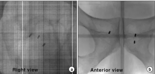
The cylindrical gold markers were 3 mm long and 1.2 mm in diameter, as shown in Fig. 1, and their physical properties are listed in the Table 1.
In proton therapy, bilateral beams are used for patients with prostate cancer, and markers are implanted in an inferior direction. Therefore, the gold markers were positioned in the prostate with their cylindrical axes perpendicular to the beam direction as shown in Fig. 2.
2. Proton beam
Treatments at the Proton Therapy Center, National Cancer
Center (NCC), Korea started on March 19, 2007. This proton
treatment facility is equipped with a cyclotron (Proteus 235,
IBA, Belgium) that produces a 230 MeV proton beam at cur-
rent of 300 nA. A beam delivery system directs proton beams
to two gantry treatment rooms, one fixed beam treatment
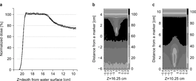
Fig. 2. (a) Normalized depth dose curve without any marker (b, c) 2-Dimensional dose distributions showing isodose contours for
the different depths of markers. The position of markers was set to zero in y-axis of b and c which is correspond to a depth Z in x-axis of a. The data were obtained from the water phantom using a small diode detector.room, and 1 experimental site. The proton-beam energies are controlled to range from 70 MeV to 230 MeV in the treatment rooms. Currently, double-scattering mode was commissioned for the two gantry rooms, and single-scattering mode and ocu- lar mode are being commissioned in the fixed beam room.
Proton-beam fields with a depth range of 22∼24 g/cm
2and a SOBP region of 6∼8 g/cm
2have been used for clinical prostate cancer treatments. For the present study, the proton beam had a depth range of 20 g/cm
2and an SOBP region of 5 g/cm
2. The beam has been configured with the open block of the 100 mm-sized snout in double scattering mode. The ki- netic energy of a proton with a 20 g/cm
2range is 172 MeV.
3. Dosimeter with a water phantom
Measuring doses in a small volume requires small steps in the movement of the phantom and a detector with a small ac- tive volume. A stereotactic diode detector (SFD, p-type silicon chip type, IBA, Belgium) with an effective diameter of 0.5 mm was installed in a three-dimensional moving water phan- tom (RFA-300, IBA, Belgium). The system was operated in a stepping mode using OmniPro-Accept 8.0 software. The step size was set to 0.3 mm, which is more than the 0.2 mm movement resolution of the phantom. The lateral range of pro- files was 2 cm. We took the lateral profiles by 2.5 mm step in depth to cover all of the affected beam field downstream of a marker. Gold markers were attached to the downstream side of
a 30×30 cm
2PMMA plate. The thickness of the plate was 3 cm. The origin of the coordinate frame in the water phantom was set to be the most-downstream surface of a gold marker.
4. Film measurement
Gafchromic External Beam Treatment (EBT) film is a very convenient tool for verifying the dose distribution of a proton beam. However, the proton stopping power of EBT film dif- fers from that of water, which contributes to dose distortion in the parallel exposure condition. We checked the upstream in- fluence of the markers using the EBT film before performing measurements with a three-dimensional water phantom. After placing markers inside small holes on an EBT film, the proton beam was irradiated.
5. Monte-Calro simulation
“Geant4” is a toolkit for the simulation of the passage of
particles through matter. Its areas of application include high
energy, nuclear and accelerator physics, as well as studies in
medical and space science.
7)The GEANT4 simulation toolkit
was used to simulate the dosimetric influence of the gold
marker in a water phantom. The aims of the simulation were
to verify the measurements, to construct the dose-volume rela-
tion with gold markers, and to elucidate the effects of organ
motion. All geometrical structure of the proton-beam nozzle
was included in the simulation program.
8)We used a
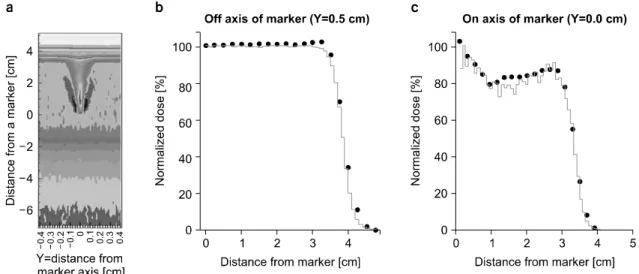
Fig. 3. (A) Simulated two-dimensional projected dose distribution (b, c) Comparisons of depth dose curves between measured data
(circles) and MC simulation (lines) for different lateral offsets.10×10×30 cm water-containing box for the water phantom and the voxel size in the MC program to be 0.1×0.1 mm
2and 0.25 mm in the transverse and axial direction of the beams, respectively.
6. Organ motion verification
Deformation and rotation of organs smears the dosimetric influence of markers. The x-ray images of 10 patients with 20 or 15 fractional treatments were used to study the interfrac- tional motion of the prostate in the presence of a rectal balloon. Two orthogonal x-ray images were taken to monitor the position of the patient in each fraction. We determined the 3-dimensional positions of three markers from the two x-ray images, and used this information to assess the deformation and the rotation of the prostate to check the smearing of the dosimetric influence of markers.
RESULTS AND DISCUSSION
The film dosimetry using an EBT film confirmed no dosi- metric influence of gold markers in the upstream region, which agrees with the Coulomb scattering distribution of a heavy charged particle according to the theory of Moliere. The nuclear scattering was too infrequent to detect the contribution of nuclear projectiles to the dose distribution upstream of the markers. Therefore, in measurements using a water phantom
and Monte-Carlo simulation, we focused on the dose dis- tribution in the region downstream of the markers.
Fig. 3 shows the depth dose curves for different marker lo- cations in the water phantom. The two-dimensional dose dis- tribution was reconstructed with the measured one-dimensional profiles for markers at two locations (at depths of 16.25 cm and 10.25 cm) in a proton beam field. The variance of the scattered angle of a charged particle is theoretically propor- tional to the atomic number of target atoms, and hence the di- vergence of the proton beam is high for interactions with a gold marker. This is responsible for the dose shadow down- stream of the gold markers. The degree of underdosing and the position of the minimum dose are related to the mo- mentum of protons at the interaction position with a marker.
The minima were 2 cm and 1cm down stream for markers that were 10.25 cm and 16.25 cm deep, respectively.
MC simulations with the GEANT4 toolkit were used to ob- tain the does-volume relation under the influence of markers.
The dose distributions downstream of the gold marker were obtained using 100 million proton events for each data set.
Fig. 3 shows that the simulated depth dose curves of proton beams with and without a marker were consistent with the measurements. The simulated three-dimentional dose dis- tributions were used to construct the dose-volume relation (Fig.
4a). Usually, the variance of less than 5% of a prescribed dose
is recommended. Bi-lateral beam fields are generally adapted
Fig. 4. (a) Normalized dose-volume histograms without any marker (dotted line) and with a single marker (solid line) (b)
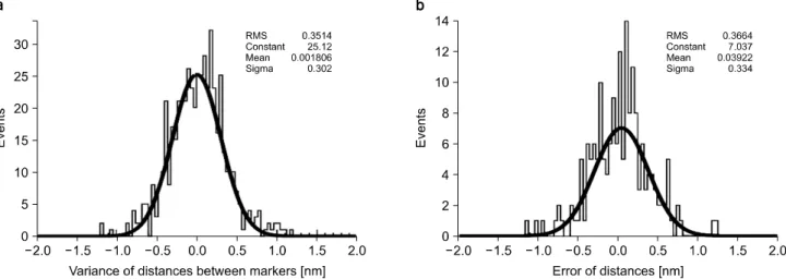
Dose-volume histogram for 1mm of organ movement against markers (hatched area).Fig. 5. (a) Distribution of the distances between markers. (b) Error of distances evaluated by comparing the different methods of
determining the marker position. Also this amount is consistent with 0.346 mm which is three dimensional variance of the pixel size of the plat panels, 0.2 mm.in prostate cancer treatment using proton beam. The volume that received less than 95% of the prescribed dose was calcu- lated to be 0.15 cc in the DVH of Fig. 4a. The minimal dose inside tumor was calculated to be 85% level of prescription dose in bi-lateral beams while the minimal dose is the 70%
level in the single beam field. This underdose is invariant un- der intra- and inter-fractional variation of translational tumor position, but not under the rotation and deformation of tumor.
Also the migration of gold marker in tumor can change the dosimetric influence of gold markers. If we assume the 1 mm of a transversal migration of marker in the prostate, the under-
dosed volume is halved as shown in Fig. 4b.
We examined the deformation and rotation of the prostate
from the position data of 10 patients with implanted markers
and a rectal balloon. Such movements can smear the dosi-
metric influence over the greater volume. In following simple
calculation, the type of inter-factional movement could be
identified. The variance of distances between markers indicates
the inter-fractional deformation of the prostate. However the
variance was measured at 0.30±0.33 mm (Mean±Standard
Deviation), which is within the statistical error of the measure-
ments as shown in Fig. 5. We also confirmed that there is lit-
of no translation, the variation was evaluated by assuming no independence between the position variances. The sum of variances is corresponds to the value in the case of no rotation.
Table 3. Assuming bilateral beams for treatment involving markers, the distal edges of fields are listed as R90, which corresponds to the distal edge receiving 90% of prescribed dose. Dmin is the minimum dose at a position inside R90.
The R90 of an unperturbed beam was 20 cm.
Marker depth [cm] R90 [cm] Dmin [%] @ cm
14.25 19.99 84.0 @ 14.94
15.44 19.95 86.5 @ 16.47
16.52 19.90 86.2 @ 17.38
17.65 19.59 84.4 @ 18.73
18.76 19.00 90.0 @ 19.00
19.92 19.92 90.0 @ 19.92
tle rotational movement by considering the vectors of the posi- tion variances. The sum of variances for anterior-posterior and superior-inferior marker positions was equivalent of the var- iance of the sum of positions as listed in Table 2. That means that the variation directions of the three markers are same. If the variance directions of positions are same, it means the prostate mainly have translational movement. Such translational movement has no effect on the variation in the dose-volume relation induced by implanting markers. The intra-fractional motion of the prostate in the presence of a rectal balloon has been reported to be trivial.
9)CONCLUSION
It is difficult to draw definitive conclusions on the clinical influence of gold markers purely from our physical measure- ment, since biological and clinical factors were not considered.
However, we are able to draw the following, tentative con- clusions:
1. The presence of markers in proton beams partially re- duces the distal range in patients, as indicated in Table 3, with the reduction being dependent on the distance between the marker and the distal edge of the beam. The marker position- ing of more than 2.5 cm in the interior direction of Clinical Target Volume and 0.4 mm increase in the distal margin would remove uncertainty the distal edge due to the presence of gold markers.
2. An underdosed volume (i.e, receiving less than 95% of
the prescribed value) of less than 0.45 cc is expected when us- ing three markers, with the maximum reduction being to about 85% of the prescribed dose.
3. There was no measurable effect of organ motion, indicat- ing that the positioning with the rectal balloon and the im- planted marker is very stable. The variation in the prostate po- sition along the beam was less than 1 mm in all directions.
REFERENCE
1. Stopping Powers and Ranges for Protons and Alpha Particles.
ICRU Report No.49 (1993); Tables and graphs of these data are available at http://physics.nist.gov/PhysRefData/.
2. Crook JM, Raymond Y, Salhani D, Yang H, Esche B:
Prostate motion during standard radiotherapy as assessed by fi- ducial markers. Radiother Oncol 37:35-42 (1995)
3. Patrick A. Kupelian, Alan Forbes, Twyla R. Willoughby,
전립선암에 대한 양성자치료에서 금마커에 의한 방사선 선량분포의 영향
*서울아산병원 방사선종양학과,
†국립암센터 양성자 치료센터,
‡삼성의료원 방사선종양학과,
§
한림대학교 강남성심병원 방사선종양학과,
∥경상대학병원 방사선종양학과,
¶
강동경희대학교병원 방사선종양학과
곽정원*ㆍ신정욱
†ㆍ김진성
‡ㆍ박성용
†ㆍ신동호
†ㆍ윤명근
†ㆍ박소아
§ㆍ김동욱
¶ㆍ임영경
∥ㆍ이세병
†본 연구에서는 양성자 치료에서 치료부위에 금 마커를 삽입하였을 때 양성자 빔의 선량과 그 분포에 미치는 영향을 평 가하였다. 1.2 mm 직경에 3 mm 길이의 원통형 금 마커가 사용되었고 양성자 빔의 여러 위치에서 각각의 선량에 미치는 영향을 조사하였다. 3차원 moving 팬텀과 유효 반경이 0.5 mm인 정위적 방사선 치료용 다이로드 검출기를 이용하여 금 마커의 방사선 선량으로의 영향을 결정하였다. Gafchromic film을 이용한 필름에 의한 선량 평가도 시행하였다. 그리고 선량-체적관계 히스토그램을 통한 분석을 위하여 GEANT4 전산 모의 도구 모음을 이용한 전산모의를 실시하였다. 마커 의 움직임에 대한 영향성을 평가하기 위하여 직장 풍선을 적용하고 금 마커를 삽입한 10명의 환자에 대한 MV 영상을 이용하였다. 하나의 금 마커에 의하여 처방선량의 95% 이하가 들어간 체적은 0.15 cc로 작지만 그 체적은 치료부위 안쪽 에 위치하고 평행이동에 대하여 발려져서 없어지지 않았다. 금 마커의 위치와 양성자 빔의 상태에 따라서 선량분포에 각각 다른 영향을 미치고 이는 임상치료에서 충분히 고려되어야 할 것으로 사료된다.
중심단어: 금 마커, 양성자 치료, 선량적인 영향
Marco van Vulpen, Uulke A. van der Heide. Intrafraction Motion of the Prostate During External-Beam Radiation Therapy:Analysis of 427 Patients with Implanted Fiducial Markers.
International Journal of Radiation Oncology Biology Physics 69:419-425 (2007)
6. Wayne Newhauser, Jonas Fontenot, Nicholas Koch, et
apeutic Proton Beam Delivery System Using the GEANT4 Code.
Journal of Korean Medical Physics 18:226 (2007)
9. John. E. McGary, Bin S. Teh, E. Brian Butler, Walter Grant, III: Prostate immobilization using a rectal balloon.
Journal of Applied Clinical Medical Physics, 3: (2002)