125
Miniature Schnauzer에서 발생한 장간막 염전 1증례
최지혜1·김현욱1·김진경1·장재영1·김준영2·윤정희2,*
1해마루 이차진료동물병원, 2서울대학교 수의과대학 BK21 수의과학연구인력양성사업단 (게재승인: 2008년 1월 30일)
Mesenteric torsion in a Miniature Schnauzer
Jihye Choi
1, Hyunwook Kim
1, Jinkyung Kim
1, Jaeyoung Jang
1, Junyoung Kim
2, Junghee Yoon
2,*
1
Haemaru Referral Animal Hospital, Seongnam 463-050, Korea
2
College of Veterinary Medicine and BK21 Program for Veterinary Science, Seoul National University, Seoul 151-742, Korea
(Accepted: January 30, 2008)
Abstracts : Mesenteric torsion was diagnosed in a 2-year-old, spayed female Miniature Schnauzer. The patient was presented with acute depression, vomiting, lethargy and hematochezia. On physical examination, severe dehydration, tachycardia, tachypnea, weak femoral pulse, delayed capillary refill time and pale mucous membrane were found and the dog was in shock. Radiography and ultrasonography revealed intestines distended with gas, ascites and the “C” shaped distended intestine. Medical treatments including fluid therapy, analgesics, antibiotics and lidocaine for reducing reperfusion injury were applied. And then, the mesenteric torsion was definitively diagnosed through exploratory laparotomy and intestinal resection and anastomosis were performed. The dog made an uneventful recovery and was free of clinical sign one week after surgery.
Mesenteric torsion is an unusual and life-threatening disease in dogs. It has usually been described in the middle and large breed dogs, especially German Shepherds. However, the mesenteric torsion should be included in the differential diagnostic lists for acute abdomen even in small breed dog. The mortality rate of mesenteric torsion can be reduced through prompt diagnosis, proper preventive therapy for shock and reperfusion injury and emergency surgery.
Keywords : acute abdomen, dog, mesenteric torsion, reperfusion injury
서 론
장간막염전
(Mesenteric torsion)
은소장분절이장간막뿌리
(mesenteric root)
를중심으로회전하여장분절의내강이 폐색되는것을말한다
[1, 6].
개에서흔하지는않지만주로수컷의성견
,
중대형견종에서발생하며,
대부분
German Shepherds
에서 발생이보고되었다[1, 2,
4, 6, 10, 12, 13, 18].
하지만,
고양이와소형견종의증례도보고되어있어
,
급성복증(acute abdomen)
을보이는환자에서품종에관계없이감별진단목록에포함시
켜야한다
[3, 7, 19, 21].
장간막염전의원인은정확히알려져있지않지만
,
기존위장관질환이나외인성췌장부전
(exocrine pancreatic insufficiency; EPI)
과관련이있다는주장이제기되고있다
[7, 10, 12, 13, 15].
장간막염전은임상증상이비특이적이고
,
환자상태가급격하게악화되어진단에어려움이있다
.
또한,
신속한쇼크 처치와수술적인교정이이루어지지않을경우높은치 사율을보인다.
따라서,
급성복증으로내원한환자에서방사선검사상장폐색소견을보이는경우탐색적개
복술을통한확진및수술적교정이추천된다
[13].
본증례에서는장간막염전으로진단된
Miniature Schnauzer
가신속한진단및치료를통해성공적으로회복된예
*Corresponding author: Junghee Yoon
College of Veterinary Medicine and BK21 Program for Veterinary Science, Seoul National University, Seoul 151-742, Korea [Tel: +82-2-880-1265, Fax: +82-2-880-8662, E-mail: heeyoon@snu.ac.kr]
126 최지혜·김현욱·김진경·장재영·김준영·윤정희
에대해살펴보고자한다
.
증 례
2
살령의중성화한암컷Miniature Schnauzer
가내원하루전날부터식욕감소와침울
,
급성의심한분출성구 토,
혈변,
이급후증을보여응급내원하였다.
평소이식증이있었으나이물로 인해수술을 받은병력은 없었 다
.
내원시체온은39.1
oC,
심박수는분당164
회,
호흡수는
46
회로빈맥과빈호흡을보였다.
피부긴장도(skin
turgor)
가감소하고안구가함몰된심한탈수상태였고,
점막이 창백하고 모세혈관 재충진시간
(cafillary refill
time)
이2
초이상지연되고대퇴동맥압(femoral pulse)
이미약하게촉진되었다
.
복부를촉진할때마다악취가나 는갈색의수양성구토물을지속적으로분출하여복통 여부를평가하기어려웠다.
분변은혈액이그대로흘러나왔고
,
환자는 외부자극에 반응을보이지않는쇼크 상태였다.
혈액검사상백혈구가
3.5 K/µl(
정상범위; 6.0~17.0),
혈소판이
114 K/µl(
정상범위; 160~430)
로감소하였고,
HCT
는68.8%(
정상범위37.0~54.0)
로증가하였다.
혈액도말검사상
band cell
이14%
로증가하고,
호중구의퇴행성변화가확인되었다
.
혈청화학검사로ALT, ALKP, BUN, creatinine, glucose, total protein
을측정한결과경 미한BUN(45 mg/dl;
정상범위7.0~27.0), creatinine(2.3
mg/dl;
정상범위0.5~1.8)
증가와 저혈당증(Glucose =
45.0 mg/dl;
정상범위77.0~125.0)
이확인되었다.
호중구의퇴행성변화와저혈당증을바탕으로환자가패혈증 상태에있는것으로판단되었다
.
패혈증의원인을찾기위해복부방사선검사와초음 파검사를실시하였다
.
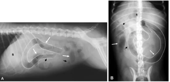
방사선검사상복수로인해복강내대비도가감소하였고
,
좌측중복부에가스가찬소장분절이확장되어
C
자형태를이루고있었으며,
그외 에도확장된소장분절이우측상복부에서관찰되었다(Fig. 1).
소장분절이국소적으로확장되어물리적인장폐색이의심되었고
,
이물,
장중첩,
장간막염전등에대 한감별진단이필요하였다.
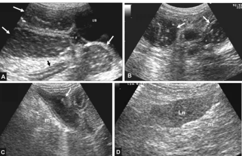
초음파검사시장간막부종과복수가관찰되었고좌측상복부소장분절이확장되
어내강에액체가차있었다
(Fig. 2).
하지만,
장분절내에이물이나장중첩소견은확인되지않았고
,
장분절의폐색을일으킬만한다른원인도보이지않아장간막염 전으로잠정진단후응급수술을결정하였다
.
심각한탈수와심혈관계쇼크상태를개선하기위해
hydroxyethylstarch(
헤모타솔; CJ
제약사업,
한국) 20 mg/kg
과
0.9% saline(0.9%
멸균생리식염수;
대한약품,
한국)
을
90 ml/kg/h
으로2
시간동안주입하였다.
진통제로tramadol(
염산트라마돌;
신풍제약,
한국) 2 mg/kg
과항생 제로cephradine(
세푸딘;
구주제약,
한국), metronidazole (
메트리날;대한약품공업주식회사,
한국), enrofloxacin(
바이트릴
;
바이엘코리아,
한국)
을주사하였다. Propofol
(Pofol inj;
동국 제약,
한국) 6 mg/kg
으로도입마취한후
, isoflurane(Rhodia Organique Fin, UK)
으로유지마취하였고
,
수액은0.9% saline
를10 ml/kg/h
속도로감량하 여주입하였다.
수술중발생할수있는재관류손상Fig. 1. Lateral (A) and ventrodorsal (B) abdominal radiographs. Abdominal detail is decreased due to ascites. Distended
small intestinal loops (long arrows) compared to normal intestines (short arrows) are observed and intestinal obstruction
is suspected. Abnormal “C” shape, distended, and gaseous intestine (dotted line) occupied in left middle abdomen and
displacing stomach (s) cranially.
Fig. 2. Abdominal ultrasonographs. In A, B, C, segmental small intestine (long arrows) is distended and filled with fluid and gas. Anechoic ascites (F) and swollen mesentery (m) are observed. In D, a hypoechoic, enlarged lymph node (Ln) is surrounded by hyperechoic, swollen mesentery (m). There is no evidence compatible to foreign body and intussusception.
Fig. 3. Gross surgical observation of the dog during exploratory laparotomy. (A) The jejunum (white arrow) was grossly
distended, and dark and congested. The small intestine (short arrow) cranial to rotated site was distended, however the
intestinal wall showed only mild congestion. (B) The rotation of the jejunum around mesenteric root (arrow) was identified
and mesenteric torsion was confirmed. The rotated jejunum appeared uniformly necrotic and a jejunal resection and
anastomosis was performed. (C) During exploration, adhesion (a) between segments of small intestine was found and
considered as an evidence of previous intestinal disease.
128 최지혜·김현욱·김진경·장재영·김준영·윤정희
(reperfusion injury)
를예방하기위해Lidocaine(
대한염산리도카인
;
대한약품공업,
한국)
을1 mg/kg
용량으로초기정맥주사한후
,
수술중에는50 µg/kg/min
의속도로연속주입하였다
.
수술중산소포화도(SpO
2)
가80
이하로떨어져
dobutamine(
염산도부타민;
하나제약,
한국)
을5 µg/kg/min
속도로연속 주입하고, 1 ml
의vasopressin (20 IU/ml;
바소프레신;
한림제약,
한국)
을20 ml
의0.9%
saline
에섞어1.2 ml/h
의속도로연속주입하였다.
이후수술을실시하는동안안정된체온
,
심박수,
호흡수를보 였고SpO
2도90
이상으로유지되었다.
개복시출혈성복수가다량관찰되었고
,
공장중간 부터회장까지소장분절이장간막뿌리를기점으로염 전되어내강이 폐색되고확장되었으며,
소장벽이심하게충혈되어검붉은색을띠고있었다
(Fig 3).
염전부위의공장을약
40 cm
정도절제하고장문합을실시하였다
.
염전된부위앞쪽으로도소장분절이확장되고충혈되었으나 이부분은 제거하지않았고 회맹장판막
(ileocecal valve)
도보존하였다.
탐색시일부소장분절사이의유착이확인되어이전에소화기계이상이있었 음을의심할수있었다
.
문합부위를장간막으로덮어준 후복강을여러번세척한뒤폐복하였다.
수술후환자는심한오심과구토를
2-3
회보였고혈액성설사를했지만
,
마취에서무사히회복하였다.
이후 혈관내파종성응고(disseminated intravascular coagulo-
pathy)
의발생을예방하기위해heparin(
신풍헤파린나트륨
;
신풍제약,
한국)
을200 IU/kg
용량으로하루3
회 피하주사하였고,
항생제와진통제는수술전과같은방법으로
8
시간간격으로투여하였다.
전해질과혈당,
혈압
, SpO
2를12
시간간격으로측정하며교정하였다. 36
시간뒤심박수와대퇴동맥압이안정되어
vasopressin
과dobutamine
의연속주입을중단하였고,
분변은점액성의흑색변으로호전되고구토횟수도감소하였다
.
술후3
일째더이상구토를보이지않아
i/d (Canine i/d; Hill's
Pet Nutrition, USA)
를유동식으로급이하였고, 6
일째는기립이가능하였고전해질을포함한혈액검사결과도 정상범위로회복되어술후
7
일째퇴원하였다.
퇴원당시정상배변과식욕을보였고
,
술후2
달까지모니터링 한결과특이적인이상은보이지않았다.
절제한장조 직을한국동물임상연구소(
한국)
에의뢰하여조직검사를실시한결과허혈성장괴사이외다른병변은보이 지않았다
.
고 찰
장간막염전은개에서드물게발생하는질환이지만
,
발생시대부분수시간내에급성으로진행하여폐사하
는 응급 질환이다
[6, 12].
이전 연구에서는German
Shepherds
에서의발생이주로보고되었으나,
그외Ber-
nese Mountain Dog, Bloodhound, Great Dane, Labrador Retriever, Mastino Napolitano, Komondorok, Irish Water
Spaniel
등에서도보고되어있어,
중대형견종에서주로발생하는것으로보인다
[2, 4, 10, 12, 13, 16, 18].
하지 만,
본증례와같이소형견종에서의발생과암컷잡종고양이에서의발생도보고되어있어품종만으로질환을
배제하기어려울것으로생각된다
[3, 7, 19, 21].
본환자는
2
살령의암컷개였으나,
이전연구에의하면수컷에서의발생률이높으며
,
평균2.6-3.1
살인어린성견에서발생하는것으로보고되었다
[13, 17, 21, 22].
장간막염전은소장
,
대장,
혹은둘다에서발생할수있다
[1, 2, 4, 10, 11, 15, 16, 17].
그중소장에서발생한
9
마리를대상으로실시한연구[21]
에의하면공장에서가장흔히발생하였고 이는본증례에서장간막염 전이발견된부위와일치하였다
.
그러나,
장간막염전이 발생하는원인이정확히알려져있지않아공장에서호 발하는원인을추정하기는어렵다.
장간막염전에대한이전보고에의하면위염전
(Gastric
dilation-volvulus; GDV),
장중첩,
장염,
위장관이물,
회 충,
구충제사용,
과도한운동,
복부의둔성외상과관련있다
[4, 10, 12, 13, 15, 17, 21].
본환자는위장관이물 등으로수술한경력은없으나,
지속적인이식증증상을 보여왔으며,
수술중이전장질환의증거로생각되는유착이소장분절사이에서확인되어
,
기존위장관질환과장간막염전의관련성을의심해볼수있다
. GDV
나장중첩이발생했던환자에서장간막염전의발생이많 았던점을바탕으로위장관운동성이상이소인이라는 주장이있으나
,
운동성장애혹은이물,
장염같은위장관질환과장간막염전의관련성에대해서는지속적인
연구가필요할것으로보인다
[10, 12].
또한,
많은보고에서
EPI
를기왕력으로가진다수의개와고양이에서장간막염전이진단되어
EPI
도장간막염전의원인으로제시되고있다
[7, 10, 22].
이는EPI
가소장운동성장애나과도한가스형성과관련이있다는기존연구결
과
[20]
와도 일치하여, EPI
의 호발 품종인German
Shepherds
에서장간막염전이주로보고된점은이러한사항과도관련이있는것으로추정된다
[22].
장간막염전은심한구토
,
설사,
복통,
침울,
식욕부진
,
쇼크등급성복증의증상을보인다[4, 10, 21, 22].
방사선검사시가스가찬소장분절이국소적으로확
장되고
,
복수로 인해복강내대비도가저하된다[22].
국소적으로소장분절이중등도이상확장되면물리적 인폐색을의심할수있으며
,
이물,
장중첩,
소장벽유래종괴
,
장간막염전등여러원인에대한감별진단이필요하다
.
이러한소견은본환자에서도관찰되었고,
그외확장된장분절이
C
자형으로주행하는이상소견이확인되었다
.
이는장간막염전의전형적인소견으로알려져 있지는않지만,
장간막염전이발생하는경우회전된장분절이
C
자형으로관찰될수있는지 추가적인고찰이필요하다
.
방사선검사와초음파검사상물리적인폐색 의소견이확인되고다른원인들이배제된경우,
신속한개복술을통해장간막염전에대한확진이필요하다
[10].
수술전쇼크에대한내과적인치료가중요하다
.
저 혈량성쇼크에대해즉시colloid
와crystalloid fluid
를이용한탈수교정이필요하며
,
본환자에서도hydroxylstarch
와
0.9% saline
을투여하였다[4, 10].
또한,
수술방법으 로는염전된장분절을재위치하는derotation
방법과장분절을절제하고문합하는방법이있다
[1, 10, 12, 22].
수술방법은생존률과는관련이없는것으로알려져있 지만
,
본환자에서는염전된장분절을재위치시키지않고절제하여조직의재관류손상을줄이는방법을이용
하였다
[1, 9, 10].
장이완전히폐색되고8-12
시간지나면장이확장되고색깔이검푸른색으로 변하게되며
,
이후장벽의저산소증으로인해점막장벽이파괴되어 장내세균과독소가복강내로유출되고전신순환계로 들어가결국저혈량성쇼크
,
패혈증및독성쇼크로인해폐사한다
[6, 14].
따라서,
수술시기가매우중요한데
,
기존에장간막염전의치사률이거의100%
에달하던것
[2, 15, 17, 21, 23]
과는달리,
내원후1
시간이내수술을실시한
12
마리환자중5
마리가생존한보고[13]
가있어즉각적인수술적교정이중요하다
.
본환자는임상증상을보인후하루정도의시간이경과하였고백 혈구감소증
,
저혈당증등패혈증의소견을보였으나내 원후3
시간이내에수술을실시하여환자의예후에좋 은영향을미친것으로판단된다.
장간막염전은앞쪽장간막동맥을폐색시켜
,
원위십 이지장,
공장,
회장,
맹장,
오름결장,
근위내림결장의허혈을일으켜
,
장괴사와독소의배출,
쇼크를야기한다
[8, 13].
이러한허혈상태뿐아니라허혈이개선되고조직에다시재관류되는초기
5
분내에도산소유래free
radical
에의해조직이손상되는재관류손상이나타나게된다
.
본환자에서는이러한 재관류손상을막기위해수술전
lidocaine
을주사하고수술중에도연속주입하여좋을예후를얻을수있었다고판단된다
[5, 13].
결 론
장간막염전은중대형견종에서호발하는질환이나
,
소형견종이나고양이에서도발생가능하므로 급성복 증의임상적특징을보이는환자가방사선검사와초음
파검사상물리적인폐색소견을보이고장중첩이나이 물등원인이확인되지않을경우탐색적개복술로확 진및원인교정이필요하다
.
또한,
수술전쇼크에대 한수액 치료와진통제,
항생제,
재관류 손상에대한lidocaine
치료,
심혈관계쇼크에대한inotropic drug
의투약이필요하며
,
이러한적극적인치료를통해환자의 생존률을높일수있을것이다.
장간막염전은매우치사율이높은질환으로기존에 발표된대부분의보고에서수술중혹은술후수시간 이내에사망하였다
.
본보고에서는쇼크상태로내원한소형견종에서발생한장간막염전증례를쇼크에대한 적극적인내과치료와탐색적개복으로장간막염전을 확인하고신속한수술로성공적으로치료할수있었다
.
참고문헌
1. Bentley AM, O'Toole TE, Kowaleski MP, Casale SA, McCarthy RJ. Volvulus of the colon in four dogs. J Am Vet Med Assoc 2005, 227 , 253-256.
2. Cairó J, Font J, Gorraiz J, Martin N, Pons C.
Intestinal volvulus in dogs: a study of four clinical cases. J Small Anim Pract 1999, 40 , 136-140.
3. Camble PJ, Page CA. Mesenteric torsion in a toy dog.
Vet Rec 1992 , 130, 166-167.
4. Carberry CA, Harvey HJ, Blackburn H. Small intestinal volvulus in a dog. Compend Contin Educ Pract Vet 1989, 11 , 1322-1325.
5. Cassutto BH, Gfeller RW. Use of intravenous lidocaine to prevent reperfusion injury and subsequent multiple organ dysfunction syndrome. J Vet Emerg Crit Care 2003, 13 , 137-148.
6. Cosenza SF. Recognizing and treating mesenteric torsion in dogs. Vet Med 1996, 91 , 929-933.
7. Drobatz KJ, Hughes D, Hill C, Walker L. Volvulus of the colon in a cat. J Vet Emerg Crit Care 1996, 6 , 99-102.
8. Evans HE, Christensen GC. The Digestive apparatus and abdomen. In: Evans HE (ed.). Miller’s Anatomy of the Dog. 3rd ed. pp. 385-462, Saunders, Philadelphia, 1993.
9. Fossum TW. Surgery of the small intestine. In: Fossum TW, Hedlund CS, Hulse DA, Johnson AL, Seim HB III, Willard MD, Carroll GL (eds.). Small Animal Surgery. 1st ed. pp. 292-318, Mosby, St. Louis, 1997.
10. Halfacree ZJ, Beck AL, Lee KC, Lipscomb VJ.
Torsion and volvulus of the transverse and descending
colon in a German shepherd dog. J Small Anim Pract
130 최지혜·김현욱·김진경·장재영·김준영·윤정희