https://doi.org/10.12750/JET.2017.32.4.297
Effect of Monosodium Glutamate on In Vitro Oocyte Maturation and Embryonic Development after Parthenogenesis in Pigs
Minji Kim1, Hyeji Shin2, Joohyeong Lee3, Seung Tae Lee1 and Eunsong Lee2,3,*
1Division of Applied Animal Science, College of Animal Life Science, Kangwon National University, Chuncheon 24341, Korea
2College of Veterinary Medicine and 3Institute of Veterinary Science, Kangwon National University, Chuncheon 24341, Korea.
ABSTRACT
This study was designed to determine the effect of monosodium glutamate (MSG) on in vitro maturation (IVM) of oocytes and early development of parthenogenesis (PA) embryos in pigs. Each IVM and IVC medium was supplemented with various concentrations (0, 0.1, 0.5 and 5 mM) of MSG and non-essential amino acids (NEAA) depending on the experimental design. Immature pig oocytes were matured for 44 h and then oocytes reached metaphase II (MII) stage were electrically activated to induce parthenogenesis (PA). When immature oocytes were treated with MSG in the absence of NEAA during IVM, nuclear maturation (83.1-87.1%), intra-oocyte glutathione content, cumulus expansion, and cleavage (91.4-93.4%) of PA embryos were not influenced by MSG treatment at all concentrations. However, blastocyst formation of PA embryos was significantly increased by 5.0 mM MSG (45.3 ± 6.2%) compared to control (25.6 ± 3.4%). MSG treatment during IVM in the presence of NEAA did not show significant effect on nuclear maturation of oocytes and blastocyst formation after PA while 0.5 mM MSG (89.3 ± 1.9%) decreased (P < 0.05) cleavage of PA embryos compared to 0.1 mM MSG (94.6 ± 1.1%). When PA embryos were treated for 7 days with MSG during IVC, 5.0 mM MSG significantly decreased blastocyst formation (27.8 ± 4.9%) compared to no treatment (41.4 ± 1.9%) while no decrease in blastocyst formation was observed in 0.1 and 0.5 mM (37.4 ± 3.4% and 34.4 ± 2.6%, respectively). Our results demonstrated that 5 mM MSG in a NEAA-free chemically defined maturation medium showed positive effect on PA embryonic development while 5 mM MSG treatment during IVC was deleterious to PA embryonic development in pigs.
(Key Words : Monosodium glutamate, Oocyte maturation, Embryonic development, Parthenogenesis, Pig
†Correspondence: Eunsong Lee (ORCID: 0000-0001-9654-7788) Phone: +82-33-250-8670, Fax: +82-33-259-5625
E-mail: [email protected]
INTRODUCTION
Recent progress in in vitro maturation of pig oocytes makes it possible to produce piglets by transferring in vitro-produced (IVP) pig embryos derived from IVM oocytes. However, despite of successful production of piglets, the developmental competence of IVP embryos derived from IVM oocytes is still inferior to their in vivo-developed counterparts which is mainly attributed to the lower quality of IVM oocytes compared to in vivo ones (Dobrinsky et al. 1996; Kang et al. 2009). For this reason, a wide variety of studies have been attempted to improve and optimize IVM system by determining effect of various factors such as energy substrate, amino acids, and various cytokines and growth factors on oocyte maturation and later embryonic development after in vitro fertilization (IVF)
and parthenogenesis (PA) in pigs (Abeydeera et al. 2000;
Sturmey and Leese, 2003; Hong and Lee, 2007; Mito et al.
2013).
Monosodium glutamate (MSG), a naturally present non- essential amino acid, is well known as a taste enhancer that is added frequently to foods, soups, and processed meats (Chang and Cha, 2001; Freeman, 2006; Ali et al., 2014).
Although MSG has been recognized as safe as a food ingredient for a long time, there are several studies reporting side effects of MSG such as MSG symptom complex including headache, sweating, chest pain, and nausea (Baad- Hansen et al., 2010; Shimada et al., 2013). MSG consists of 78% of glutamic acid and 22% of sodium (Samuels et al, 1999) and is an ingredient contained in meats, fishes, milk, and some vegetables (Winkel et al., 2008). It has been
of MSG, is used as a substrate for protein synthesis, involves in acid-base balance in kidney, and also acts as a precursor for neurotransmitter synthesis, nucleotide and nucleic acids, and glutathione (Newsholme et al., 2003). Despite there are still controversy surrounding the safety or side effects of MSG as a flavor enhancer, dietary supplementation with MSG is generally recognized as safe in human, pig, and other animals (Boutry et al., 2011; Brosnan and Brosnan, 2013; Rezaei et al., 2014).
Effect of MSG on the reproductive function has been studied by several workers. Eweka and Om’Iniabohs (2011) reported that MSG at high doses might have some harmful effects on the oocytes of the ovaries and cause female infertility in adult Wistar rats. Oladipo et al. (2015) reported that MSG induced considerable changes in ovarian structure including degeneration of follicles, oocytes and medulla with vacuoles having blood congestion in Sprague-dawley rats. In another study, Spinacci et al. (2017) reported in pigs a beneficial effect of MSG on oocyte-sperm binding and fertilization in vitro. To date, a few studies are available on the effect of MSG on oocyte maturation and embryonic development in pigs. The objective of this study was to determine the effect of MSG during IVM and IVC on in vitro oocyte maturation and parthenogenetic development of PA embryos in pigs. To this end, immature pig oocytes were cultured under MSG treatment during in vitro maturation and nuclear maturation of oocytes, intraoocyte glutathione content, and developmental competence of parthenogenesis embryos were determined.
MATERIALS AND METHODS
1. Culture Media and Reagents
All reagents used in this study were purchased from Sigma-Aldrich (St. Louis, MO, USA) unless specified otherwise. For IVM of immature pig oocytes, medium-199 (M-199) (Invitrogen, Grand Island, NY, USA) containing 10%
(v/v) pig follicular fluid (Experiment 4) or porcine zygote medium (PZM)-4 containing 5.5 mM glucose and 0.1% (w/v) polyvinyl alcohol (PVA) were used (Experiments 1-3). These media were supplemented further with 0.91 mM pyruvate, 0.6
of MSG according to the experimental design. The medium for in vitro culture (IVC) of PA and SCNT embryos was PZM-3 containing 0.3% (w/v) bovine serum albumin (BSA). The IVC medium was modified in this study by adding 0.34 mM tri-sodium citrate, 2.77 mM myo-inositol, and 10 μM β -mercaptoethanol (Lee et al., 2014).
2. Oocyte Collection and IVM
Prepubertal gilt ovaries were obtained at a local abattoir.
Follicular contents containing cumulus-oocyte complexes (COCs) were aspirated from superficial follicles of 3-8 mm in diameter. The COCs had multiple layers of cumulus cells were selected and washed in HEPES-buffered Tyrode’s medium containing 0.05% (w/v) PVA (TLH-PVA). The COCs were placed into each well of a four-well culture dish (Nunc, Roskilde, Denmark) containing 500 μl of IVM medium enriched with 80 μg/ml follicle stimulating hormone (Antrin R-10, Kyoritsu Seiyaku, Tokyo, Japan) and 10 IU/ml human chorionic gonadotrophin (Intervet International BV, Boxmeer, Holland). The COCs were cultured at 39°C under humidified atmosphere of 5% CO2 and 95% air. After 22 h in the maturation culture, the COCs were washed properly and then cultured in hormone-free IVM medium for an additional 22 h and 20 h for PA and SCNT, respectively.
3. Experimental Design
In experiment 1, effects of various concentrations (0, 0.1, 0.5, and 5.0 mM) of MSG in a non-essential amino acids-free chemically defined PZM-4 on oocyte maturation and PA embryonic development were examined. Effect on MSG treatment during IVM as in Experiment 1 on intra-oocyte GSH contents and cumulus expansion of IVM oocytes were determined in Experiment 2. In Experiment 3, 0.1, 0.5, and 5.0 mM MSG were added to a chemically defined PZM-4 in the presence of 1% (v/v) minimum essential medium NEAA and effects of MSG on oocyte maturation and PA embryonic development were evaluated. Finally, effects of MSG treatment during IVC on PA embryonic development was assessed in Experiment 4.
Fig. 1. Photomicrographic images of in vitro-matured porcine oocytes. Oocytes were matured in a non-essential amino acids-free porcine zygote medium-4 that was supplemented with 0.1% (w/v) polyvinyl alcohol, 5.5 mM glucose, and various concentrations of monosodium glutamate (MSG). Oocytes matured in maturation medium containing 0 (control, A and E), 0.1 (B and F), 0.5 (C and G), or 5.0 mM (D and H) MSG were examined for intra-oocyte glutathione (GSH) contents (A-D) and cumulus expansion (E-H). Oocytes were stained with CellTracker Blue to detect GSH contents.
4. Parthenogenetic Activation (PA) and In Vitro Culture of PA embryos After IVM, oocytes were denuded from their cumulus cells by gentle pipetting in IVM medium containing 0.1% (w/v) hyaluronidase. Oocytes extruded the first polar body were selected and allocated to PA. Oocytes were placed in an electrode chamber and PA was induced by applying two pulses of 120 V/mm direct current for 60 μsec in a 280 mM mannitol solution that was supplemented with 0.05 mM MgCl2
and 0.1 mM CaCl2. Then, activated oocytes were washed properly in IVC medium and incubated for 4 h in IVC medium containing 7.5 μg/ml. PA embryos were washed three times, placed in 30-μl IVC medium droplets under mineral oil, and cultured for 7 days at 39°C in a humidified atmosphere of 5% CO2, 5% O2 and 90% N2.
5. Determination of Intra-Oocyte GSH content and Cumulus Cell Expansion after IVM
GSH content in IVM oocytes was determined as previously described (Sakatani et al., 2007) using Cell-Tracker Blue CMF2HC (4-chloromethyl-6.8-difluoro-7-hydroxycoumarin; Invitrogen) to detect GSH as blue fluorescence (Fig. 1). A group of 10 to 12 oocytes per replicate were incubated for 30 min in TLH-PVA supplemented with 10 μM Cell-Tracker, washed properly and incubated for 30 min in a PZM-3 containing 0.3% (w/v) BSA
at 39°C in the dark. Then, oocytes were washed, placed into 2-μL droplets of Dulbecco’s phosphate-buffered saline (D-PBS) 0.1% (w/v) PVA, and observed for blue fluorescence under an epifluorescence microscope (TE-300; Nikon). The fluorescence intensities of oocytes were analyzed with the ImageJ software and normalized to control oocytes.
The extent of cumulus cell expansion after IVM was evaluated subjectively as previously reported (Vanderhyden et al., 1990). Briefly, no expansion was scored as 0, minimum observable response with the cumulus cells in the outermost layer became round and glistening as 1, the expansion of outer cumulus cell layers as 2, the full expansion of all cumulus cell layers except corona radiata as 3, and the full expansion of all cumulus cell layers including corona radiata as 4 (Fig. 1).
6. Statistical Analysis
Statistical analyses were done using the Statistical Analysis System (version 9.3; SAS Institute, Cary, NC, USA). The data were analyzed by the general linear model procedure followed by the least square method when the treatments differed at P
< 0.05. The percentage data such as embryo cleavage and blastocyst formation were arcsine transformed before analysis to maintain homogeneity of variance. The results are expressed as the mean ± standard error of the mean (SEM).
MSG (mM) in maturation medium*
% of oocytes that reached MII
No. of PA oocytes cultured†
% of embryos developed to No. cells in blastocyst
≥ 2-cell Blastocyst
0 (control) 86.3 ± 3.1 139 91.4 ± 2.6 25.6 ± 3.4b 38.0 ± 2.4
0.1 83.1 ± 3.0 138 92.8 ± 1.2 32.9 ± 4.6ab 40.8 ± 1.9
0.5 83.8 ± 2.1 144 93.1 ± 1.7 37.2 ± 5.8ab 40.1 ± 1.8
5.0 87.1 ± 1.8 148 93.4 ± 1.2 45.3 ± 6.2a 38.0 ± 2.0
†Four replicates.
*The maturation medium was NEAA-free porcine zygote medium (PZM)-4 containing 0.1% polyvinyl alcohol (PVA) and 5.5 mM glucose.
abValues in the same column with different superscript letters are different (P < 0.05).
Table 2. Effect of monosodium glutamate (MSG) in a non-essential amino acids (NEAA)-free chemically defined medium on intra-oocyte glutathione (GSH) contents and cumulus cell expansion after IVM
MSG (mM) in maturation medium*
No. of MII oocytes examined for GSH
Relative level (pixels/oocyte) of GSH
No. of oocytes examined for cumulus expansion
Cumulus cell expansion score†
0 (control) 42 1.00 ± 0.06 77 1.50 ± 0.23
0.1 42 0.93 ± 0.06 77 1.25 ± 0.14
0.5 42 0.89 ± 0.06 76 1.28 ± 0.16
5.0 42 0.98 ± 0.06 82 1.20 ± 0.12
*Three replicates.
†Cumulus cell expansion was scored as 0 (no response), 1 (minimum observable response with the cells in the outermost layer of the cumulus become round and glistening), 2 (the expansion of outer cumulus cell layers), 3 (the expansion of all cumulus cell layers except corona radiata), and 4 (the expansion of all cumulus cell layers).
RESULTS
1. Effect of MSG in a Non-Essential Amino Acids (NEAA)-Free Chemically Defined Medium on Oocyte Maturation and PA Embryonic Development (Experiment 1)
When immature oocytes were matured in a chemically defined medium containing 0, 0.1, 0.5, and 5.0 mM MSG, nuclear maturation of oocytes (83.1-87.1%). After PA, blastocyst formation was significantly increased by 5.0 mM MSG treatment (45.3 ± 6.2%) during IVM compared to control (25.6 ± 3.4%) while embryo cleavage (91.4-93.4%) and mean number of cells in blastocyst (38.0-40.8 cells) were not altered by the treatment (Table 1).
2. Effect of MSG in a Non-Essential Amino Acids (NEAA)-Free Chemically Defined Medium on Intra-Oocyte GSH contents and Cumulus Expansion (Experiment 2)
The GSH contents (1.00 ± 0.06 and 0.89-0.98 pixels/oocyte for control and MSG-treated oocytes, respectively) cumulus
expansion scores (1.50 and 1.20-1.28 for control and MSG- treated oocytes, respectively) of IVM oocytes that were untreated or treated with various concentrations of MSG are shown in Table 2. The GSH contents and cumulus expansion were not influenced by MSG treatment during IVM at all concentrations tested.
3. Effect of MSG in a Chemically Defined Medium Containing Non-Essential Amino Acids (NEAA) on oocyte maturation and PA embryonic development (Experiment 3)
As shown in Table 3, MSG treatment during IVM in the presence of NEAA did not alter nuclear maturation of oocytes (93.0-96.9%). After PA, embryo cleavage was significantly lower in 0.5 mM MSG group (89.3%) than in 0.1 mM MSG (94.6%). However, there was no difference in blastocyst formation (47.1 ± 2.3, 43.5 ± 4.0, and 50.4 ± 3.1% for 0.1, 0.5, and 5.0 mM MSG, respectively) and mean number of cells in blastocyst (32.2-33.7 cells) among the concentrations of MSG examined.
Table 3. Effect of monosodium glutamate (MSG) in a chemically defined medium containing non-essential amino acids (NEAA) on oocyte maturation and embryonic development parthenogenesis (PA)
MSG (mM) in maturation medium*
% of oocytes that reached MII
No. of PA embryos cultured†
% of embryos developed to No. cells in blastocyst
≥ 2-cell Blastocyst
0.1 96.2 ± 1.2 149 94.6 ± 1.1a 47.1 ± 2.3 32.6 ± 1.3
0.5 96.9 ± 1.5 151 89.3 ± 1.9b 43.5 ± 4.0 33.7 ± 1.3
5.0 93.0 ± 1.9 145 93.8 ± 0.7ab 50.4 ± 3.1 32.2 ± 1.3
†Four replicates.
*The maturation medium was porcine zygote medium (PZM)-4 containing 0.1% polyvinyl alcohol (PVA), 0.1% (v/v) NEAA, and 5.5 mM glucose.
ab Values in the same column with different superscript letters are different (P < 0.05).
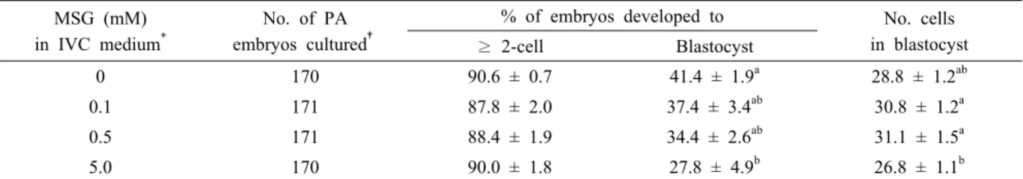
Table 4. Effect of monosodium glutamate (MSG) in in vitro culture (IVC) medium on embryonic development after parthenogenesis (PA) MSG (mM)
in IVC medium*
No. of PA embryos cultured†
% of embryos developed to No. cells in blastocyst
≥ 2-cell Blastocyst
0 170 90.6 ± 0.7 41.4 ± 1.9a 28.8 ± 1.2ab
0.1 171 87.8 ± 2.0 37.4 ± 3.4ab 30.8 ± 1.2a
0.5 171 88.4 ± 1.9 34.4 ± 2.6ab 31.1 ± 1.5a
5.0 170 90.0 ± 1.8 27.8 ± 4.9b 26.8 ± 1.1b
†Five replicates.
*The IVC medium was NEAA-free porcine zygote medium (PZM)-4 containing 0.1% (w/v) polyvinyl alcohol (PVA) and 5.5 mM glucose.
ab Values in the same column with different superscript letters are different (P < 0.05).
4. Effect of MSG during IVC in a Non-Essential Amino Acids (NEAA)-Free Chemically Defined Medium on PA Embryonic Development (Experiment 4)
PA embryos were cultured in a NEAA-free chemically defined PZM-4 medium that was further supplemented with 0 (control), 0.1, 0.5, and 5.0 mM MSG to examine the effect of MSG on embryonic development. Treatment of embryos with 5.0 mM MSG (27.8 ± 4.9%) during IVC significantly decreased (P < 0.05) blastocyst formation of PA embryos compared to no treatment control (41.4 ± 1.9%). In addition, means cell number of blastocyst was also decreased in 5.0 mM MSG group (26.8 ± 1.1 cells/blastocyst) compared to 0.1 and 0.5 mM MSG groups (30.8 ± 1.2 and 31.1 ± 1.5 cells/blastocyst, respectively) (Table 4).
DISCUSSION
Reproductive biotechnologies such as IVF, intracytoplasmic sperm injection, and somatic cell nuclear transfer have been widely used to produce animals with specific purposes. In the
utilization of these technologies, IVM oocytes are routinely used. Thus, the quality of IVM oocytes are essential factor for normal development of IVP mammalian embryos. In this study, we evaluated the effect of MSG, a routinely used taste enhancer in many kinds of foods, on oocyte maturation and subsequent in vitro development of PA embryos in pigs. We found that MSG showed differential effect on PA embryonic development depending on the time of treatment. MSG treatment at 5 mM concentration during IVM was beneficial for blastocyst formation of PA embryos whereas treatment of PA embryos with the same concentration of MSG for 7 days during IVC showed a deleterious effect on embryonic development.
In the pig follicular fluid, various amino acids including glutamate, glutamine, aspartate, glycine, arginine, alanine, leucine, lysine, proline, and valine (Jimena et al., 1993; Hong and Lee, 2007). Of those, glutamate is the most plentiful amino acid irrespective of the follicular size and its concentration is approximately twice as much as those of other amino acids. In a previous study (Hong and Lee, 2007) examined the effect of various amino acids in a maturation
formation of IVF embryos. In this study, MSG treatment during IVM did not affect nuclear maturation of oocytes while 5 mM MSG improved blastocyst formation of PA embryos.
This result was inconsistent with the previous finding 0.439 mM glutamic acid did not show beneficial effect on both oocyte maturation and IVF embryonic development. This discrepancy might be due to the different concentration of glutamate used. Hong and Lee (2007) determined the glutamate concentration as 0.439 mM in the pig follicular fluid and used that concentration of glutamate.
The extent of cumulus cell expansion and intra-oocyte GSH contents have been routinely used as an indicator to assess the quality of IVM oocytes (Eppig, 1982; Abeydeera et al., 1998;
De Matos and Furnus, 2000; Lee et al., 2014) because in vivo oocytes shows full expansion of cumulus cell layer (Motlik et al., 1986). It is well known that GSH protects oocytes from harmful effects of reactive oxygen species (Yoshida et al., 1993; Maedomari et al., 2007). In this study, although 5 mM MSG showed a beneficial effect on embryonic development, no effect of MSG treatment during IVM was observed in these parameters. There are several studies reported that the good cumulus cell expansion and high intra-oocyte GSH contents are important criteria of high quality of oocytes but does not guarantee better embryonic development (Marei et al., 2012;
Park et al., 2016).
In contrast to the beneficial effect of MSG treatment during IVM, treatment of PA embryos with 5 mM MSG for 7 days during IVC inhibited blastocyst formation, especially at the 5 mM concentration. It was not clear how MSG acted and affected embryonic development in this study. Considering the concentration of glutamate in pig follicular was around 0.439 mM, the concentration of MSG used in this study (5.0 mM) was approximately 10 times higher than the follicular concentration. In addition, in contrast to IVM treatment for 44 h, PA embryos were treated for longer period (7 days) during IVC. Relatively longer period of treatment with high concentration of MSG might showed a deleterious effect. In summary, our results demonstrate that 5 mM MSG in a NEAA-free chemically defined maturation medium has positive effect on PA embryonic development while 5 mM MSG treatment during IVC is deleterious to PA embryonic development in pigs. Further studies would be needed to clarify
fluid, and oviductal or uterine fluid and on the function of reproductive organs including follicular development, oocyte maturation, ovulation, and early embryonic development.
ACKNOWLEDGMENTS
This research was supported by Basic Science Research Program through the National Research Foundation of Korea (NRF) funded by the Ministry of Science, ICT and Future Planning (Grant No. 2015R1A2A2A01005490) and 2016 Research Grant from Kangwon National University (No. 520160228).
REFERENCES
Abeydeera LR, Wang WH, Cantley TC, Prather RS and Day BN. 1998. Presence of β-mercaptoethanol can increase the glutathione content of pig oocytes matured in vitro and the rate of blastocyst development after in vitro fertilization.
Theriogenology 50: 747-756.
Abeydeera LR, Wang WH, Cantley TC, Rieke A, Murphy CN, Prather RS and Day BN. 2000. Development and viability of pig oocytes matured in a protein-free medium containing epidermal growth factor. Theriogenology 54:787-797 Ali AA, El-Seify GH, El Haroun HM and Soliman MAEMM.
2014. Effect of monosodium glutamate on the ovaries of adult female albino rats and the possible protective role of green tea. Menoufia Med. J. 27:793-800.
Baad-Hansen L, Cairns BE, Ernberg M and Svensson P. 2010.
Effect of systemic monosodium glutamate (MSG) on headache and pericranial muscle sensitivity. Cephalalgia 30:68–76.
Boutry C, Matsumoto H, Airinei G, Benamouzig R, Tome´ D, Blachier F and Bos C. 2011. Monosodium glutamate raises antral distension and plasma amino acid after a standard meal in humans. Am. J. Physiol. 300: G137–G145.
Brosnan JT and Brosnan ME. 2013. Glutamate: a truly functional amino acid. Amino Acids 45: 413-418.
Chang KJ and Cha W. 2001. Studies on flavor enhancer products used in Korean households in the Inchon area.
Nutr. Sci. 4:47-54.
Dobrinsky JR, Johnson LA and Rath D. 1996. Development of a culture medium (BECM-3) for porcine embryos: effects of bovine serum albumin and fetal bovine serum on embryo development. Biol. Reprod. 55: 1069-1074.
De Matos DG and Furnus CC. 2000. The importance of having high glutathione (GSH) level after bovine in vitro maturation on embryo development: effect of β -mercaptoethanol, cysteine and cystine. Theriogenology 53:
761-771.
Eppig JJ. 1982. The relationship between cumulus cell-oocyte coupling, oocyte meiotic maturation, and cumulus expansion.
Dev. Biol. 89:268-272.
Eweka AO and Om’Iniabohs FAE. 2011. Histological studies of the effects of monosodium glutamate on the ovaries of adult Wistar rats. Ann. Med. Health Sci. Res. 1:37-44.
Freeman M. 2006. Reconsidering the effects of monosodium glutamate: a literature review. J. Am. Acad. Nurse Pract.
18: 482-486.
Hong J and Lee E. 2007. Intrafollicular amino acid concentration and the effect of amino acids in a defined maturation medium on porcine oocyte maturation, fertilization, and preimplantation development. Theriogenology 68: 728-735.
Jimena P, Castilla JA, Peran F, Ramirez JP, Gil T, Mozas J, Martinez L and Herruzo A. 1993. Distribution of free amino acids in human preovulatory follicles. Horm. Metab.
Res. 25: 228-230.
Kang J, Koo O, Kwon D, Park H, Jang G, Kang S and Lee B. 2009. Effects of melatonin on in vitro maturation of porcine oocyte and expression of melatonin receptor RNA in cumulus and granulosa cells. J. Pineal Res. 46:22-28.
Lee J, Lee Y, Park B, Elahi F, Jeon Y, Hyun SH and Lee E.
2014. Developmental competence of IVM pig oocytes after SCNT in relation to the shrinkage pattern induced by hyperosmotic treatment. Theriogenology 81: 974-981.
Maedomari N, Kikuchi K, Ozawa M, Noguchi J, Kaneko H, Ohnuma K, Nakai M, Shino M, Nagai T and Kashiwazaki N. 2007. Cytoplasmic glutathione regulated by cumulus cells during porcine oocyte maturation affects fertilization and embryonic development in vitro. Theriogenology 67:
983-993.
Marei WF, Ghafari F and Fouladi-Nashta AA. 2012. Role of hyaluronic acid in maturation and further early embryo development of bovine oocytes. Theriogenology 78: 670-677.
Mito T, Yoshioka K, Noguchi M, Yamashita S and Hoshi H.
2013. Recombinant human follicle-stimulating hormone and transforming growth factor-alpha enhance in vitro maturation of porcine oocytes. Mol. Reprod. Dev. 80:
549-560.
Motlik J, Fulka J and Fléchon JE. 1986. Changes in intercellular coupling between pig oocytes and cumulus cells during maturation in vivo and in vitro. J. Reprod.
Fert. 76: 31-37.
Newsholme P, Procopio J, Lima MMR, Pithon‐Curi TC and Curi R. 2003. Glutamine and glutamate¾their central role in cell metabolism and function. Cell. Biochem. Funct. 21:
1-9.
Oladipo IC, Adebayo EA and Kuye OM. 2015. Effects of monosodium glutamate in ovaries of female Sprague-Dawley rats. Int. J. Curr. Microbiol. App. Sci. 4: 737-745.
Park B, Lee H, Lee Y, Elahi F, Lee J, Lee ST, Park CK, Hyun SH and Lee E. 2016. Cilostamide and forskolin treatment during pre-IVM improves preimplantation development of cloned embryos by influencing meiotic progression and gap junction communication in pigs. Theriogenology 86: 757-765.
Rezaei R1, Knabe DA, Tekwe CD, Dahanayaka S, Ficken MD, Fielder SE, Eide SJ, Lovering SL and Wu G. (2013).
Dietary supplementation with monosodium glutamate is safe and improves growth performance in postweaning pigs. Amino Acids 44: 911-923.
Sakatani M, Suda I, Oki T, Kobayashi SI, Kobayashi S and Takahashi M. 2007. Effects of purple sweet potato anthocyanins on development and intracellular redox status of bovine preimplantation embryos exposed to heat shock.
J Reprod Dev 53: 605-614.
Samuels A. 1999. The toxicity/safety of processed free glutamic acid (MSG): a study in suppression of information. Account.
Res. 6: 259-310.
Shimada A, Cairns BE, Vad N, Ulriksen K, Pedersen AML, Peter Svensson P and Baad-Hansen L. 2013. Headache and mechanical sensitization of human pericranial muscles after repeated intake of monosodium glutamate (MSG). J.
Headache Pain 14: 2 (1-9).
Spinaci M, Bucci D, Gadani B, Porcu E, Tamanini C and Galeati G. 2017. Pig sperm preincubation and gamete coincubation with glutamate enhance sperm-oocyte binding and in vitro fertilization. Theriogenology 95: 149-153.
Sturmey RG and Leese HJ. 2003. Energy metabolism in pig oocytes and early embryos. Reproduction 126: 197-204.
expansion-enabling factor by mouse oocytes and the role of oocytes in promoting granulosa cell differentiation. Dev.
Biol. 140: 307-317.
Winkel C, de Klerk A, Visser J, de Rijke E, Bakker J, Koenig T, Renes H. 2008. New developments in umami (enhancing) molecules. Chem. Biodivers. 5: 1195-1203.
after fertilization in pig oocytes: relevance to the ability of oocytes to form male pronucleus. Biol. Reprod. 49: 89-94.
Received December 14 2017, Revised December 19, 2017, Accepted December 21, 2017