40(1) : 77 81 (2009)
77
분비나무 줄기 수피 추출물에 의한 HeLa Cell Line의 증식억제 효과와 RAW264.7 세포에서 Lipopolysaccharide에 의해 유도된 Nitric Oxide
생성 저해효과
배기은·정한수·김동섭·최영웅·김영숙·김영균*
국민대학교임산공학과
Inhibitory Effect on the Lipopolysaccharide-Induced Nitric Oxide Formation in RAW264.7 and on the Proliferation of HeLa Cell Lines by the
Stem Bark Extracts of Abies nephrolepis
Ki-Eun Bae, Han-Soo Chong, Dong-Sup Kim, Young-Woong Choi, Yeung-Suk Kim and Young Kyoon Kim
*Department of Forest product, Kook min University, Seoul, 136-702, Korea
Abstract −Abies nephrolepis(Pinaceae) extracts were tested for determined immune system regulating activity based on anti- inflammatory activity, antioxidant activity and anti-proliferative effect on HeLa cell line. The A. nephrolepis extracts increased dose-dependently anti-proliferation of HeLa cell line. The DM fraction of the extracts having anti-proliferatative effects of HeLa cell line was fractionalized four subfractions(D1~D4). Inflammation-induced NO production was inhibited by D2 and D4 in LPS-activated RAW264.7 macrophages. And also, this fractions showed antioxidant activity examined by DPPH radical scav- enging effects. These results suggest that the potential use of DM fraction of A. nephrolepis in chemoprevention and regulation overproduction of NO on pathogenic conditions. The mechanism of the inflammatory effects, however, must be evaluated through various parameters in the induction cascade of NO production.
Key words −Abies nephrolepis, Pinaceae, Antioxidant, Antiinflammation, Anticancer
Oxidative stress는수많은생리학과병리학현상에서중
요한역할을한다. Oxidative stress에수반되는활성산소종 (reactive oxygen species, ROS)에는 oxygen, superoxide, hydroxy radical, hydrogen peroxide 등이있으며, 이들은미
토콘드리아내의전자전달계및백혈구세포의활성화등정 상적인세포기능을유지하는데중요한역할을담당하는세 포내신호전달물질로작용한다. 하지만 ROS의조절기능에
이상이초래되면염증반응이발생하거나, 세포사멸을일으
키게된다.1)염증반응은생체나조직에어떠한기질적변화
가가해질때그손상부위를재생하려는기전으로서, 일단
자극이가해지면염증매개인자에의해세포간극활성산 소종의생성도급격히증가된다. 따라서과도한 ROS의생
성에의한세포의스트레스는궁극적으로노화, 암, 뇌졸중,
당뇨병, 관절염, 면역결핍증, 동맥경화등여러성인병의직
접적인원인으로알려져있다.2)
내독소의하나인 lipopolysaccharide (LPS)는 RAW264.7
세포와 같은 대식세포에서 tumor necrosis factor-alpha (TNF-α), Interleukin-6 (IL-6), Interleukin-1β (IL-1β)와같은 proinflammatory cytokine을증가시키며,3,4) proinflammatory gene들의유도는전사인자의활성화에의해일어나게된다.
이러한염증매개물질의형성은 PhospholipaseA2의활성으 로인해 arachidonic acid가 Prostaglandin (PG)으로바뀌는
과정및 Nitric oxide (NO)형성과정으로이어지게된다.5,6) NO는 IL-1β, TNF-α, LPS 등과같은염증유발인자 및암
발생인자에의해발현되는 inducible nitric oxide synthase (iNOS)에의해과량발생한다.7)생성된 NO는 free radical
의일종으로혈관확장, 비선택적숙주방어등의작용을하지
만, 지속적과발현은오히려점막손상을촉진하고, 그결과
일부에서는암발생등의질환을유도한다. 이처럼염증과
*교신저자(E-mail):[email protected] (Tel):82-2-910-4825
암사이의 연관성은 150년전부터 알려져왔으며, 1863년
Virchow는만성염증이발생한부위에서암이발생되는경
향성이높음을밝혔다.8)
본연구에앞서나무 150수종의각추출물에대한 HeLa
Cell line의증식억제효과에대하여 screening하였고, 그결
과 15 수종에서우수한효과가있는것으로확인되었으며,
그중하나인분비나무(A. nephrolepis, Pinaceae)를공시재
료로선택하였다. 우리나라전국산야지에분포하는분비나
무는한국을비롯하여중국북동부, 동부시베리아에널리
분포하며소나무과(Pinaceae)의상록침엽교목이다. 꽃의개
화기는4-5월이며열매는 9월에익으며위로향하고긴난
형또는난상원통형이며길이 4-5.5 cm, 직경 2-2.5 cm로서
녹갈색이고실편은녹색이다. 잎은선형이며, 전나무와유
사하나잎끝이둘로갈라지고구상나무보다얇고가늘다.9)
분비나무에관한선행연구로는분비나무잎의페놀성화 합물들이갖는암을비롯한질환의예방효능에대해보고 되어졌다.10)또한수피추출물이 N-Smase 와ceramide에억
제효과를보여면역반응이교란되는것을막는다고보고 되어져있다.11)그외에분비나무와같은과에속하는프랑
스산해송(Pinus maritime, Pinaceae)의수피로부터페놀성
물질로구성된 Pycnogenol이라는추출물에대한연구가보
고되어있다.12) Pycnogenol은 free radical을제거하여 NO생
성을조절하며, Pro-inflammatory cytokine인 Interlukin-1의
생성을저해하여항염작용을가짐을확인하였다.
따라서, 본연구는선행실험결과분비나무추출물이 HeLa
cell line에대한증식억제효과가있다는점과소나무과식
물추출물의항암, 항염있다는점에착안하여분비나무추
출물이항염증제로도응용가능성이있는지알아보고자실 시하였다.
재료 및 방법
재료 −Methanol, n-hexane, dichloromethane, and ethyl acetate는 Duksan pure chemicals (Ansan, Korea)에서구입
하였으며, silica gel는 Merck (Darmstadt, Germany)에서구
입하였다. Dulbecco’s modified Eagle’s minimum essential medium (DMEM), fetal bovine serum (FBS), penicillin, streptomycin은 Welgene Bioscience (Daegu, Korea)에서 구
입하였다. 3-(4,5-dimethylthiazol-2-yl)-2,5-diphenyltetrazolium bromide (MTT)는 AMRESCO (Solon,Ohio)에서구입하였
다. Dimethyl sulfoxide (DMSO), phosphoric acid는 Duksan pure chemicals (Ansan, Korea), naphtylethylene- diamine dihydrochloride는 ACROS (Nes jersey, USA)에서
구입하였다. L-N6-(1-iminoethyl)lysine (L-NIL), Escherichia coli. lipopolysaccharide (LPS)는 Sigma Chemical Co. (CA, U.S.A.)에서구입하였다. DPPH (1,1-diphenyl-2-picryl hydrazyl),
sulfanilamide는 Sigma-Aldrich (USA)에서구입하였다.
분비나무의 추출 및 분리 − 분비나무줄기수피 600 g을 MeOH을용매로 46oC에서 5시간씩 3회추출하였다. 추출물
을여과하고감압농축기로농축하여 MeOH추출물(75.81 g)
을얻었다. 추출물을물과 n-hexane (n-Hex)을 1:1로섞어
용매극성분리를진행하여 n-hexane fraction을얻었고, n- hexane 분획하고남은물fraction과 DM (Dichloromethane)
을 1:1로섞어 DM 층을얻었다. 이와동일한방법으로 ethyl acetate (EA) fraction 그리고 water (Aqu) fraction을순차적
으로얻었다. 각 fraction을여과한후유기용매를제거하기
위해감압농축기로농축하였고, 수율은 n-hexane (8.64 g, 11.4%), DM (21.91 g, 28.9%) EA (6.46 g, 8.5%) 그리고 Aqu (34.09 g, 51.6%)을얻었다. 이중 HeLa cell line에대
한증식억제효과와 Nitric oxide (NO)억제활성이뛰어난 DM fraction을 MeOH : DM = 15:1에서 10:1, 5:1, MeOH로
단계적으로극성을높인전개용매를이용하여 Vacuum dry chromatography로 4개의소분획 (D1~D4)으로나누었다.
세포배양 −RAW264.7 대식세포와 HeLa Cell line은한국
세포주은행에서동결상태로구입하였다. RAW264.7 대식세
포는 FBS (10%)과 penicillin (100 U/ml)와 streptomycin sulfate (100µg/ml)가함유된 DMEM배지에서습한조건의 37oC, 5% CO2 Incubator에서배양하였다. HeLa Cell line은
RAW264.7 대식세포와동일한방법으로배양하였다.
세포독성 − 본실험에서 RAW264.7 대식세포에 대한
D1~D4의세포독성및실험시처리농도를결정하고, HeLa
cell line에대한분비나무추출물과 D1~D4 fraction의세포
독성을측정하기위해 MTT assay를사용하였다.13)간단히
기술하면먼저96 well plate에 1×10⁴ cells/well로동일하게
분주하고 24시간동안배양하였다. 기존의배지를제거하고
새로운배지를넣어준후 DMSO에녹인시료를다양한농
도 (6.25~400µg/ml)로 DMEM 배지에희석하여첨가하였
다. DMSO의처리농도는배지대비 0.1%이하가되도록하
였다. 이를다시 24시간배양한후에배지를제거하고 MTT
시약 (5 mg/ml)을넣고, 4시간동안방치한후상등액을제
거하였다. 형성된 formazan의각 well에 DMSO 20µl를첨
가한후 orbital shaker를이용하여녹였다. 30분후 570 nm
에서흡광도를측정하였다. 실험은3회반복실시하여평균
값을구하였으며, Control의흡광도값을기준으로세포생
존율을비교하였다. 또한흡광도가 50% 감소할때나타내
는시료의농도(IC50)값을구하여시험물질간의세포독성
을비교하였다.
DPPH radical 소거활성에 의한 항산화활성 − 항산화활 성은 1,1-diphenyl-2-picrylhydrazyl (DPPH)을이용하여측
정하였다.14) 간단히기술하면 Methanol 1000 ml에 DPPH (M.W 394.3) 118.29 g 를넣고교반시켜 300 uM의 DPPH solution을만들었다. 300 uM DPPH 400µl에시료용액 1 ml
을넣어섞은후실온에서 30분간방치한후 UV-Vis spectro- photometer를이용하여 515 nm에서의흡광도를측정하였
다. 결과값은 DPPH의흡광도가 50% 감소할때나타나는
시료의농도(IC50)로표시하였으며, 각시료는 3회반복하여
실험을실시하여평균값을구하였으며, positive control로는 ascorbic acid를사용하였다.
Nitric oxide (NO) 측정 − 본 실험은 LPS를 이용하여 RAW264.7 대식세포에서 Nitric oxide synthase enzyme을발
현시키고생성된 NO의양을 Griess 시약으로측정하였다. RAW264.7 대식세포는 Phenol red가없는 DMEM에 10%
FBS와 penicillin (100 U/ml)와 streptomycin sulfate (100µg/ml) 를첨가하여배지로사용하였다. 24 well plate
에 RAW264.7대식세포를 2×10⁵cells/well로동일하게분주
하고 24시간동안배양한후시료 (D1~D4)를여러농도로
처리하였다. 이를1시간전처리한후 LPS (1µg/ml)를가하
고 24시간동안 배양하였다. 세포 배양상등액 100µl와 Griess reagent [1%(w/v) sulfanilamide in 5% (v/v) pho- sphoric acid 와 0.1% (w/v) naphtylethylene- diamine-HCl]
100µl를혼합하여 96 well plate에서 10분동안 반응시킨
후 540 nm에서 흡광도를측정하였다. 검량선은 sodium
nitrite 용액을이용하여작성하였고, 이를이용하여흡광도
평균을 nitrite양으로환산하여비교하였다. Positive control
로는 L-N6-(1-iminoethyl)lysine (L-NIL)을사용하였다. 결과 및 고찰
HeLa cell line에 대한 세포독성 − 먼저분비나무추출물 의각 fraction (n-Hex, DM, EA, Aqu.)에대한세포독성을
MTT assay로확인하여세포증식억제효과가가장뛰어난
fraction을확인하였다. 그후 HeLa cell line에대한세포증
식억제효과가뛰어난 DM fraction의소분획(D1~D4)의세
포독성을 MTT assay를이용하여확인하였다. 그결과 n- Hex 과 DM fraction에서세포독성효과를나타냈으며, EA
와 Aqu. fraction에서는세포독성효과를보이지않았다(Fig.
1). 세포독성효과를나타낸 n-Hex과 DM fraction의 IC50가 각각 147.80µg/ml, 100.95µg/ml으로측정되었다. 이러한
결과에근거하여 DM fraction을소분획(D1~D4)으로나누
었으며, 이번연구의시료로정하게되었다.
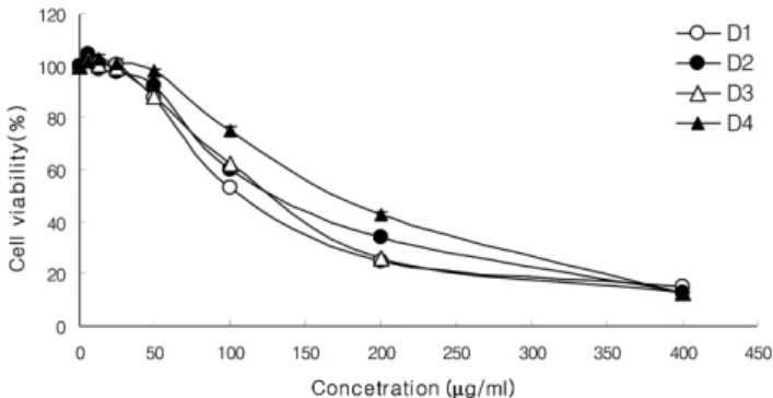
소분획으로나눈 D1~D4의모든시료에서도세포독성효
과가있는것으로나타났으며, 농도의존적으로감소하는
것을관찰할수있었다. 이중 D1의시료가 IC50 194.26µg/
ml으로가장우수한효과를보였다. 또한 D2, D3, D4의 IC50
를확인한결과각각 201.46µg/ml, 195.64µg/ml, 220.91µg/
ml으로써그효과가확인되었다(Fig. 2). 이결과로볼때,
분비나무의 DM fraction에항암효과를가지고있는성분이
있음을추측할수있었다.
DPPH radical 소거활성에 의한 항산화활성 − 항산화물 질의가장특징적인기작은유리기와반응하는것으로유 리기소거작용은활성라디칼 (free radical)에전자를공여
하여식물중의항산화효과나인체에서노화를억제하는
척도로 사용된다. DPPH는 안정한 유리기로 cysteine,
glutathione과 같은 아미노산과 ascorbic acid, aromatic
amine 등에의해환원되어탈색되므로항산화물질의항산
화측정에많이이용되고있다. 분비나무추출물과기존에
잘알려져있는항산화제인 ascorbic acid를대조군으로하
여 DPPH라디칼소거법에의한항산화활성실험을실시하
였다. 항산화활성의측정결과시료의처리농도에비례하게
항산화활성을나타내는것으로확인되었다(Table I). 그중
에서 D2와 D4가항산화효과가높았으며, positive control
인 ascorbic acid와비교하였을때, 분비나무추출물의 DM
fraction이뛰어난항산화효과를나타내지는않는것으로
Fig. 1.Cell cytotoxicity of A. nephrolepis extracts on HeLa cell lines. Cells were exposed to each fractions (from 6.25µg/ml to 200µg/ml). Cytotoxicity was assessed by MTT assay after 24 h. incubation. The proliferative response of HeLa cell lines are markedly reduced in a concentration-dependent manner in the presence of Hex and DM fractions.
Fig. 2.Cell cytotoxicity of D1~D4 on HeLa cell lines. Cells were exposed to D1~D4 (from 6.25µg/ml to 400µg/ml).
Cytotoxicity was assessed by MTT assay after 24h. incubation.
The proliferative response of HeLa cell lines are markedly reduced in a concentration-dependent manner in the presence of D1~D4 fraction.
보여진다.
Nitric oxide(NO) 생성 측정 − 활성산소중하나이며, 염
증유발에중요한역할을하는것으로알려진 Nitric oxide
(NO)생성에대한 D1~D4의효과를알아보았다. 생성된 NO
양을 D1~D4 및그유도체가 LPS에의해유도되는 iNOS
에어떠한영향을미치는지를확인하기위하여 iNOS에의
해유도되는 NO생성에대한시험물질의저해활성을 Griess
반응을이용하여확인하였다(Fig. 3). 그결과 LPS 단독처
리군에서는 NO가과량생성되는것을확인할수있었으며,
D1~D4을처리한군에서는농도의존적으로 NO생성을저
해하는것을확인할수있었다. 그중에서 D2 fraction과 D4 fraction에서 NO 생성억제효과가뛰어난것으로나타났다.
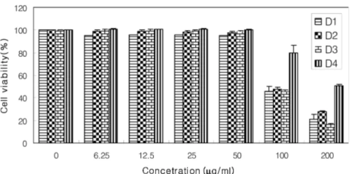
RAW264.7대식세포에 대한 세포독성 −D1~D4의세포독
성을알아보기위한 MTT assay의결과의 RAW264.7 대식
세포에 대한세포독성을평가하기 위한 MTT assay결과 D1~D4 모두 6.25~50µg/ml의처리농도에서유의성있는
세포독성을나타내지않아서실험결과에서나타난 NO의생
성량의변화가세포독성에의한영향과는무관함을확인하 였다(Fig. 4).
결 론
염증은조직의상해나파괴에의해서일어나는과정에서 상해를보호하는증상으로서최근에들어서선천성면역,
암과매우긴밀한관계를갖고있는것으로알려져있다. 활
성산소와 NO의과발현으로부터유도되는염증은세포의구
성성분인 DNA손상, 단백질의변이및신경손상등을일으
켜악성세포인암을유발할수있다. 본연구진은천연물의
성분중에항암효과가있는물질을검색하는과정에서분비 나무추출물이종양증식억제효과를나타냄을발견하였다.
이추출물의 DM fraction을 Vacuum dry chromatography를
실시하여 네 가지 fraction (D1~D4)을 얻었다. 네 가지 fraction이 HeLa cell line에대하여농도의존적으로세포증
식억제효과가나타나는것을 MTT assay로확인할수있
었다. 앞서이야기하였듯이항암, 항염증연구가활발히진
행되고있는점에착안하여, LPS로산화적스트레스를 유
발시킨 RAW264.7 대식세포로분비나무추출물이 항염증
제로도응용가능성이있는지알아보고자하였다. 염증은
생체나조직에기질적변화가일어날때손상부위를수복 재생하려는기전으로이과정에서수반되는혈관확장, 세
포막유동성증가, 부종등의현상은세포외로유리된화학
적매개인자에의해더욱증진되며염증매개인자의자극 에의해세포간극활성산소종의생성도급격히증가된다 는점에기인하여분비나무추출물 DM fraction (D1~D4)
의 DPPH radical 소거활성을측정하였다. 그결과 D2와 D4 fraction에서항산화효과가좋은것으로나타났다.
염증과정중에많은양의염증유도사이토카인 (Proinflam-
Table I. Antioxidant activity on DM fraction of A. nephrolepis extract
Treatment IC50 (µg/µl)a)
DPPH radical scavenging activity
D1 852.59±3.22
D2 435.73±4.23
D3 809.92±3.23
D4 136.55±5.13
Ascorbic acid 133.98±3.22
a)IC50 values were calculated from regression lines using three five different concentrations. Data represent the mean±S.D.
(Standard division) of triplicate experiments.
Fig. 3. Inhibitory effect of D1~D4 on the nitric oxide production in RAW 264.7 macrophages. The production of nitric oxide was assayed from culure medium of cells stimulated with LPS(1µg/ml) in the presence of D1~D4 (from 6.25µg/ml to 50µg/ml). L-N6-(1-iminoethyl) lysine (L- NIL) was used as an assay positive control at a concentration of 10µM. NO production was determined by ELISA method.
Sample concentration was determined using MTT assay.
Fig. 4. Cell cytotoxicity of D1~D4 on RAW264.7 cells. Cell viability was assessed by the MTT assay in RAW264.7 macrophages pretreated with D1~D4 for 24 h. The results demonstrated that the D1~D4 fractions had no cytotoxicity to RAW264.7 macrophages tested at dosage less than 50µg/ml.
matory cytokines) 이 생성되는데, 포유세포에서 iNOS는 interferon-γ, LPS 그리고다양한염증유도사이토카인에노
출되는경우에만발현된다. NOS중 iNOS에의한 NO 생성
이절대적으로많으며이는병리적으로중요한작용을한 다.15)이렇게활성화된대식세포가분비하는것으로알려진
NO의생성에미치는영향을알아보기위해 RAW264.7 대
식세포를이용하여 NO생성량변화를측정하였다. 그결과 D1~D4 fraction이 RAW264.7 대식세포에서 LPS에의해유
도된 NO생성을농도의존적으로뚜렷하게감소시키는것
을확인하였다. 특히 D2과D4 fraction에서 NO생성억제효
과가뛰어났다. 또한 D1~D4 fraction의세포독성이 NO생성
억제효과에 영향을 끼치진 않았는지 D1~D4 분획물의 RAW264.7대식세포에대한세포독성을 MTT assay를통해
알아보았고, 그결과 D1~D4 모두 6.25~50µg/ml의처리농
도에서유의성있는세포독성을나타내지않았다. 이는본
실험에서확인된NO의생성량의변화가세포독성에의한영
향과는무관함을나타내었다.
이러한결과들로볼때, HeLa cell line 증식억제효과를
가지고있는분비나무추출물의 DM fraction 중 NO 생성
억제효과와항산화효과모두뛰어난 D2와 D4 fraction에
항암및항염증에관련된성분임을추측해볼수있었다. 이
사실에기초하여분비나무추출물의 DM fraction은항암및
항염증물질의분리및그작용기전연구에중요한기초자 료가될것이라사료된다.
사 사
본 연구는 산림청 ‘산림과학기술개발사업(과제번호: S210707L010110 과 S120808L1101104)’의지원에의하여
이루어진것입니다.
인용문헌
1. McCord, J. (1974) Free radicals and inflammation: protection of synorial fluid by superoxide dismutase. Science. 185: 529- 2. Pharham, P. (2005) The Immune System. 531. Garland Science,
New York.
3. Lee, E. S., Ju, H. K., Moon, T. C., Lee, E., Jahng, Y., Lee S.H., Son, J. K., Baek, S. H. and Chang, H. W. (2004) Inhi- bition of nitric oxide and tumor necrosis factor-α (TNF-α)
production by propenone compound through blockade of nuclear factor (NF)-kB activation in cultured murine mac- rophage. Biol. Pharm. Bull. 27: 617-620.
4. MukIS, N., Ishikawa, Y., Ikeda, N., Fujioka, N., Watanabe, S.
and Kuno, K. (1996) Novel insight into molecular mech- anism of endotoxin shock; biochemical analysis of LPS receptor signaling in a cell-free system targeting NF-kapperB and regulation of cytokine production/action through beta2 integrin in vivo. J. Leukoc. Biol. 59: 145-151.
5. Vane, J. A. (1971) Inhibition of prostaglandin synthesis as a mechanism of action for aspirin-like durgs. Nat. New. Biol.
23: 232-235.
6. Funk, C. D., Frunk, L. B., Kennedy, M. E., Pong, A. S. and Fitzgerald, G. A. (1991) Human platelet/erythroleukemia cell prostaglandin G/H synthase: cDNA cloning, expression, and gene chromosomal assignment. FASEB J. 5: 2304-2312.
7. Richard, G. and Salvador, M. (1994) Nitric oxide synthases in mammals. Biochem. J. 298: 249-258.
8. Balkwill F., Mantovani, A. (2001) Inflammation and cancer:
back to Virchow? Lancet. 357: 537-45.
9.이창복 (1999) 대한식물도감, 233향문사, 서울.
10. Lee, S. J., Lee, K. W., Lee, H. J. (2004) Abies nephrolepis leaf phenolics prevent the inhibition of gap junction intercellular communication by hydrogen peroxide in rat liver epithelial cells. Biofactors. 21(1-4): 357-60
11. Kim, S. K., Jung, S. M., Ahn, K. H., Jeon, H. J., Lee, D. H., Jung, K. M., Jung, S. Y. and Kim, D. K. (2005) Identification of three competitive inhibitors for membrane-associated, Mg2+-dependent and neutral 60 kDa sphingomyelinase activ- ity. Arch Pharm Res. Aug: 28(8): 923-9
12. Fabio, V., Kobuchi, H. and Lester, P. (1998) Procyanidins extracted from Pinus maritima (Pycnogenol): scavengers of free radical species and modulators of nitrogen monoxide metabolism in activated murine RAW 264.7 macrophages.
Free Radic Biol Med. May: 24(7-8): 1120-9.
13. Mosmann, T. (1983) Rapid colorimetric assay for cellular growth and survival: application to proliferation and cyto- toxicity assay. Journal of Immunological Methods. 65: 55-63.
14. Yokozawa, T., Chen, D. P., Kong, E., Tanaka, T., Nonaka, G.I.
and Nishioka, I. (1998) Study on the inhibitory effect of tanins and flavonoids against the 1,1-diphenyl1-2 picrylhy- drazyl radical. Biochem Pharmacol. 56: 213-222.
15. Joydeb, K. K., Young, J. S. (2008) Inflammation: Gearing the journey to cancer. Mutation Research.659: 15-30.
(2009년 3월 10일)