Medial opening wedge high tibial osteotomy (MOWHTO) is an effective method for the treatment of osteoarthritis confined to the medial side of the knee joint with varus axial malalignment.
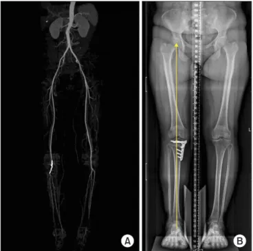
We experienced a case of pseudoaneurysm of the popliteal artery after MOWHTO inflicted by a drill bit. To our knowledge, there has been no report of injury of the popliteal artery due to drilling with a drill bit during MOWHTO for screw fixation of the lock
ing plate. We postulate the mechanism of this vascular injury and describe the clinical features and management.
Case Report
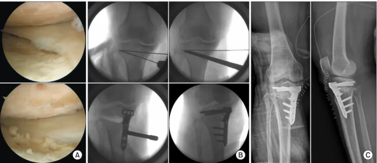
A 53yearold female patient underwent MOWHTO for medial compartment osteoarthritis. A preoperative radiograph showed degenerative osteoarthritis with medial joint space narrowing, sclerosis, and 6° of varus deformity. Magnetic resonance imaging (MRI) showed unicompartmental osteoarthritis of the medial femoral condyle and medial tibial condyle corresponding to Outerbridge grade IV; therefore, MOWHTO was planned. After confirming there was no instability of the ligaments under spinal anesthesia, the cartilage and the meniscus were assessed through arthroscopy. Arthroscopy revealed cartilage lesions in the medial femoral condyle and medial tibial condyle, and microfracture was performed (Fig. 1A).
After the arthroscopic procedure, a longitudinal skin incision of about 8 cm was performed on the medial side of the knee joint.
The pes anserinus was exposed and pulled downward, and the superficial medial collateral ligament was subperiosteally released from the tibia, and the Hohmann retractor was inserted into the posterior medial side of the tibia to protect the neurovascular structures and expose the osteotomy site.
Under fluoroscopic guidance, the Kirschner wire was inserted
Department of Orthopedic Surgery, Wonkwang University College of Medicine, Iksan; Department of Orthopedic Surgery, Mokpo Hankook Hospital, Mokpo;
3