Korean J Vet Res(2018) 58(1) : 1~7 https://doi.org/10.14405/kjvr.2018.58.1.1
1
<Original Article>
Artemisia capillaris Thunb. inhibits melanin synthesis activity via ERK-dependent MITF pathway in B16/F10 melanoma cells
Evelyn Saba
1, Mi Ju Oh
1, Yuan Yee Lee
1, Dongmi Kwak
1, Suk Kim
2, Man Hee Rhee
1,*
1
Department of Veterinary Medicine, College of Veterinary Medicine, Kyungpook National University, Daegu 41566, Korea
2
Department of Veterinary Medicine, College of Veterinary Medicine, Gyeongsang National University, Jinju 52828, Korea (Received: November 30, 2017; Revised: January 11, 2018; Accepted: January 25, 2018)
Abstract: Genus Artemisia occurs as a hardy plant and has a wide range of culinary and medicinal features. In this study, we aimed to describe the melanin inhibitory activity of one Artemisia species, i.e., Artemisia capillaris Thunb.
Ethanol extracts of fermented Artemisia capillaris (Art.EtOH.FT) and non-fermented Artemisia capillaris (Art.EtOH.CT) were tested for their ability to inhibit tyrosinase activity and melanin pigmentation. Both extracts showed dose-dependent inhibition against α-melanocyte stimulating hormone−stimulated melanin formation and tyrosinase activity, without cytotoxicity. At 100 µg/mL, both extracts showed greater inhibition than kojic acid, the positive control. Protein expressions of microphthalmia-associated transcription factor (MITF), tyrosinase (TYR), tyrosinase-related protein 1 (TRP-1), and tyrosinase-related protein 2 (TRP-2) at the transcriptional level were determined by using real-time and semi-quantitative polymerase chain reaction. To complete the mechanistic study, presences of upstream elements of MITF, the phosphorylated-extracellular signal-regulated kinase (p-ERK), and phosphorylated−mitogen-activated protein kinase kinase (p-MEK) were confirmed by using western blot analysis. Expressions of p-TYR, p-TRP-1 and p-TRP- 2, downstream factors for p-ERK and p-MITF, were translationally inhibited by both extracts. Art.EtOH.FT induced more potent effects than Art.EtOH.CT, especially signal transduction effects. In summary, Artemisia capillaris extracts appear to act as potent hypopigmentation agents.
Keywords: Artemisia capillaris, hypopigmentation, melanin inhibition, tyrosinase
Introduction
Variety in the skin colour of different human races is attrib- uted to the amount of melanin pigment present in the skin.
Melanin is basically the compound that gives colour to our skin in various shades depending upon its concentration [2].
It is formed by a process called melanogenesis in which α- melanocyte stimulating hormone ( α-MSH) binds to its receptor i.e., melanocortin 1 receptor (MC1R) which elevates the lev- els of cyclic adenosine monophosphate (cAMP) that acti- vates microphathalamia-associated transcription factor (MITF) via different pathways like extracellular signal-regulated kinase (ERK), protein kinase B and cAMP response element binding protein causing its degradation. This affects the rate limiting step of this process i.e., tyrosinase (TYR). TYR catalyses the conversion of tyrosine to L-3,4-dihydroxyphe- nylalanine (L-DOPA) that forms melanin later. There are other two factors that are affected down stream of MITF and TYR like tyrosinase-related protein 1 (TRP-1) and tyrosi- nase-related protein 2 (TRP-2) whose activation leads to mel- anogenesis [12-14, 34].
Genus Artemisia is one of the most important genera in Asteraceae family and is widely found throughout the world.
Artemisia capillaris Thunb., which is one of the species of genus Artemisia is widely used traditional medicinal plant and extensively used food supplement. Artemisia capillaris ( A. capillaris) that is also called ‘Yin Chen Hao’ is exten- sively used in China for over 2,000 years particularly for its remarkable hepatoprotective effects like in jaundice, hepati- tis and gall bladder diseases [1, 5, 7, 10, 16, 19, 30]. Many biologically active metabolites in it like coumarins, fla- vonoids, sterol glycosides, essential oils and polyacetylenes have been reported to exhibit a wide range of biological activities like, antioxidant, anti-malarial, anti-cancerous, anti- viral and anti-fungal [3, 17, 20, 23, 24, 32]. Moreover, it is also applied externally on the head for relief from headaches [4].
Up till now there has been no study made to elucidate the anti-melanogenic effects of this A. capillaris. Therefore, we checked the TYR inhibition and melanin production inhibi- tion via the mechanistic study of pathway involved in this process. Our results have shown that the two sample extracts of A. capillaris i.e., fermented and non-fermented type showed
*Corresponding author
Tel: +82-53-950-5967, Fax: +82-53-950-5955
E-mail: [email protected]
remarkable inhibition in TYR activity and also showed decreased melanin content via the ERK activated MITF deg- radation pathway especially in the fermented type. Further studies in future on this extract can guarantee its usage in the cosmetic industry as a skin whitening agent.
Materials and Methods
Chemicals and reagents
Dulbecco’s modified Eagle’s medium (DMEM) (Korea), fetal bovine serum (FBS) (WelGene, Korea), streptomycin and penicillin (Lonza, USA), TRIzol reagent (Invitrogen, USA), oligo dT (Bioneer, Korea), MITF, TYR, TRP-1, TRP-2 and β-actin primers were obtained from Bioneer. 3-(4,5-dimeth- ylthiazol-2-yl)-2,5-diphenyltetrazoliumbromide (MTT) was purchased from Sigma-Aldrich (USA). Specific antibodies used against phospho- and/or total form of mitogen-acti- vated protein kinase kinase (MEK), ERK, β-actin and sec- ondary antibody rabbit HRP linked were purchased from Cell Signalling Technology (USA). Antibodies for phospho- MITF, TYR, TRP-1 and TRP-2 were obtained from Santa Cruz Bio- technology (USA). TYR from mushroom and L-DOPA were purchased from Sigma (USA). All other reagents were of local analytical grade.
Sample preparation
A. capillaris fermented and non-fermented dried samples were obtained from enzyme LAPA (Korea) and fermented according to Saba et al. [29]. They were then extracted with 70% ethanol and filtered using a Whatman filter paper (pore size 185 mm; Sigma-Aldrich). The filtrate was then con- verted to powder form using freeze drying. Later they were dissolved in dimethyl sulfoxide (DMSO) and used according to experimental design dosages.
Gas chromatography mass spectrometry (GC-MS) GC-MS was carried out for the fermented and non-fer- mented Artemisia samples using 100 mg of powered sample.
Agilent technology 7890A Gas chromatograph systems (Agi- lent Technologies, USA) attached to XLMSD-5975C instru- ment with electrospray ionization mode was harvested. The component’s percentage was calculated by comparing its average peak to the total area under curve. The component with highest percentage was acetic acid (40%) in fermented sample with the next being deoxy arteminisin (7%). The sim- ilar components were also elevated in non-fermented sample but their concentrations were lower than fermented type.
Cell line
Murine melanoma cell line B16/F10, originating from American Type culture collection was cultured in DMEM supplemented with 8% FBS (WelGene) and 100 IU/mL pen- icillin and 100 µg/mL streptomycin sulfate (Lonza). The incu- bating conditions were humidified in 5% CO
2at 37
oC.
Cell-free TYR inhibition assay
The assay was performed with slight modifications as pre- viously described [33]. Briefly 10 µL of A. capillaris fer- mented and non-fermented samples was put in 96-well in triplicates and mixed with 60 µL of 50 mmol/L phosphate buffer on ice (pH 6.8). Then 20 µL of 0.9 mg/mL L-DOPA was added to each well. Finally 10 µL of mushroom TYR was added in each well and the plate was incubated at 27
oC for 10 min. After incubation, the amount of dopachrome pro- duction was determined spectrophotometrically at 450 nm by a microplate reader (Versamax microplate reader; Molecular devices, USA). Kojic acid in this experiment was taken as a positive control.
Cell viability assay
To determine the cytotoxic effects of A. capillaris extracts, cell viability assay was done using MTT reagent which was added to culture medium at a final concentration of 0.1 mg/
mL. After 4 h of incubation at 37
oC in 5 % CO
2, the result- ing violet coloured crystals were dissolved in DMSO 100 µL/ well and absorbance values were measured at 560 nm.
Melanin inhibition assay
The B16/F10 cells were seeded in the 6-well culture plate at a density of 2.5 × 10
3cells/well and then incubated for 5 days. After the cells reached the desired confluency, they were treated with A. capillaris samples and then stimulated with α-MSH. The cells were then incubated again for 3 days and then harvested using 0.25% trypsin-EDTA solution and transferred to 1.5 mL microcentrifuge tubes. The tubes were then centrifuged at 10,000 × g for 10 min and pellet was dis- solved in 2 mol/L NaOH for 15 min at 60
oC. Later this dis- solved mixture was transferred to 96-well plates and absorbance was measured at 450 nm with a microplate reader (Versamax microplate reader; Molecular devices). The absorbance was compared to the synthetic standard melanin curve (Sigma).
RNA extraction and real-time polymerase chain reac- tion (PCR)
Total RNA was extracted from the B16/F10 cells after they
were treated with A. capillaris extracts and stimulated with
α-MSH using TRIzol according to Manufacturer’s instruc-
tions. Total RNA (2 µg) was annealed with Oligo dt (Bion-
eer) for 10 min at 70
oC and cooled for 5 min on ice, reverse
transcribed using reverse transcriptase pre-mix (Bioneer) in
20 µL of reaction mixture and run for 90 min at 42.5
oC using
thermal cycler. The reactions were terminated at 95
oC for 5
min to inactivate the reverse transcriptase. The reverse tran-
scription polymerase chain reaction was performed using ali-
quots of cDNA obtained from RT reaction in a PCR premix
(Bioneer). The PCR products were then electrophoresed on
1% agarose gel stained with ethidium bromide and visual-
ized using ImageQuant LAS 500 (GE health care life sci-
ences, Korea). The intensity of band densities was normalized
for corresponding GAPDH, which is housekeeping gene used
as an RNA internal standard and ratios were compared.
Moreover, PCR product was also analysed via real-time PCR (Bio-Rad CFX96 Real-Time systems; Bio-Rad Laboratories, USA) using primers sequence is given in Table 1.
Western blot analysis
B16/F10 cells were treated or left untreated with both A.
capillaris extracts (25–100 µg/mL) in the presence or absence of α-MSH (10 µM). Cytosolic and nuclear proteins were extracted according to the instructions of NE-PER Nuclear and cytosolic extraction reagents (Thermo Scientific, Korea).
Proteins were then measured using PRO-MEASURE Protein Measurement Solution (iNtRON Biotechnology, Korea). They were then separated by 10% polyacrylamide gels through SDS-PAGE and transferred onto polyvinylidene fluoride (PVDF) membranes (Immobilion-P; Millipore, USA). Nonspecific binding on the PVDF membranes was minimized with a blocking buffer containing 5% non-fat dry milk and 0.1%
Tween-20 in tris-buffered saline. The membranes were then incubated with specific primary antibodies overnight at 4
oC followed by 1 h incubation with horseradish peroxidase-con- jugated anti-rabbit antibody (1:3,000 dilution). Bound anti- bodies were visualized using enhanced chemiluminescence (Supex, Korea) and images were analysed using ImageJ soft- ware. β-actin was taken as an internal control.
Statistical analysis
Data were presented as mean ± SEM. One way ANOVA and Dunnett’s test were applied for the statistical evaluation of data. Statistical analyses with p < 0.01, p < 0.005 and p <
0.001 were considered significant.
Results
A. capillaris extracts inhibited TYR activity in cell-free system
The rate limiting step in melanogenesis processes is the enzyme TYR [27, 36], therefore we geared to investigate if A.
capillaris extracts have effect on this enzyme. Using the cell- free system with mushroom TYR, we found that both the fer- mented and non-fermented types of A. capillaris extracts
inhibited TYR activity in dose dependent manner as shown in Figure 1A. Moreover, since many herbal extracts possess a variety of compounds in them, that can incur cytotoxicity, for this purpose we checked the cytotoxicity of doses used in our study for inhibition of melanogenesis, and none of the dos- age used showed any toxic effects on B16/F10 cells (Fig. 1B).
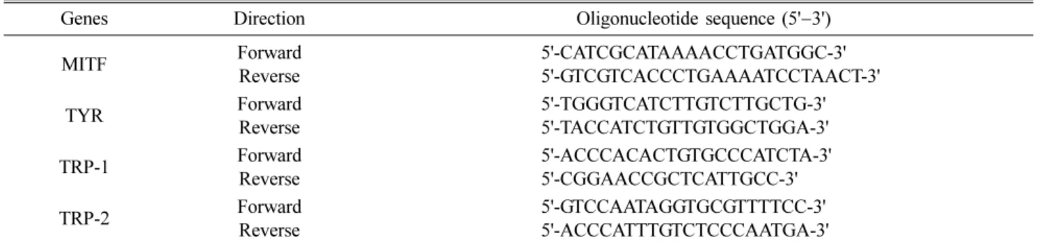
Melanin content suppression by A. capillaris extracts Melanin is the natural pigment that gives colour to our skin. However, if it is excreted in higher levels than the threshold by melanocytes, it can lead to benign or malignant Table 1. Oligonucleotide sequences of primers used for real-time polymerase chain reaction
Genes Direction Oligonucleotide sequence (5' −3')
MITF Forward
Reverse
5'-CATCGCATAAAACCTGATGGC-3' 5'-GTCGTCACCCTGAAAATCCTAACT-3'
TYR Forward
Reverse
5'-TGGGTCATCTTGTCTTGCTG-3' 5'-TACCATCTGTTGTGGCTGGA-3'
TRP-1 Forward
Reverse
5'-ACCCACACTGTGCCCATCTA-3' 5'-CGGAACCGCTCATTGCC-3'
TRP-2 Forward
Reverse
5'-GTCCAATAGGTGCGTTTTCC-3' 5'-ACCCATTTGTCTCCCAATGA-3'
MITF, microphathalamia-associated transcription factor; TYR, tyrosinase; TRP-1, tyrosinase-related protein 2; TRP-2, tyrosinase-related protein 2.
Fig. 1. Inhibition of mushroom TYR activity by Artemisia cap-
illaris (A. capillaris) extracts. Both A. capillaris extracts inhib-
ited the mushroom TYR activity ,
***p < 0.001 when compared
with untreated group (A). No cytotoxicity was observed for
both extracts in B16/F10 cells (B).
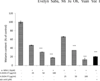
form of cancers [15]. Considering this matter we checked the effect of A. capillaris extracts on B16/F10 cells when they were stimulated with α-MSH for melanin secretion. As shown in Figure 2, both extracts when compared with con- trol, suppressed the melanin contents in the cells in a dose dependent manner.
Effects of A. capillaris extracts on the mRNA expres- sion of MITF and TYR related proteins
Above results elaborated the effects of A. capillaris extracts on TYR and melanin contents. However, it is impor- tant to understand the underlying mechanisms both at tran- scriptional and translational levels by which these extracts are elucidating their inhibitory effects. The mRNA expressions as shown in Figure 3A and B by reverse transcriptase PCR and real-time PCR for MITF, TYR, TRP-1 and TRP-2 showed a dose dependent decrease with the increase in extracts concentration.
A. capillaris extracts exerted their anti-melanogenic effects via the mitogen activated protein kinase (MAPK)/
ERK dependent MITF pathway
There have been many reports in past that indicate ERK pathway’s involvement in the melanogenesis inhibition through degradation of MITF [37]. This led us to explore the upstream pathways that might be involved in the degradation of MITF leading to inhibition of melanin production. The results of immunoblotting showed that A. capillaris extracts activated the phospho-MEK that is the upstream regulator of ERK. In addition, they also activated the levels of phospho-ERK in a dose dependent manner (Fig. 4A). Furthermore the results also showed that both extracts exerted their anti-melano- genic activities via the ERK dependent MITF pathway since they also inhibited the phosphorylation of all four factors for melanin synthesis i.e., p-MITF, p-TYR, p-TRP-1 and p-TRP- 2 with fermented type being more potent than non-fer-
mented type (Fig. 4B).
Discussion
The basal layer of epidermis contains specialized cell Fig. 2. Suppression in melanin content by A. capillaris extracts.
Melanin content in crude lysates was suppressed by both A.
capillaris extracts when B16/F10 cells were stimulated with α- melanocyte stimulating hormone ( α-MSH).
***p < 0.001 when compared with control.
Fig. 3. Depressed expression of the MITF pathway genes by A.
capillaris extracts. B16/F10 cells were seeded in 6 well plates and treated with indicated concentrations of Artemisia samples and then stimulated with α-MSH (10 µM). Later RNA was extracted and MITF, TYR, TRP-1 and TRP-2 expression levels were checked by real-time polymerase chain reaction (RT-PCR) (A) and also by quantitative RT-PCR (B). GAPDH was taken as internal control and all values were compared against it.
***