Regulation of Hepatic Gluconeogenesis
by an ER-Bound Transcription Factor, CREBH
Min-Woo Lee,1,9Dipanjan Chanda,2,9Jianqi Yang,4Hyunhee Oh,5Su Sung Kim,5Young-Sil Yoon,1Sungpyo Hong,1 Keun-Gyu Park,8In-Kyu Lee,7Cheol Soo Choi,5,6Richard W. Hanson,4Hueng-Sik Choi,2,3,*and Seung-Hoi Koo1,*
1Department of Molecular Cell Biology, Sungkyunkwan University School of Medicine, 300 Chunchun-dong, Jangan-gu, Suwon, Gyeonggi-do 440-746, Korea
2Hormone Research Center, School of Biological Sciences and Technology, Chonnam National University, Gwangju 500-757, Korea
3Research Institute of Medical Sciences, Department of Biomedical Sciences, Chonnam National University Medical School, Gwangju 501-746, Korea
4Department of Biochemistry, Case Western Reserve University School of Medicine, Cleveland, OH 44106-4935, USA
5Lee Gil Ya Cancer and Diabetes Institute
6Division of Endocrinology
Gil Medical Center, Gachon University of Medicine and Science, Incheon 405-760, Korea
7Departments of Internal Medicine and Biochemistry and World Class University Program, Research Institute of Aging and Metabolism, Kyungpook National University School of Medicine, Daegu 700-422, Korea
8Department of Internal Medicine, Keimyung University School of Medicine, Daegu, Korea
9These authors contributed equally to this work
*Correspondence:[email protected](H.-S.C.),[email protected](S.-H.K.) DOI 10.1016/j.cmet.2010.02.016
SUMMARY
Endoplasmic reticulum (ER)-bound transcription fac- tor families are shown to be involved in the control of various metabolic pathways. Here, we report a critical function of ER-bound transcription factor, CREBH, in the regulation of hepatic gluconeogen- esis. Expression of CREBH is markedly induced by fasting or in the insulin-resistant state in rodents in a dexamethasone- and PGC-1a-dependent manner, which results in the accumulation of active nuclear form of CREBH (CREBH-N). Overexpression of con- stitutively active CREBH activates transcription of PEPCK-C or G6Pase by binding to its enhancer site that is distinct from the well-characterized CREB/
CRTC2 regulatory sequences in vivo. Of interest, knockdown of CREBH in liver significantly reduces blood glucose levels without altering expression of genes involved in the ER stress signaling cascades in mice. These data suggest a crucial role for CREBH in the regulation of hepatic glucose metabolism in mammals.
INTRODUCTION
Glucose homeostasis is tightly regulated to meet the fuel requirement in mammals. Under fasting, secretion of pancreatic hormone glucagon and adrenal hormone glucocorticoid is induced to enhance hepatic glucose production (Pilkis et al., 1988a, 1988b). This process is accomplished, in part, via activa- tion of gluconeogenesis, resulting from the transcriptional acti- vation of gluconeogenic genes such as cytosolic isoform of phosphoenolpyruvate carboxykinase (PEPCK-C) or glucose 6 phosphatase (G6Pase) (Hall and Granner, 1999; Hanson and
Reshef, 1997). Activation of cAMP-dependent transcriptional program is mainly mediated by CREB/CREB-regulated tran- scriptional coactivator 2 (CRTC2)-dependent transcriptional machinery, whereas glucocorticoid signal is conveyed via the action of glucocorticoid receptor (GR), a member of nuclear hor- mone receptor (NR) superfamilies (Herzig et al., 2001; Koo et al., 2005; van Schaftingen and Gerin, 2002). Indeed, both cAMP response element (CRE) and GR response element (GRE) have been found in gluconeogenic gene promoters, underscoring the importance of these transcriptional machineries in this path- way (Hanson and Reshef, 1997; van Schaftingen and Gerin, 2002). As demonstrated in Cushing’s syndrome, excessive glu- cocorticoid could promote insulin resistance by opposing the action of insulin in peripheral tissues, including liver, suggesting that the increased GR activity might be responsible for the pro- gression of type II diabetes (Andrews and Walker, 1999; Ross and Linch, 1982; Zinker et al., 2007). Of interest, both GR and CREB/
CRTC2 were shown to induce the NR coactivator peroxisome proliferator-activated receptor g coactivator 1 alpha (PGC-1a), which regulates activities of various transcription factors, includ- ing GR, hepatic nuclear factor 4 (HNF4), or FOXO1a (Herzig et al., 2001; Koo et al., 2005; Puigserver et al., 2003; Yoon et al., 2001).
Elevation of PGC-1a expression is displayed in mouse models of type II diabetes, and its liver-specific ablation markedly affects glycemic profiles in mice, showing the importance of this factor in the regulation of hepatic glucose metabolism (Herzig et al., 2001; Koo et al., 2004; Leone et al., 2005; Lin et al., 2004; Puig- server et al., 2003).
Recently, regulated intramembrane proteolysis (RIP) has emerged as a new mechanism to influence energy metabolism, differentiation, and endoplasmic reticulum (ER) stress response/
unfolded protein response (UPR) (Brou et al., 2000; Brown et al., 2000; Haze et al., 1999). Members of the ER membrane-bound basic leucine zipper (bZIP) transcription factor family, such as ATF6, Luman, and OASIS, constitute a novel class of factors that are regulated by ER stress-dependent mechanisms (Haze et al., 1999; Kondo et al., 2005; Raggo et al., 2002). ATF6,
a founding member of this family, is an ER resident transcription factor and is activated following UPR-dependent translocation to Golgi, where the proteolytic cleavage of this factor releases its N-terminal transcription factor moiety into the nucleus.
Activated ATF6 is responsible for UPR-mediated activation of target genes such as GRP78/Bip, CHOP, and XBP-1 (Chen et al., 2002; Shen et al., 2002; Ye et al., 2000).
cAMP response element-binding protein H (CREBH), a liver- specific bZIP transcription factor that belongs to this family, is shown to be regulated by UPR-dependent proteolytic cleavage (Chin et al., 2005; Omori et al., 2001) and regulates the transcrip- tional process of genes such as serum amyloid P-component (SAP) and C-reactive protein (CRP) in response to systemic inflammatory signals in liver (Zhang et al., 2006). Of interest, CREBH was also shown to transcriptionally activate PEPCK-C promoter, suggesting a potential link between this factor and the hepatic glucose metabolism (Chin et al., 2005). In this study, we show that CREBH expression is induced during fasting or insulin-resistant state, which, in turn, results in the accumula- tion of the active nuclear form of CREBH (CREBH-N). Nuclear CREBH enhances hepatic gluconeogenesis by activating transcription of PEPCK-C or G6Pase via a unique regulatory sequence in a CRTC2-dependent manner. Furthermore, acute depletion of hepatic CREBH results in the reduction of blood glucose levels both in wild-type and diabetic mice. These data support that CREBH is an important physiological regulator of hepatic gluconeogenesis.
RESULTS
CREBH Expression Is Induced during Fasting and by Insulin Resistance
Previously, nuclear CREBH (CREBH-N) was shown to enhance PEPCK-C promoter activity in hepatic cells without further delin- eation in its physiological relevance in gluconeogenesis (Chin et al., 2005). To assess the potential involvement of CREBH in hepatic gluconeogenesis, we measured its expression levels in mouse liver. Of interest, CREBH expression was significantly induced during fasting conditions and was reduced upon refeeding, a characteristic regulatory pattern known for genes in the gluconeogenesis (Figure 1A). Furthermore, mRNA levels for hepatic CREBH were also induced in mouse models of diet- induced or genetic insulin resistance (Figure 1B andFigure S1A available online), showing a strong correlation between CREBH expression and gluconeogenic potential in liver. Indeed, we observed increased appearance of both full-length and nuclear CREBH under fasting or by insulin resistance (1.9-fold [FL] or 4.9-fold [N] induction under fasting over refeeding, 6.3-fold [FL]
or 1.8-fold [N] induction in db/db over WT mice;Figures 1A and 1B), suggesting that CREBH could be involved in the tran- scriptional process of hepatic gluconeogenesis.
To identify a mechanism for CREBH regulation, we treated hepatocytes with stimuli known to mimic fasting signals. Treat- ment of cells with dexamethasone, but not with cAMP agonist forskolin, enhanced CREBH expression significantly (Figure 1C and data not shown), suggesting an involvement of GR in the process. Indeed, transfection analysis, as well as chromatin immunoprecipitation (ChIP) assay, revealed a functional involve- ment of GR in the regulation of CREBH transcription (Figures
S1B and S1C). The involvement of GR in the regulation of CREBH was further supported by recent microarray analysis showing that GR knockout mice displayed reduced dexametha- sone-dependent expression of CREBH (ArrayExpress: http://
www.ebi.ac.uk; ID: E-MEXP-1816). PGC-1a functions as a tran- scriptional coactivator for NRs (Rhee et al., 2003; Yoon et al., 2001). We thus tested whether PGC-1a was also involved in the transcriptional regulation of hepatic CREBH. Expression of PGC-1a significantly enhanced expression of CREBH in hepato- cytes (Figure 1D). Conversely, knockdown of PGC-1a in the mouse liver resulted in reduction of mRNA and protein levels for CREBH (Figure 1E). Dexamethasone-dependent activation of CREBH promoter activity was induced synergistically with cotransfection of PGC-1a expression vector; the effect was blunted by coexpression of either constitutively active AKT or small heterodimer partner (SHP), a known inhibitor for PGC- 1a-NR interaction (Chanda et al., 2008) (Figures S1D–S1F).
These data suggest the presence of a mechanism for PGC-1a/
GR-dependent activation of CREBH that may contribute to the regulation of hepatic glucose metabolism.
CREBH-N Promotes Hepatic Gluconeogenesis
To test the functional significance of CREBH expression, we prepared adenovirus for nuclear CREBH (Ad CREBH-N). Trans- duction of hepatocytes with Ad CREBH-N increased mRNA levels for gluconeogenic genes (Figure 2A). In addition, hepatic glucose production was significantly induced by CREBH-N expression in hepatocytes, suggesting that CREBH-N indeed promotes hepatic gluconeogenesis (Figure 2B). To verify the effect of CREBH-N on glucose homeostasis in vivo, we injected Ad CREBH-N or Ad GFP control virus into the wild-type mice. Ad CREBH-N infection led to elevations in hepatic mRNA levels of PEPCK-C and G6Pase (Figures 2C and 2D). Furthermore, blood glucose levels were significantly higher in mice with Ad CREBH- N compared with those in control mice (Figure 2E), showing that acute overexpression of CREBH promoted hepatic gluconeo- genic gene expression in vivo, without altering body weight (Figure S2A). The insulin levels were generally higher in mice with Ad CREBH-N without reaching the statistical significance (Figure S2B). No changes were shown for the phosphorylation status of CRTC2, excluding a potential indirect regulation of glu- coneogenic genes by CREBH-N (Figure S2B). CREBH was shown to regulate genes involved in the acute phase response (APR) such as CRP and SAP (Zhang et al., 2006). We thus measured the mRNA levels for such genes in livers with Ad CREBH-N. Though CRP expression was slightly elevated in animals infected with Ad-CREBH-N, we were not able to observe any changes in SAP expression (Figure 2F). Furthermore, no detectable differences were noted in GRP78 or ATF6 expression between two groups of mice. We therefore speculated that, whereas expression of CREBH alone was sufficient to induce expression of gluconeogenic program, additional components such as ATF6 might be required for the complete activation of hepatic genes in the APR.
CREBH Activates Transcription of Gluconeogenic Genes via a CRTC2-Dependent Manner
Next, we wanted to delineate the mechanism for CREBH-medi- ated transcriptional process. A previous study suggested that
Cell Metabolism
Role of CREBH in Hepatic Gluconeogenesis in Mammals
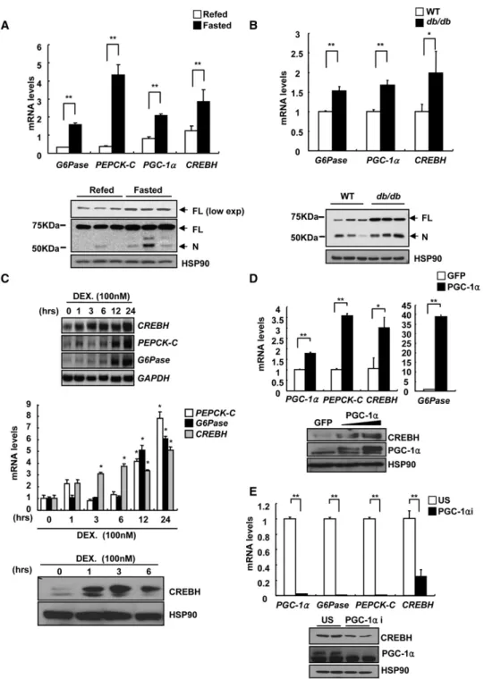
Figure 1. Induction of CREBH Expression during Fasting or by Insulin Resistance
(A) (Top) Q-PCR analysis showing hepatic CREBH or gluconeogenic gene expression in mice less under 16 hr fasted or 16 hr fasted/4 hr refed conditions (**p < 0.01, t test; n = 5). (Bottom) Western blot analysis showing hepatic CREBH protein levels in mice as indicated above (FL, full-length; N, nuclear).
(B) (Top) Q-PCR analysis showing hepatic CREBH or gluconeogenic gene expression in either wild-type or db/db mice under 16 hr fasted condition (**p < 0.01,
*p < 0.05, t test; n = 3). (Bottom) Western blot analysis showing hepatic CREBH protein levels in either wild-type or db/db mice as indicated above.
(C) Effects of dexamethasone on CREBH expression. Northern blot (top) or Q-PCR (middle) analysis of G6Pase, PEPCK-C, and CREBH expression using rat primary hepatocytes treated with 100 nM dexamethasone for an indicated time period (*p < 0.05 compared to 0 hr, t test; n = 3). (Bottom) Western blot analysis showing hepatic CREBH-N protein levels by dexamethasone treatment.
(D) Effects of Ad-PGC-1a on CREBH expression. (Top) Q-PCR analysis of G6Pase, PEPCK-C, and CREBH expression using RNAs from rat primary hepatocytes infected with either Ad-GFP or Ad-PGC-1a (**p < 0.01,*p < 0.05, t test; n = 3). (Bottom) Western blot analysis showing hepatic CREBH-N protein levels by Ad-GFP or Ad-PGC-1a.
(E) Effects of PGC-1a knockdown on CREBH expression. (Top) Q-PCR analysis of G6Pase, PEPCK-C, and CREBH expression using RNAs from livers of mice infected with either Ad-US or Ad-PGC-1a RNAi (**p < 0.01,*p < 0.05, t test; n = 5). (Bottom) Western blot analysis showing hepatic CREBH-N protein levels by Ad-US or Ad-PGC- 1a RNAi.
Data represent the mean ± SD.
CREBH could enhance PEPCK-C promoter activity by binding to the well-characterized CRE at 125 from the transcriptional start site (Chin et al., 2005). Contrary to the previous report, our mapping studies revealed a unique putative CREBH response
element (CREBHRE) in the promoters of PEPCK-C and G6Pase that are distinct from CRE ( 452 versus 125 for PEPCK-C, 91 versus 161/ 136 for G6Pase) (Figure 3A); the occupancy of CREBH over putative CREBHRE on each promoter was Figure 2. CREBH Promotes Hepatic Gluconeogenesis
(A) Effects of CREBH on gluconeogenic gene expression. Northern blot (top, left) or Q-PCR (bottom) analysis of G6Pase, PEPCK-C, and CREBH expression using RNAs from rat primary hepatocytes infected with either Ad-GFP or Ad-CREBH-N (*p < 0.05, t test; n = 3). A representative western blot analysis showing hepatic nuclear CREBH (CREBH-N) protein levels was also shown (top, right).
(B) (Top) Glucose output assay showing effects of CREBH on glucose production in rat primary hepatocytes. Forskolin (10 mM) was used as a positive control.
(Bottom) Western blot assay shows the expression level of Flag-tagged CREBH-N (*p < 0.05, t test; n = 3).
(C and D) Effects of Ad-CREBH-N infection on hepatic gene expression. Q-PCR analysis and western blot analysis of CREBH (C) and Q-PCR analysis of gluco- neogenic genes (D) from mouse liver infected with either Ad-GFP or Ad-CREBH-N (**p < 0.01, t test; n = 5).
(E) Sixteen hour fasting glucose levels in mice expressing Ad-GFP or Ad-CREBH-N (*p < 0.05; t test; n = 5).
(F) Effects of Ad-CREBH-N infection on ER stress genes using RNAs from mouse liver infected with either Ad-GFP or Ad-CREBH-N (**p < 0.01, t test; n = 5).
Data represent the mean ± SD.
Cell Metabolism
Role of CREBH in Hepatic Gluconeogenesis in Mammals
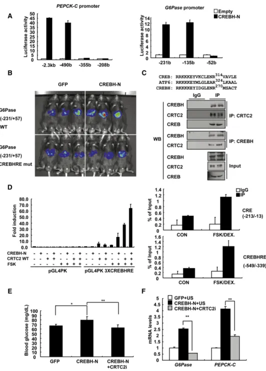
Figure 3. CREBH Regulates Gluconeogenic Genes via a CREBH Response Element
(A) Luciferase assay using HepG2 cells transiently transfected with PEPCK-C (left) or G6Pase (right) luciferase construct together with expression vector for CREBH-N. Representative data were shown from three independent experiments.
(B) Live imaging of hepatic G6Pase-luciferase (Ad-WT G6Pase [ 231/+57]-Luc or Ad- CREBHRE mutant G6Pase [ 231/+57]-Luc) activity in response to CREBH-N in C57BL/6 mice.
(C) (Top) Amino acid sequence comparison among mouse CREB, ATF6, and CREBH showing conservation at Arg314 in CREB, Arg324 in ATF6, and Arg270 in CREBH. (Bottom) Coimmunoprecipitation assay showing endogenous interaction between CRTC2 and CREB or CREBH in rat primary hepatocytes. A represen- tative western blot analysis was shown.
(D) (Left) Luciferase assay using HepG2 cells transiently transfected with basal pGL4 PK or pGL4 PK 3XCREBHRE construct together with expression vector for CREBH-N or wild-type CRTC2. Representative data were shown from three independent experiments with triplicate conditions. (Right) Q-PCR analysis of chro- matin immunoprecipitation experiments using anti-CRTC2 antibody showing specific occupancy of CRTC2 over CRE (between 213 and 13, top) or CREBHRE (between 549 and 339, bottom) on rat PEPCK promoter.
(E) Sixteen hour fasting glucose levels from wild-type mice (**p < 0.01, *p < 0.05, t test; n = 6–7) injected with Ad-GFP + Ad-US, Ad-CREBH-N + Ad-US, or Ad-CREBH-N + Ad-CRTC2 RNAi.
confirmed by ChIP assay and electrophoretic mobility shift assay (Figures S2C and S2D). Furthermore, CREBHRE muta- tions significantly blunted cAMP/DEX-mediated induction of PEPCK-C/G6Pase promoter activity, suggesting that CREBH mediates fasting signals to induce gluconeogenic genes (Fig- ure S2E). To confirm the transcriptional regulation of G6Pase by CREBH in vivo, we generated reporter adenovirus bearing either wild-type or CREBHRE mutant G6Pase promoter and injected it into the wild-type mice. Optical in vivo imaging anal- ysis revealed a normal fasting-dependent induction of wild- type G6Pase promoter (Figure S3A), which was ablated in mice with CREBHRE mutant promoter (data not shown). Coinfection of Ad CREBH-N or Ad CREBH-FL enhanced wild-type G6Pase promoter activity (more than 3- to 4-fold) compared with Ad GFP control (Figure S3B). However, the stimulatory effect of CREBH-N was largely blunted in mice with CREBHRE mutant promoter, showing that, indeed, CREBHRE is critical in medi- ating CREBH-dependent G6Pase transcription in vivo (Figures 3B andS3C).
Recently, ATF6, a closely related bZIP factor of CREBH, was shown to interact with CRTC2 and inhibit CREB/CRTC2-depen- dent transcriptional regulation (Wang et al., 2009). Sequence comparison revealed a conservation of critical amino acids for CRTC2 interaction among CREB, ATF6, and CREBH, prompting us to investigate whether CREBH would interact with CRTC2 (Figure 3C, top). Indeed, Flag-tagged CRTC2 interacted with either HA-tagged CREB or HA-tagged CREBH similarly in a coimmunoprecipitation study (Figure S3D). Furthermore, we observed the physical interaction between endogenous CREBH and CRTC2 in primary hepatocytes as well as in db/db mouse liver (Figures 3C, bottom, andS3E), which would synergistically activate CREBHRE-dependent transcription (Figure 3D, left).
The occupancy of CRTC2 over both CREBHRE and CRE was enhanced upon FSK treatment, showing that both sites could be under the further control by cAMP-dependent transcriptional mechanism (Figure 3D, right). On the other hand, a closely related bZIP factor ATF6 did not alter CREBH-dependent activation of G6Pase promoter activity (Figure S3F). Instead, we observed that nuclear ATF6 repressed FSK-dependent G6Pase promoter activity in a CRE-dependent manner, in accordance with the recent report regarding the inhibitory role of ATF6 on CREB/CRTC2-dependent transcription (Wang et al., 2009).
To further confirm the dependency of CRTC2 on CREBH- mediated induction of gluconeogenic program, we monitored the effects of Ad CREBH-N on blood glucose levels upon CRTC2 knockdown. As shown inFigure 2E, mice with CREBH-N expression displayed higher glucose levels compared with control groups. The CREBH-N-dependent elevations in blood glucose levels were largely blunted by knockdown of CRTC2 by Ad CRTC2 RNAi coinjection, indicating that CRTC2 is required for the CREBH-dependent process in vivo (Figures 3E andS4A). Pyruvate challenge test demonstrated that CREBH- dependent activation of hepatic gluconeogenesis appeared to be impaired upon CRTC2 knockdown (Figure S4B). Indeed,
acute depletion of CRTC2 significantly reduced CREBH-depen- dent activation of PEPCK-C and G6Pase mRNA levels in mouse liver (Figure 3F). Similar results were obtained with experiments performed in primary hepatocytes (Figures S4C and S4D). These data support our hypothesis that CRTC2 could function as a coactivator for CREBH.
Knockdown of CREBH Improves Fasting Hyperglycemia in Diabetic Mice
To verify the physiological role of CREBH in glucose homeo- stasis, we produced adenovirus-expressing small hairpin RNA for CREBH (Ad CREBH RNAi). Transduction of hepatocytes with Ad CREBH RNAi significantly reduced expression of gluco- neogenic genes, as well as glucose production (Figure S4E).
Indeed, mice with reduced hepatic CREBH expression displayed lower fasting blood glucose levels compared with controls in the normal context (Figure 4A, left). Hepatic mRNA levels for gluco- neogenic genes were reduced accordingly upon depletion of CREBH (Figure 4B, left). We then wanted to investigate whether acute reduction of hepatic CREBH would also affect glucose metabolism in pathological conditions. Of interest, hepatic CREBH knockdown significantly lowered blood glucose levels and reduced gluconeogenic gene expression in db/db mice (Figures 4A, right, and 4B, right). In both wild-type and db/db mice, no significant changes in plasma insulin levels or CRTC2 phosphorylation status were observed (data not shown). Unlike the changes in gluconeogenic genes, no significant changes were shown for mRNA levels of known ER stress regulators such as ATF6 or GRP78 upon CREBH knockdown (Fig- ure S4F). Although no changes were shown in SAP expression, mRNA levels for CRP was significantly reduced by depletion of CREBH in mouse liver, in accordance with its increase by Ad CREBH-N (Figure 2F for comparison) or the previous report describing the role of this factor in the regulation of CRP tran- scription (Zhang et al., 2006). Finally, in order to further verify whether CREBH is involved in the hepatic glucose production in vivo, we performed euglycemic-hyperinsulinemic clamp studies. Indeed, hepatic glucose production was significantly reduced upon CREBH knockdown both at the basal condition and during the clamp period, without changes in glucose disposal rate, showing that reduced blood glucose levels by CREBH knockdown were largely due to the decreased glucose production from liver in vivo (Figure 4C).
DISCUSSION
Previously, CREBH was shown to be activated by APR via enhanced expression and increased proteolytic processing to produce active nuclear moiety (CREBH-N) (Zhang et al., 2006).
Following its activation, CREBH works in conjunction with ATF6, another ER resident bZIP factor that is regulated similarly, to induce transcription of genes in the APR pathway such as SAP, CRP, or APOB. In our hands, acute knockdown or overexpres- sion of CREBH in liver only affected expression of CRP, but not of SAP or APOB (Figures 2F andS4F and data not shown). The
(F) Q-PCR analysis showing effect of adenoviruses as in (E) on hepatic expression of gluconeogenic genes in wild-type mice fasted for 16 hr (**p < 0.01, t test;
n = 5–7).
Data in (A) and (D–F) represent the mean ± SD.
Cell Metabolism
Role of CREBH in Hepatic Gluconeogenesis in Mammals
discrepancy might stem from the fact that we utilized a transient system, whereas Zhang et al. employed chronic CREBH-knock- down animals. Alternatively, regulation of APR genes by CREBH
requires additional factors such as ATF6 that are also induced by proinflammatory signals. Because we studied the role of CREBH in more physiological settings, we may not be able to recapitulate Figure 4. Knockdown of CREBH Relieves Hyperglycemia
(A) Sixteen hour fasting glucose levels from wild-type mice (left; **p < 0.01, t test; n = 5) or db/db mice (right; *p < 0.05, t test; n = 5) injected with either Ad-US or Ad-CREBH RNAi.
(B) Q-PCR analysis showing effect of Ad-US or Ad-CREBH RNAi infection on hepatic expression of gluconeogenic genes in wild-type mice (top-left; **p < 0.01,
*p < 0.05, t test; n = 5) or db/db mice (top-right; **p < 0.01, t test; n = 5) fasted for 16 hr. A representative western blot analysis of CREBH-FL and CREBH-N protein levels from mouse liver infected with either Ad-US or Ad-CREBH RNAi (bottom-left, wild-type mice; bottom-right, db/db mice).
(C) Peripheral and hepatic insulin sensitivity were assessed by means of hyperinsulinemic-euglycemic clamps. From left to right, basal hepatic glucose produc- tion, clamp hepatic glucose production, whole-body glucose infusion rate, and percent inhibition of insulin-dependent hepatic glucose production are shown (*p < 0.05; n = 5–7).
(D) A proposed model for the role of fasting-dependent activation of CREBH in hepatic gluconeogenesis.
Data in (A–C) represent the mean ± SD.
such conditions that would fully activate CREBH-containing complex required for the induction of APR pathway.
We noticed that hepatic CREBH was also induced during fast- ing via a cortisol-dependent mechanism at the transcriptional level. It will be of great interest to verify whether there is an involvement of a yet-to-be defined mechanism to activate proteolytic cleavage of CREBH in response to fasting. We did not observe changes in blood glucose levels upon CREBH-N expression or CREBH knockdown during feeding conditions, underscoring the potential importance of CREBH in the regula- tion of fasting metabolism in vivo (data not shown). We are currently investigating whether specific fasting signals such as cAMP or glucocorticoid are involved in the regulated processing of CREBH in liver.
In summary, our data provide an alternative fasting-mediated transcriptional route to modulate hepatic gluconeogenesis (Fig- ure 4D). PGC-1a, a major regulator of hepatic glucose metabo- lism (Herzig et al., 2001; Yoon et al., 2001), was also involved in the regulation of CREBH by inducing its expression. Unlike the previous report (Chin et al., 2005), we identified that CREBH utilized a unique CREBHRE in the promoter of PEPCK-C or G6Pase to transcriptionally modulate hepatic gluconeogenesis in a CRTC2-dependent manner (Figures S3A–S3F). It will be interesting to delineate the relative contribution between pre- existing mechanisms and the new transcriptional machinery proposed in this report to hepatic glucose metabolism. Regula- tion of hepatic glucose production is an important therapeutic strategy to alleviate hyperglycemia in type II diabetes. Identifica- tion of CREBH as a critical regulator for hepatic gluconeogenesis would expand our knowledge to understand the intricate mech- anisms for proper glycemic control and help to develop a poten- tial treatment for such disease.
SUPPLEMENTAL INFORMATION
Supplemental Information includes Supplemental Experimental Procedures and four figures and can be found with this article online atdoi:10.1016/
j.cmet.2010.02.016.
ACKNOWLEDGMENTS
We would like to thank Sun Myung Park and Yo-Na Kim for technical assis- tance. We would also like to thank Dr. Seok-Yong Choi for critical reading.
This work was supported by a grant of the Korea Healthcare technology R&D Project, Ministry for Health, Welfare, and Family Affairs, Republic of Korea (A080150) (S.-H.K.) and by the NRF through the National Research Laboratory program (NRL-ROA-2005-000-10047-0) (H.-S.C.).
Received: June 29, 2009 Revised: December 1, 2009 Accepted: February 26, 2010 Published: April 6, 2010
REFERENCES
Andrews, R.C., and Walker, B.R. (1999). Glucocorticoids and insulin resis- tance: old hormones, new targets. Clin. Sci. (Lond.) 96, 513–523.
Brou, C., Logeat, F., Gupta, N., Bessia, C., LeBail, O., Doedens, J.R., Cumano, A., Roux, P., Black, R.A., and Israe¨l, A. (2000). A novel proteolytic cleavage involved in Notch signaling: the role of the disintegrin-metalloprotease TACE. Mol. Cell 5, 207–216.
Brown, M.S., Ye, J., Rawson, R.B., and Goldstein, J.L. (2000). Regulated intra- membrane proteolysis: a control mechanism conserved from bacteria to humans. Cell 100, 391–398.
Chanda, D., Park, J.H., and Choi, H.S. (2008). Molecular basis of endocrine regulation by orphan nuclear receptor Small Heterodimer Partner. Endocr. J.
55, 253–268.
Chen, X., Shen, J., and Prywes, R. (2002). The luminal domain of ATF6 senses endoplasmic reticulum (ER) stress and causes translocation of ATF6 from the ER to the Golgi. J. Biol. Chem. 277, 13045–13052.
Chin, K.T., Zhou, H.J., Wong, C.M., Lee, J.M., Chan, C.P., Qiang, B.Q., Yuan, J.G., Ng, I.O., and Jin, D.Y. (2005). The liver-enriched transcription factor CREB-H is a growth suppressor protein underexpressed in hepatocel- lular carcinoma. Nucleic Acids Res. 33, 1859–1873.
Hall, R.K., and Granner, D.K. (1999). Insulin regulates expression of metabolic genes through divergent signaling pathways. J. Basic Clin. Physiol. Pharma- col. 10, 119–133.
Hanson, R.W., and Reshef, L. (1997). Regulation of phosphoenolpyruvate carboxykinase (GTP) gene expression. Annu. Rev. Biochem. 66, 581–611.
Haze, K., Yoshida, H., Yanagi, H., Yura, T., and Mori, K. (1999). Mammalian transcription factor ATF6 is synthesized as a transmembrane protein and activated by proteolysis in response to endoplasmic reticulum stress. Mol.
Biol. Cell 10, 3787–3799.
Herzig, S., Long, F., Jhala, U.S., Hedrick, S., Quinn, R., Bauer, A., Rudolph, D., Schutz, G., Yoon, C., Puigserver, P., et al. (2001). CREB regulates hepatic gluconeogenesis through the coactivator PGC-1. Nature 413, 179–183.
Kondo, S., Murakami, T., Tatsumi, K., Ogata, M., Kanemoto, S., Otori, K., Iseki, K., Wanaka, A., and Imaizumi, K. (2005). OASIS, a CREB/ATF-family member, modulates UPR signalling in astrocytes. Nat. Cell Biol. 7, 186–194.
Koo, S.H., Flechner, L., Qi, L., Zhang, X., Screaton, R.A., Jeffries, S., Hedrick, S., Xu, W., Boussouar, F., Brindle, P., et al. (2005). The CREB coactivator TORC2 is a key regulator of fasting glucose metabolism. Nature 437, 1109–1111.
Koo, S.H., Satoh, H., Herzig, S., Lee, C.H., Hedrick, S., Kulkarni, R., Evans, R.M., Olefsky, J., and Montminy, M. (2004). PGC-1 promotes insulin resistance in liver through PPAR-alpha-dependent induction of TRB-3. Nat. Med. 10, 530–534.
Leone, T.C., Lehman, J.J., Finck, B.N., Schaeffer, P.J., Wende, A.R., Boudina, S., Courtois, M., Wozniak, D.F., Sambandam, N., Bernal-Mizrachi, C., et al.
(2005). PGC-1alpha deficiency causes multi-system energy metabolic derangements: muscle dysfunction, abnormal weight control and hepatic steatosis. PLoS Biol. 3, e101.
Lin, J., Wu, P.H., Tarr, P.T., Lindenberg, K.S., St-Pierre, J., Zhang, C.Y., Mootha, V.K., Ja¨ger, S., Vianna, C.R., Reznick, R.M., et al. (2004). Defects in adaptive energy metabolism with CNS-linked hyperactivity in PGC-1alpha null mice. Cell 119, 121–135.
Omori, Y., Imai, J., Watanabe, M., Komatsu, T., Suzuki, Y., Kataoka, K., Wata- nabe, S., Tanigami, A., and Sugano, S. (2001). CREB-H: a novel mammalian transcription factor belonging to the CREB/ATF family and functioning via the box-B element with a liver-specific expression. Nucleic Acids Res. 29, 2154–2162.
Pilkis, S.J., Claus, T.H., and el-Maghrabi, M.R. (1988a). The role of cyclic AMP in rapid and long-term regulation of gluconeogenesis and glycolysis. Adv.
Second Messenger Phosphoprotein Res. 22, 175–191.
Pilkis, S.J., el-Maghrabi, M.R., and Claus, T.H. (1988b). Hormonal regulation of hepatic gluconeogenesis and glycolysis. Annu. Rev. Biochem. 57, 755–783.
Puigserver, P., Rhee, J., Donovan, J., Walkey, C.J., Yoon, J.C., Oriente, F., Ki- tamura, Y., Altomonte, J., Dong, H., Accili, D., and Spiegelman, B.M. (2003).
Insulin-regulated hepatic gluconeogenesis through FOXO1-PGC-1alpha inter- action. Nature 423, 550–555.
Raggo, C., Rapin, N., Stirling, J., Gobeil, P., Smith-Windsor, E., O’Hare, P., and Misra, V. (2002). Luman, the cellular counterpart of herpes simplex virus VP16, is processed by regulated intramembrane proteolysis. Mol. Cell. Biol. 22, 5639–5649.
Cell Metabolism
Role of CREBH in Hepatic Gluconeogenesis in Mammals
Rhee, J., Inoue, Y., Yoon, J.C., Puigserver, P., Fan, M., Gonzalez, F.J., and Spiegelman, B.M. (2003). Regulation of hepatic fasting response by PPAR- gamma coactivator-1alpha (PGC-1): requirement for hepatocyte nuclear factor 4alpha in gluconeogenesis. Proc. Natl. Acad. Sci. USA 100, 4012–4017.
Ross, E.J., and Linch, D.C. (1982). Cushing’s syndrome—killing disease:
discriminatory value of signs and symptoms aiding early diagnosis. Lancet 2, 646–649.
Shen, J., Chen, X., Hendershot, L., and Prywes, R. (2002). ER stress regulation of ATF6 localization by dissociation of BiP/GRP78 binding and unmasking of Golgi localization signals. Dev. Cell 3, 99–111.
van Schaftingen, E., and Gerin, I. (2002). The glucose-6-phosphatase system.
Biochem. J. 362, 513–532.
Wang, Y., Vera, L., Fischer, W.H., and Montminy, M. (2009). The CREB coac- tivator CRTC2 links hepatic ER stress and fasting gluconeogenesis. Nature 460, 534–537.
Ye, J., Rawson, R.B., Komuro, R., Chen, X., Dave´, U.P., Prywes, R., Brown, M.S., and Goldstein, J.L. (2000). ER stress induces cleavage of membrane- bound ATF6 by the same proteases that process SREBPs. Mol. Cell 6, 1355–1364.
Yoon, J.C., Puigserver, P., Chen, G., Donovan, J., Wu, Z., Rhee, J., Adelmant, G., Stafford, J., Kahn, C.R., Granner, D.K., et al. (2001). Control of hepatic gluconeogenesis through the transcriptional coactivator PGC-1. Nature 413, 131–138.
Zhang, K., Shen, X., Wu, J., Sakaki, K., Saunders, T., Rutkowski, D.T., Back, S.H., and Kaufman, R.J. (2006). Endoplasmic reticulum stress activates cleavage of CREBH to induce a systemic inflammatory response. Cell 124, 587–599.
Zinker, B., Mika, A., Nguyen, P., Wilcox, D., Ohman, L., von Geldern, T.W., Opgenorth, T., and Jacobson, P. (2007). Liver-selective glucocorticoid receptor antagonism decreases glucose production and increases glucose disposal, ameliorating insulin resistance. Metabolism 56, 380–387.