344
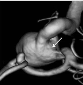
wide neck involving the parent artery circumferentially at the right paraclinoid internal carotid artery (ICA) (Fig. 1). The rup- tured AcoA aneurysm was treated successfully by clipping. Endo- vascular coil embolization was performed for the paraclinoid INTRODUCTION
Stent-assisted coiling is a safe and effective method for treating wide-necked aneurysms
1), but ischemic stroke related to stent thrombosis is a major potential drawback. All thromboses occur within one year after the initial procedure, very late stent throm- bosis (VLST), which is defined as stent thrombosis occurring one year after implantation
2), following sole stent-assisted coil- ing has not been previously reported. Here, we report the first case of VLST, which occurred 14 months after a typical sole stent-as- sisted coiling procedure.
This case highlights the possible mechanism of VLST after sin- gle stent-assisted coiling and raises concerns about the duration of prophylactic antiplatelet therapy (APT).
CASE REPORT History and procedure
A 56-year-old woman presented with subarachnoid hemor- rhage from a ruptured aneurysm in the anterior communicating artery (AcoA) and an unruptured large aneurysm with an ultra-
Very Late Stent Thrombosis after Sole Stent-Assisted Coiling at the Paraclinoid Giant Aneurysm :
Could Prophylactic Antiplatelet Therapy Be Ceased at the Only 1 Year after Procedure?
Jung-Hoon Shin, M.D., Seong-Ho Park, M.D., Chang-Hyun Kim, M.D., Chang-Young Lee, M.D.
Department of Neurosurgery, Keimyung University School of Medicine, Daegu, Korea
Stent thrombosis is a major limitation of stent-assisted coiling, which is an effective method for treating wide-necked aneurysms. Although early in- stent thrombosis has been reported, very late stent thrombosis (VLST) (>1 year) has not been reported following implantation of a single self-ex- pandable stent designed for coiling. Herein, the authors present a case of VLST that occurred 14 months after single stent implantation in a large paraclinoid aneurysm with an ultra-wide neck involving the parent artery circumferentially. This case indicates the need for establishing guidelines regarding the optimal duration of prophylactic antiplatelet therapy following stent-assisted coiling, which remains undefined in the neuroendovascu- lar field.
Key Words : Aneurysm · Antiplatelet · Stent · Stent-assisted coiling · Very late stent thrombosis.
Case Report
•
Received : November 9, 2013
•Revised : February 21, 2014
•Accepted : September 29, 2014
•
Address for reprints : Chang-Young Lee, M.D.
Department of Neurosurgery, Keimyung University School of Medicine, 56 Dalseong-ro, Jung-gu, Daegu 700-712, Korea Tel : +82-53-250-7732, Fax : +82-53-250-7356, E-mail : [email protected]
•
This is an Open Access article distributed under the terms of the Creative Commons Attribution Non-Commercial License (http://creativecommons.org/licenses/by-nc/3.0) which permits unrestricted non-commercial use, distribution, and reproduction in any medium, provided the original work is properly cited.
J Korean Neurosurg Soc 56 (4) : 344-347, 2014
http://dx.doi.org/10.3340/jkns.2014.56.4.344
Copyright © 2014 The Korean Neurosurgical Society
Print ISSN 2005-3711 On-line ISSN 1598-7876www.jkns.or.kr
Fig. 1. Three-dimensional angiography showing an aneurysm at the an- terior communicating artery and a large aneurysm with a wide circum- ferential neck (arrow) at the paraclinoid internal carotid artery.
online © ML Comm
345
VLST after Sole Stent-Assisted Coiling | JH Shin, et al.
aneurysm 4 weeks after clipping. Coiling was performed using a stent-assisted technique for wide neck remodeling. The pa- tient received oral aspirin (300 mg/d) and clopidogrel (75 mg/d) for 7 days before the coiling procedure, which was performed using a transfemoral approach under full anticoagulation with intravenous heparin (activated clotting time >250 s). First, a closed-cell Enterprise stent (4.5×37 mm; Cordis, Miami Lakes, FL, USA) was navigated in the ICA and implanted across the wide neck of the aneurysm, and coiling then was performed us- ing 19 bare platinum coils (Orbit; Cordis, Miami Lakes, FL, USA) via a SL-10 microcatheter (Boston Scientific, Natick, MA, USA), which was jailed outside the stent. The final angiogram showed near complete obliteration of the aneurysm with mini- mal neck remnant (Fig. 2A).
The patient had an uneventful postoperative course and was neurologically normal at the out-patient department follow-up.
Furthermore, follow-up angiography performed at 12 months af- ter the coiling procedure depicted no change in the small amount of residual filling of the aneurysmal neck and a well-preserved parent artery without any thrombosis or stenosis (Fig. 2B).
Antiplatelet therapy
The patient was maintained on dual antiplatelet therapy (APT) for 12 months after the initial treatment. We used 300 mg aspi- rin and 75 mg clopidogrel as the dual antiplatelet regimen for 4 weeks followed by 200 mg aspirin and 75 mg clopidogrel for 5 months and 100 mg aspirin and 75 mg clopidogrel for the re- maining 6 months. After the 1-year follow-up angiographic eval- uation, dual APT was switched to aspirin monotherapy.
Very late thrombosis
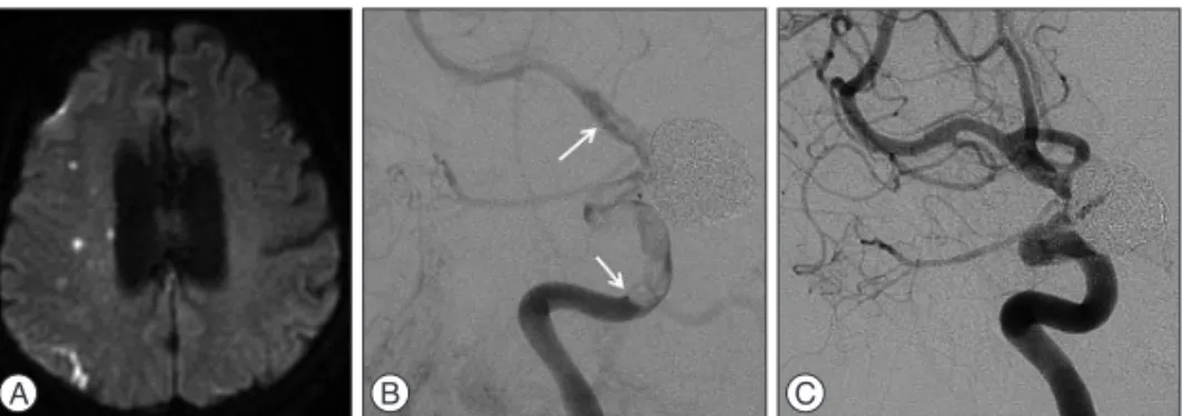
Two months after switching to aspirin monotherapy, the pa- tient experienced transient ischemic attack symptoms. Comput- ed tomography angiography revealed occlusion of the right ICA at the level of stent implantation. Diffusion weighted imaging taken for evaluating ischemic symptoms demonstrated multi- ple embolic infarctions in the right hemisphere (Fig. 3A). Digi- tal subtraction angiography (DSA) revealed in-stent thrombosis extending from the cavernous ICA to the distal ICA (Fig. 3B). The patient admitted stopping her aspirin
medication prior to a dental procedure conducted 3 days before the ischemic event.
Management of thrombosis The patient was managed conserva- tively, because the potential risk of throm- bus distal migration was considered to be high for interventional therapy. Clopi- dogrel 75 mg was added to aspirin 200 mg and low molecular heparin was ad- ministrated for 10 days. A follow-up an- giography showed completely resolution
of the thrombosis with good flow through the parent artery (Fig.
3C). The patient was discharged neurologically intact on 200 mg aspirin and 75 mg clopidogrel.
DISCUSSION
Stent-assisted coiling is increasingly being used to treat aneu- rysms
6,11), but, although this is an effective and relatively safe technique
1), it has inherent problems such as thromboembolic complications and in-stent stenosis
10,18). The timing of stent thrombosis can be classified as acute (<24 h post procedure), ear- ly or subacute (24 h to 30 days post procedure), late (31 days to 1 year post procedure), or very late (>1 year post procedure)
2). In-stent thrombosis within 1 year after stenting has been re- ported to occur at a rate of around 3% after stent-assisted coil-
ing
10,14,15,19). However, no report has previously described VLST
following implantation of a sole stent, which is self-expanding intracranial stent designed for coiling assistance. The mecha- nisms of VLST previously proposed are; progressive in-stent ste- nosis, premature discontinuation of dual APT, delayed atypical response to adjacent polyglycolic acid-loaded “bioactive” coils, spontaneous occlusion of the parent artery, stenting across ma- jor arterial side branches, stent mal-apposition, stent fracture, stent strut penetration of the necrotic core, disruption of vulner- able plaque near the stent, radiation therapy, and hypersensitiv-
Fig. 2. A : Angiography taken immediately after coiling showing a minor amount of residual filling in the aneurysm neck and a preserved parent artery without any thrombosis. B : Same angle at 1-year follow-up angi- ography demonstrating no change in the small amount of residual filling of the aneurismal neck and no stenosis or thrombosis.
A B
Fig. 3. A : Diffusion weighted image taken for evaluating the patient’s ischemic symptoms demon- strating multiple embolic infarctions in the right hemisphere. B : Angiography obtained to evaluate the patient’s ischemic symptoms showing in-stent thrombosis (arrows) extending from the cavern- ous internal carotid artery to the distal internal carotid artery. C : Follow-up angiography showing a completely resolved thrombosis after management with antiplatelet agents and systemic heparin- ization.
A B C
346
J Korean Neurosurg Soc 56 | October 2014ity vasculitis
4,7). In our case, no in-stent stenosis or stenosis around the stented vessel was noted by 1-year follow-up angiography.
Furthermore, we used only bare-metal coils. Thus, premature discontinuation of antiplatelet medication was the likely cause of VLST in our case. The prevention of thromboembolic com- plications after stenting is currently achieved with prophylactic dual APT
12), but the regimens and durations of prophylactic APT are controversial
9,21). It is recognized intracranial stents are throm- bogenic until stent endothelialization occurs, and that the dura- tion of APT should be based on the completion of endothelializa- tion. It has been reported by some that endothelialization is complete within 6 months after BMS implantation
5,8,16), but no human data is available regarding the rate of stent endothelial- ization after stent-assisted coiling. In the interventional cardiol- ogy field, current guidelines recommend dual APT for at least 1 month for BMSs, but up to 1 year for drug-eluting stents, which cause delayed endothelialization
22). However, in the neuroendo- vascular field, the optimal duration of prophylactic APT follow- ing stent-assisted coiling remains undefined, and clinical prac- tices are highly discrepant; some authors have recommended only 3 or 6 months of antiplatelet therapy after stent placement for coiling
17,20). These recommendations seem to be based on the facts that the target vessel for stent placement is usually nonath- erosclerotic and that stents being used for coiling assistance are of bare-metal and have self-expandable properties, and thus, do not delay endothelialization over stent struts or cause endothe- lial injury
16).
However, a much longer duration of APT may be required when stent endothelialization is only partially achieved. The oc- currence of VLST may mean that formation of a complete neo- intimal layer incorporating stent struts has failed despite the passage of considerable time after stent implantation. Further- more, it has been established that exposed stent struts are prone to thrombosis. Our patient had a large aneurysm with a circum- ferential wide neck, which suggests a large portion of the stent might have unopposed the parent vessel. In addition, it is likely that stent struts facing a circumferential wide aneurysmal neck are endothelialized at a far slower rate than that of a similar sized stents placed across a smaller aneurysmal neck. Another possi- ble pathophysiology of incomplete endothelialization involves mechanical factors of closed-cell stents. These stents can be kinked or flattened in tortuous curves, which can result in incomplete stent apposition, causing incomplete endothelialization
3,13). Thus, the ultra-wide neck involving the parent artery circumferential- ly and the closed-cell stent deployed in the tortuous carotid si- phon probably contributed to the incomplete stent endothelial- ization in our case. Although we present only a single case of VLST after sole stent-assisted coiling, our observation suggest that APT is needed for much longer period after sole stent-assisted coil- ing for a large aneurysm with an ultra-large circumferential wide neck. A study is required to determine optimal APT regimens ac- cording to stent type, number, and aneurysm size, shape after stent-assisted coiling.
CONCLUSION
We present a case of a VLST after sole stent-assisted coiling.
Although VLST is extremely rare, it is a serious event with poten- tially devastating consequences. This case demonstrates that long-term APT and patient strict adherence are required to pos- sibly prevent this complication when a large portion of the stent is anticipated to be unopposed to the vessel wall.
References
1. Amenta PS, Dalyai RT, Kung D, Toporowski A, Chandela S, Hasan D, et al. : Stent-assisted coiling of wide-necked aneurysms in the setting of acute subarachnoid hemorrhage : experience in 65 patients. Neurosurgery 70 : 1415-1429; discussion 1429, 2012
2. Cutlip DE, Windecker S, Mehran R, Boam A, Cohen DJ, van Es GA, et al. : Clinical end points in coronary stent trials : a case for standardized definitions. Circulation 115 : 2344-2351, 2007
3. Ebrahimi N, Claus B, Lee CY, Biondi A, Benndorf G : Stent conformity in curved vascular models with simulated aneurysm necks using flat- panel CT : an in vitro study. AJNR Am J Neuroradiol 28 : 823-829, 2007 4. Farb A, Burke AP, Kolodgie FD, Virmani R : Pathological mechanisms
of fatal late coronary stent thrombosis in humans. Circulation 108 : 1701- 1706, 2003
5. Farb A, Sangiorgi G, Carter AJ, Walley VM, Edwards WD, Schwartz RS, et al. : Pathology of acute and chronic coronary stenting in humans. Cir- culation 99 : 44-52, 1999
6. Fargen KM, Hoh BL, Welch BG, Pride GL, Lanzino G, Boulos AS, et al. : Long-term results of enterprise stent-assisted coiling of cerebral aneu- rysms. Neurosurgery 71 : 239-244; discussion 244, 2012
7. Fiorella D, Hsu D, Woo HH, Tarr RW, Nelson PK : Very late thrombosis of a pipeline embolization device construct : case report. Neurosurgery 67 (3 Suppl Operative) : onsE313-onsE314; discussion onsE314, 2010 8. Grewe PH, Deneke T, Machraoui A, Barmeyer J, Müller KM : Acute and
chronic tissue response to coronary stent implantation : pathologic find- ings in human specimen. J Am Coll Cardiol 35 : 157-163, 2000 9. Grines CL, Bonow RO, Casey DE Jr, Gardner TJ, Lockhart PB, Moliterno
DJ, et al. : Prevention of premature discontinuation of dual antiplatelet therapy in patients with coronary artery stents : a science advisory from the American Heart Association, American College of Cardiology, Soci- ety for Cardiovascular Angiography and Interventions, American Col- lege of Surgeons, and American Dental Association, with representation from the American College of Physicians. J Am Coll Cardiol 49 : 734- 739, 2007
10. Kanaan H, Jankowitz B, Aleu A, Kostov D, Lin R, Lee K, et al. : In-stent thrombosis and stenosis after neck-remodeling device-assisted coil em- bolization of intracranial aneurysms. Neurosurgery 67 : 1523-1532; dis- cussion 1532-1533, 2010
11. Kim DJ, Suh SH, Lee JW, Kim BM, Lee JW, Huh SK, et al. : Influences of stents on the outcome of coil embolized intracranial aneurysms : com- parison between a stent-remodeled and non-remodeled treatment. Acta Neurochir (Wien) 152 : 423-429, 2010
12. King SB 3rd, Smith SC Jr, Hirshfeld JW Jr, Jacobs AK, Morrison DA, Williams DO, et al. : 2007 focused update of the ACC/AHA/SCAI 2005 guideline update for percutaneous coronary intervention : a report of the American College of Cardiology/American Heart Association Task Force on Practice guidelines. J Am Coll Cardiol 51 : 172-209, 2008 13. Krischek O, Miloslavski E, Fischer S, Shrivastava S, Henkes H : A com-
parison of functional and physical properties of self-expanding intracra-
nial stents [Neuroform3, Wingspan, Solitaire, Leo+, Enterprise]. Minim
Invasive Neurosurg 54 : 21-28, 2011
347
VLST after Sole Stent-Assisted Coiling | JH Shin, et al.