추간판내 Prostaglandin E 2 양의 임상적 의의 *
아주대학교 의과대학 신경외과학교실
김형석·조기홍·김기용·안영환·안영민·윤수한·조경기
= Abstract =
Clinical Implication of Prostaglandin E 2 Content in Lumbar Disc Disease
Hyung Seok Kim, M.D., Ki Hong Cho, M.D., Ki Young Kim, M.D., Young Hwan Ahn, M.D., Young Min Ahn, M.D.,
Soo Han Yoon, M.D., Kyung Gi Cho, M.D.
Department of Neurosurgery, School of Medicine, Ajou University, Suwon, Korea
bjective:A prospective biochemical assay of prostaglandin E
2content in symptomatic lumbar disc materials was done in order to clarify the pathogenesis of lumbar radiculopathy.
Patients and Methods:Forty-eight disc specimens were purified by a standard solid-phase extraction pro- cedure and analyzed by an enzymelinked immunosorbent assay for prostaglandin E
2. Clinical and anatomic correlations were evaluated with analysis of variance and t-test.
Results:Acute herniated lumbar disc diseases tended to be associated with a higher prostaglandin E
2content than degenerative lumbar disc disease. Sequestered discs tended to be associated with a higher prostaglandin E
2content than extruded discs, which also showed higher prostaglandin E
2content than protruded ones. A sciatica and positive straight leg raising test appeared to be associated with a higher prostaglandin E
2content than a negative test.
Conclusion:This result suggests that the level of prostaglandin E
2would be correlated with clinical symptom and sign in the inflammatory process of lumbar disc herniation.
KEY WORDS:Intervertebral disc herniation・Radiculopathy・Prostaglandin E
2.
서 론
요추, 특히 하부요추는 체중부하를 많이 받을 뿐 아니라 많은 운동을 담당하고 있는 곳으로, 추간판에 다양한 병적 상태에 의하여 굳어진 수핵이 약해진 섬유륜을 밀고 돌출되 거나 섬유륜을 뚫고 수핵의 일부가 밖으로 빠져나오는 것을 요추간판 탈출증이라고 한다.
요추간판 탈출증에 의한 요통 및 척추신경근증상은 신체 장애를 유발시키는 주요 원인중의 하나이나 아직 원인 및 병 태생리가 명확히 알려져 있지 않다
2). 1934년에 Mixter와 Barr는 추간판이 탈출되면서 척수신경을 압박하여 하지 방 사통이 유발된다는‘mechanical compression theory’를
발표하였다
11). 그후에 이 개념이 폭넓게 받아들여졌으나 척 추신경근증상을 설명하기에는 부족한 점이 많다. 왜냐하면, 추간판 탈출증이 심하나 척추신경증상이 없는 경우가 있는 가 하면, 추간판 탈출증이 심하지 않더라도 척추신경증상이 심한 경우가 있기 때문이다
1)7)2). 즉, 척추신경근증상과 추간 판 탈출의 정도가 항상 일치하지 않는다는 사실이다
18)21)22).
최근 연구에 의하면 추간판 탈출증 증상의 발현에 있어 서 튀어나온 추간판과 신경근의 상호작용에서 생화학적인 부분에 대한 관심이 높아지고 있다. 탈출된 추간판 조직은 일련의 조직반응을 일으키는 것으로 알려져 있고, 이는 기 계적 신경근 압박 이외에 국소 염증이 유발된 후 이것들 이 복합적으로 작용하여 증상을 일으키는 것으로 생각된 다
1)10)11)14)16).
본 연구에서는 급성 추간판 탈출증 환자와 퇴행성 질환 환
OOOO
*2000년 제 18 차 춘계학술대회에 구연 포스터로 발표된 내용임.
자의 추간판 물질을 섬유륜, 수핵 및 파열절편으로 분류하 여 각각의 prostaglandin E
2양을 측정하여 이를 토대로 임 상적 및 해부학적인 관계를 밝혀 보고자 한다.
대상 및 방법
1. 대 상
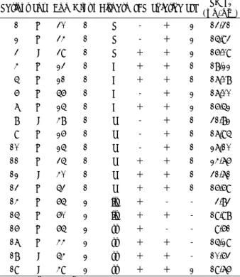
1996년 3월부터 1999년 1월 본원에서 요추 척추신경근 증상이 있는 환자에서 추간판 제거술을 시행한 후 영하 70 도에서 냉동 보관중인 총 350예의 추간판 조직검체 중, 방 사선 소견과 수술소견을 비교하여 group 1과 group 2로 분 류하였다. Group 1은 급성 추간판 탈출증(돌출형, 탈출형, 유리형)으로 총 350예 중 205예였고 group 2는 퇴행성 척 추질환(척추관 협착증, 척추 전방전위증)으로 145예였다. 이 중 각각 group 1은 13예, group 2는 6예, 총 19예를 무작 위 추출을 시행하였고 group 1은 돌출형에서는 추간판를 섬 유륜과 수핵으로 그리고 탈출형과 유리형에서는 각 예마다 섬유륜, 수핵, 파열절편으로 세분하여 36개의 조직검체를, group 2는 각 예마다 섬유륜, 수핵으로 세분하여 12개의 조 직검체를 얻어 전체 48개의 추간판 조직검체에서 prosta- glandin E 2를 측정하였다(Table 1).
2. 방 법
냉동 추간판 조직검체에서 prostaglandin E
2의 추출은 Powell이 기술한 solid phase extraction 과정을 사용하 였다
17). 이 방법은 추간판 조직검체를 mechanical tissue mincer를 사용하여 자르고 공이와 막자사발에서 잘게 부순 후 100μM indomethacin이 들어 있는 phosphate buffer solution에 녹여서 원심분리하여 상층액을 구하고 1cc의 상 층액과 4cc의 100% ethyl alcohol을 섞어 30분뒤 다시 원 심분리하여 상층액 1cc를 구한다. 구한 상층액 1cc를 N
2gas 를 사용하여 ethyl alcohol을 증발시킨후 남은 상층액을 oc- tadecylsilyl silica column(Sep-Pak cartridges, Waters)
에 여과시킨후 3cc methyl alcohol을 다시 여과시켜 얻은 용 액을 N
2gas로 증발을 시켜 prostaglandin이 농축되어 있는 용액을 얻는다.
농축액에서 prostaglandin E
2의 측정은 ELISA kit(Bio- grak
TM, Prostaglandin E
2competitive enzyme-immu- noassay system, Amersham Pharmacia Biotech, Buc- kinghamshire, England)를 사용하여 정량화 하였다.
측정된 prostaglandin E
2수치와 환자의 술전 임상증상 (요통, 하지 방사통, 하지 직거상 검사)과 비교 분석하였다.
결과는 ANOVA(analysis of variance)와 independent t test를 이용하여 통계처리하였다.
결 과
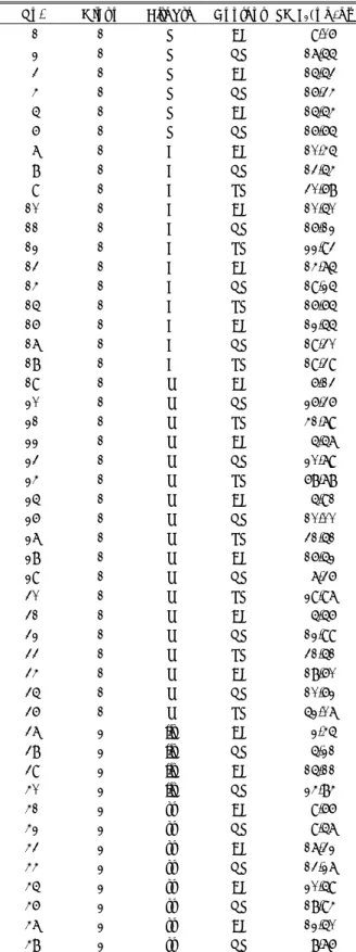
전체 19예에서 남자는 14명 여자는 5명이었고 평균나이 는 40세였다. Group 1의 13예 중에서 3예는 돌출형, 4예는 탈출형, 6예는 유리형이었고, group 2의 6예 중에서 4예는 척추관 협착증, 2예는 척추 전방전위증이었다. 요통이 있는 예는 19예 중 13예였고 하지 방사통이 있는 예는 19예 중 17예였다. Group 1에서 하지 직거상 검사상 35° 미만이 8예, 35° 이상이 5예였다(Table 2).
Table 1. Disease entities vs. intervertebral disc specimens Disc specimens(No.) Disease entities
AF NP RP Total (No. ) Protrusion(3 cases) 3 3 - 6 Extrusion(4 cases) 4 4 4 12 Group 1
(13 cases)
Sequestration(6 cases) 6 6 6 18 36 Stenosis(2 cases) 2 2 - 4 Group 2
(6 cases) Listhesis(4 cases) 4 4 - 8 12 Total(No.) 19 cases 19 19 10 48
Listhesis:Spondylolisthesis
AF:annulus fibrosus, NP:nucleus pulposus, RP:ruptured particle
Table 2. Summary of results
Patient Sex Age Group Disease LBP Sciatica SLR PG E
2(pg/gm) 1 M 30 1 P - + 2 13.31 2 M 34 1 P - + 2 15.93 3 F 39 1 P + + 2 16.09 4 M 23 1 E + + 1 18.22 5 M 21 1 E + + 1 17.08 6 M 56 1 E - + 2 17.00 7 M 25 1 E + + 2 16.52 8 F 48 1 S - + 1 31.82 9 M 26 1 S - + 1 17.95 10 M 25 1 S - + 1 27.10 11 M 35 1 S + + 1 24.76 12 F 40 1 S + + 1 31.71 13 M 53 1 S + + 1 16.69 14 M 65 2 lis + - - 3.83 15 M 60 2 lis + + - 19.98 16 M 65 2 st + - - 9.61 17 M 44 2 st + + - 15.29 18 F 54 2 st + + - 10.63 19 F 49 2 st + + 2 19.76 1:<35’, 2:>35’
P:Protrusion, E:Extrusion, S:Sequestration, lis:Spondylo- listhesis, st:Spinal stenosis, LBP:Low back pain, Sciatica:
Radiating leg pain, LR:Straight leg raising test
19예에서 얻은 48개의 추간판 검체에서 측정된 prosta- glandin E
2의 수치는 2.45~68.78pg/gm(평균수치:17.85 pg/gm)이었다. Prostaglandin E
2의 혈장내 평균수치는 5 pg/gm이다(Table 3).
Group 1에서 group 2보다 prostaglandin E
2의 수치가 20.32±6.2pg/gm과 13.18±6.3pg/gm로 통계적으로 유의 하게 높았다(Fig. 1)(p<0.05). Group 1에서 추간판의 탈출 된 형태에 따라 돌출형, 탈출형, 유리형에서 각각 15.11±
1.5pg/gm:17.18±0.7pg/gm:22.34±6.5pg/gm로 돌출 형보다는 탈출형에서, 탈출형보다는 유리형에서 수치가 높 았고(Fig. 2)(p<0.05), 추간판의 부위에 따라 섬유륜:수 핵:파열절편에서 각각 11.28±4.6:15.91±5.0:34.53
±15.8pg/gm으로 섬유륜보다 수핵에서, 수핵보다 파열절편 에서 수치가 높았다(Fig. 3)(p<0.05). 하지 방사통이 있는 예와 없는 예에서 prostaglandin E
2의 평균수치는 19.4±
6.0:6.72±4.0pg/gm으로 하지 방사통이 있는 예에서 높았 고(Fig. 4)(p<0.05), group 1에서 하지 직거상 검사상 35°
미만과 35°이상인 환자군에서 각각 23.17±6.5:15.77±
Table 3. Disc materials and prostaglandin E
2content No. Group Disease Content PG E
2(pg/gm)
1 1 P AF 9.06 2 1 P NP 17.55 3 1 P AF 15.53 4 1 P NP 16.34 5 1 P AF 15.54 6 1 P NP 16.65 7 1 E AF 10.45 8 1 E NP 13.54 9 1 E RP 30.68 10 1 E AF 10.50 11 1 E NP 16.12 12 1 E RP 22.93 13 1 E AF 14.75 14 1 E NP 19.25 15 1 E RP 16.65 16 1 E AF 12.55 17 1 E NP 19.30 18 1 E RP 19.39
19 1 S AF 6.13
20 1 S NP 26.36 21 1 S RP 41.79
22 1 S AF 5.57
23 1 S NP 20.79 24 1 S RP 68.78
25 1 S AF 5.91
26 1 S NP 10.00 27 1 S RP 31.51 28 1 S AF 16.52
29 1 S NP 7.36
30 1 S RP 29.97
31 1 S AF 5.56
32 1 S NP 12.99 33 1 S RP 31.51 34 1 S AF 18.60 35 1 S NP 10.62 36 1 S RP 52.07
37 2 lis AF 2.45
38 2 lis NP 5.21
39 2 lis AF 15.11 40 2 lis NP 24.84
41 2 st AF 9.66
42 2 st NP 9.57
43 2 st AF 17.32 44 2 st NP 13.27 45 2 st AF 20.59 46 2 st NP 18.94 47 2 st AF 12.50
48 2 st NP 8.76
P:Protrusion, E:Extrusion, S:Sequestration, lis:Spondylolis- thesis, st:Spinal stenosis, AF:Annulus fibrosus, NP:Nucleus pulposus, RP:Ruptured intervertebral disc
Fig. 2. Prostaglandin E
2content in different types of interverte- bral disc herniation among Group 1(P:Protrusion, E:
Extrusion, S:Sequestration).
Fig. 1. Prostaglandin E
2content in disc materials in each group.
Group 1:Patients with simple herniated intervertebral
disc(n=13). Group 2:Patients with chro-nic degene-
rative spinal disease(n=6).
2.0pg/gm로 35°미만인 예에서 높았다(Fig. 5)(p<0.05). 요 통이 있는 예와 없는 예에서 prostaglandin E
2수치의 차이 는 통계적 유의성이 없었다.
고 찰
최근 여러 연구를 통하여 척추신경근증상의 유발원인으로 염증반응이 중요한 역할을 한다는 것이 알려졌다
6)10)19). 이러 한 연구 결과들은 탈출된 추간판에서 phospholipase A
2효 소의 활성이 증가되어 있는 것과 추간판 조직에서 여러가지
염증세포가 다수 관찰된다는 사실로 증명되었다
6)19). 1987년 McCarron등은 실험동물에서 추간판의 수핵을 요추의 경막 외 공간에 넣었을 경우 생리식염수를 넣은 대조군보다 염증 반응이 더 심하였다고 발표하여 수핵이 염증반응을 유발시킨 다는 것을 밝혔다
10). 정상적으로 혈관이 분포하지 않는 수핵 은 면역체계로부터 격리되어 있기 때문에 추간판이 경막외 공간으로 탈출시 항원으로 작용하여 자가면역반응이 유발되 고 만성염증으로 진행을 하게된다
12). 염증을 유발시키는 매개 체로는 phospholipase A
2, cytokines, prostaglandin E
2, nitric oxide, immunoglobulins 등이 알려져 있다
16).
1990년에 Saal 등은 튀어나온 추간판에서 phospho- lipase A
2의 활동성이 다른 부위보다 20~10,000배 높다 고 발표하였고
19)1992년에 Franson 등은 사람의 튀어나온 추간판에서 추출한 phospholipase A
2가 실험동물에서 심한 염증반응을 유발한다고 발표하였다
4). Phospholipase A
2는 통증을 유발시키는데 중요한 역할을 하는데 그 기전으로는 첫째, 추간판의 섬유륜 또는 경막외공간에 분포하고 있는 통 각수용체를 자극하고 둘째, 신경근을 구성하는 인산화지질 을 손상시키며 셋째, 염증매개체인 arachidonic acid me- tabolites, 즉 prostaglandin과 leukotriens 등을 생성하여 2차적으로 염증반응을 일으킨다
16). 1992년에 Saal 등은 동 물실험을 통하여 phospholipase A
2가 신경에서 탈수초화 및 지질침착 그리고 신경원의 축삭돌기에 손상을 주는 신경 독성이 있음을 증명하였다
20).
1994년에 Willburger와 Wittenberg 등은 추간판 탈출증 환자의 추간판과 요추관절조직에서 prostaglandin과 leu- kotriene이 증가되어 있음을 발표하였고, 1996년에 Kang 등은 탈출된 추간판에서 prostaglandin E
2의 수치가 현저히 증가되어 있다고 발표하였다
23)24).
1996년에 John 등은 추간판 탈출증에서도 탈출형과 유리 형에서 돌출형보다 prostaglandin E
2수치가 더 높고 하지 방 사통 및 하지 직거상 검사상 양성반응과 관계가 있음을 밝혀 척추신경근증상과 염증이 밀접한 관계가 있음을 알아내었다
13).
Phospholipase A
2에 의하여 2차적으로 생성되는 pros- taglandin과 leukotriene은 중요한 염증반응의 매개체로서 조직손상을 유발하고, 통각수용체의 역치를 조절하고 신경섬 유중 C-fiber를 자극하여 통증 유발에 중요한 역할을 하는 것으로 알려져 있다
3)5)8)9)15).
본 실험에서는 다양한 염증반응을 유발시키는 매개체 중 prostaglandin E
2의 수치를 측정하여 임상적 관계와 해부학 적인 관계를 밝혀 보고자 하였다. Prostaglandin E
2는 가장 강력한 염증 반응 매개체로 급성 척추질환에서 만성 퇴행성 질환 보다 높은 수치를 보였고, 추간판 구조상 섬유륜보다는
Fig. 5. Prostaglandin E
2content by the degree of straight leg raising test.
Fig. 3. Prostaglandin E
2content by the site of herniated inter- vertebral disc in Group 1(AF:annulus fibrosus, NF:nu- cleus pulposus, RP:ruptured intervertebral disc).
Fig. 4. Prostaglandin E
2content by presence of sciatica.
수핵과 파열된 수핵검체에서 높은 수치를 보였다. 하지 방사 통 및 하지 직거상 검사상 양성으로 나타나는 척수신경근증 상과 prostaglandin E
2의 수치는 상관관계를 보였다. 이는 증상의 병인에 염증반응이 중요한 역할을 한다는 것을 시사 한다. 실험 결과상 파열된 수핵에서 통계학적으로 유의하게 prostaglandin E
2의 수치가 증가된 것으로 보아 수핵의 파 열이 주위 염증반응을 심하게 유발시키고 이로 인하여 척추 신경근증상이 유발되는 것으로 생각된다.
결 론
염증반응이 요추 추간판 탈출증 환자의 증상발현에 미치 는 역할을 알아보기 위하여 추간판에서 prostaglandin E
2의 양을 측정하여 임상증상과의 연관 관계를 비교 분석하였다.
Prostaglandin E
2는 가장 강력한 염증 반응 매개체로 급 성 척추질환인 경우, 특히 파열된 수핵 검체에서 높은 수치 를 보였는데, 이는 증상의 병인에 염증반응이 중요한 역할을 한다는 것을 시사한다.
본 실험을 통하여 만성 척추질환보다는 급성 척추질환에서 그리고 파열절편에서 높은 prostaglandin E
2수치를 보였다.
이는 파열절편이 주위에 염증반응을 유발시킴으로써 신경근을 자극하여 척수신경근증상을 더욱 심화시키는 것으로 사료된다.
•
논문접수일:2000년 6월 7일•
심사완료일:2000년 8월 29일•
책임저자:조 기 홍442-721 경기도 수원시 팔달구 원천동 산 5번지 아주대학교 의과대학 신경외과학교실
전화:031) 219-5232, 5662, 전송:031) 216-6658 E-mail:[email protected]
References
1) Boden SD, Davis DO, Dina TS:Abnormal magnetic resonance
scans of the lumbar spine in asymptomatic subjects
:a pros- pective investigation. J Bone Joint Surg Am 72
:403-408, 1990
2) Deyo RA, Tsui-Wu YJ:Descriptive epidemiology of low backpain and its related medical care in the United States. Spine 12
:264-268, 1987
3) Franson RC:Isolation and characterization of a phosph-
olipase A2 from an inflammatory exudate. J Lipid Res 19
:18-23, 1978
4) Franson RC, Saal JS, Saal JA:Human disc phospholipase A
2 is inflammatory. Spine 17
(suppl
):S129-S132, 1992
5) Greaves MW, Camp RD:Prostaglandins, leukotrienes phos-pholipase, platelet activation factor, and cytokines
:an inte- grates approach to inflammation of human skin. Arch Dermatol Res 280
(suppl
):33-41, 1988
6) Grnblad M, Virri J, Tolonen J, Seitsalo S, Kpa E, Kankare J, et al:A controlled immunohistochemical study of inflammatory
cells in disc herniation tissue. Spine 19
:44-51, 1994
7) Kang JD, Georgescu HI, Stefanovic-Racic M, McIntyre-La-rkin L, Donaldson WF, Evans CH:Toward a biochemical
understanding of human intervertebral disc degeneration and herniation. Spine 22
:1065-1073, 1997
8) Levine JD, Lau W, Kiniat G, Goetzl EJ:Leukotriene B4 pro-
duces hyperalgesia that is dependent on polymorphonuclear leukocytes. Science 225
:743-745, 1984
9) Levine JD:Hyperalgesic properties of 15-lipoxygenase prod-
ucts of arachidonic acid. Proc Natl Acad Sci USA 83
:5331- 5334, 1986
10) McCarron RF, Wimpee MW, Hudkins PG, Laros GS:The in-
flammatory effect of nucleus pulposus. Spine 12
:760-764, 1987
11) Mixter WJ, Barr JS:Rupture of the intervertebral disc with in-volvement of the spinal canal. N Engl J Med 211
:210-215, 1932
12) Naylor A:The biophysical and biochemical aspects of int-ervertebral disc herniation and degeneration. Ann R Coll Surg Engl 31
:91-114, 1962
13) O'Donnel JL, O'Donnel AL:Prostaglandin E2 content in
herniated lumbar disc disease. Spine 21
:1653-1656, 1996
14) Oystein P, Nygaard, Svein IM, Bjarne O:The inflammatoryproperties of contained and noncontained lumbar disc her- niation. Spine 22
:2484-2488, 1997
15) Pateomichelakis S, Rood JP:Prostaglandin E2 increases
mechanically evolved potentials in the peripheral nerve. Exp- erientia 37
:282-284, 1981
16) Philippe Goupille, Malcolm IV Jayson, Jean-Pierre Valat, Anthony J Freemont:The role of imflammation in disk her-
niationassociated radiculopathy. Seminars in Arthritis and Rheumatism 28
:60-71, 1998
17) Powell WS:Rapid extraction of oxygenated metabolites of
arachidonic acid from biological samples using octadecylsilyl silica. Prostaglandins 20
:947-957, 1980
18) Saal JA, Saal JS, Herzog RJ:The natural history of lumbar
intervertebral disc extrusions treated nonoperatively. Spine 15
:683-686, 1990
19) Saal JS, Franson RC, Dobrow R, Saal JA, White AH, Gold- thwaite N:High levels of inflammatory phospholipase A2