Interaction of CLIP-170, a Regulator of Microtubule Plus End Dynamics, with Kinesin 1 via KIF5s
Won Hee Jang1, Young Joo Jeong1, Won Hee Lee2, Mooseong Kim2, Sang-Jin Kim3, Sang-Hwa Urm4 and Dae-Hyun Seog1*
1Department of Biochemistry, Inje University College of Medicine, Busan 614-735, Korea
2Department of Neurosurgery, Inje University College of Medicine, Busan 614-735, Korea
3Department of Neurology, Inje University College of Medicine, Busan 614-735, Korea
4Department of Preventive Medicine, Inje University College of Medicine, Busan 614-735, Korea Received February 2, 2017 /Revised March 15, 2017 /Accepted March 16, 2017
Microtubules are long rods in the cytoplasm of cells that plays a role in cell motility and intracellular transport. Microtubule-based transport by motor proteins is essential in intracellular transport. Kinesin 1 is a molecular motor protein that mediates the intracellular transport of various membranous vesi- cles, mRNAs, and proteins along microtubules. It is comprised of two heavy chains (KHCs, also called KIF5s) and two light chains (KLCs). KIF5s bear a motor domain in their amino (N)-terminal regions and interact with various cargoes through the cargo-binding domain in their carboxyl (C)-terminal regions. To identify proteins interacting with KIF5B, yeast two-hybrid screening was performed, and a specific interaction with the cytoplasmic linker protein 170 (CLIP-170), a plus end microtubule-bind- ing protein, was found. The coiled-coil domain of CLIP-170 is essential for interactions with KIF5B in the yeast two-hybrid assay. CLIP-170 bound to the cargo-binding domain of KIF5B. Also, other KIF5s, KIF5A and KIF5C, interacted with CLIP-170 in the yeast two-hybrid assay. In addition, gluta- thione S-transferase (GST) pull-downs showed that KIF5s specifically interacted with CLIP-170. An an- tibody to KIF5B specifically co-immunoprecipitated CLIP-170 associated with KIF5B from mouse brain extracts. These results suggest that kinesin 1 motor protein may transport CLIP-170 in cells.
Key words : Cargo, CLIP-170, kinesin 1, microtubule, microtubule motor
*Corresponding author
*Tel : +82-51-890-6974, Fax : +82-51-894-5801
*E-mail : [email protected]
This is an Open-Access article distributed under the terms of the Creative Commons Attribution Non-Commercial License (http://creativecommons.org/licenses/by-nc/3.0) which permits unrestricted non-commercial use, distribution, and reproduction in any medium, provided the original work is properly cited.
Journal of Life Science 2017 Vol. 27. No. 6. 673~679 DOI : https://doi.org/10.5352/JLS.2017.27.6.673
Introduction
Some proteins necessary for cell function must be trans- ported to the appropriate compartments or organelles after synthesis in the cell body. Motor proteins transport various kinds of membranous vesicles and protein complexes along microtubules [9, 11]. In the cells, microtubules are the major longitudinal cytoskeletal filament. Microtubules form through the polymerization of α-tubulin and β-tubulin [8].
In the cells, microtubule polymerization is initiated at micro- tubule organizing centers (MTOCs) and microtubules have two structurally distinct ends, plus-end and minus-end [8].
The minus-end of microtubule is located at MTOC and the plus-end is oriented cell peripherally [8]. Kinesins are plus-
end-directed molecular motor proteins that move along mi- crotubule tracks and involve in the transport of various car- goes, including membrane vesicles, organelles, proteins and their complexes, and mRNAs [8, 9]. Kinesin 1 is the first identified member of kinesin superfamily proteins (KIFs) as a major motor protein in cells [2]. It is a heterotetrameric protein of two heavy chains (KHCs, also called KIF5s), which contain the amino (N)-terminal motor domain, and two light chains (KLCs), which bind to the stalk region of KHCs [8]. KHCs have been revealed to consist of three close- ly related subtypes: KIF5A, KIF5B, and KIF5C [13]. KIF5B is expressed ubiquitously in many tissues, whereas KIF5A and KIF5C are specifically to nervous tissue [13]. KIF5A, KIF5B, and KIF5C form homodimers or heterodimers [13].
Mice with a kif5B null mutation die during early embryonic development because of impaired transport of multiple es- sential organelles, including mitochondria and lysosomes, but the mitochondrial phenotype in yolk sac-derived cul- tured cells from kif5B null mice could be rescued by exoge- nous expression of KIF5A or KIF5C [22]. Thus, the members of KIF5s are thought to have functionally similar role(s) in
cells.
Many different types of cargoes moved by kinesin 1 have been identified, including mitochondria, neurotransmitter receptor-containing vesicles, virus particles, and mRNA granules [9, 12]. The amino-acid sequence of the coiled-coil cargo-binding domain is highly homologous between KIF5s [12]. The cargo-binding domain in KIF5s is thought to bind directly to some cargoes [9, 12]. For example, mitochondria [9], glutamate receptor-interacting protein 1 (GRIP1) [21], mRNA/protein (mRNP) complex [12], and γ-aminobutyric acid receptor-associated protein (GABARAP) interact di- rectly with the cargo binding-domain of KIF5s [16]. Also, other cargoes and several binding proteins including c-jun NH2-terminal kinase (JNK)-interacting proteins (JIPs) and amyloid-β precursor protein (APP) bind to the KLCs of kine- sin 1 [9, 15]. Although many cargoes including organelles moved along microtubules by kinesin 1 have been identified, it is still unclear whether kinesin 1 transports the micro- tubule components and regulates the microtubule assembly.
In this study, we screened for the binding-proteins that inter- act specifically with the cargo-binding domain of KIF5s and found cytoplasmic linker protein 170 (CLIP-170), which binds to microtubule plus ends with high affinity. CLIP-170 interacts with the carboxyl (C)-terminal tails of α-tubulin as well as end-binding protein 1 and regulates the ends of growing microtubules [17]. The interaction between KIF5s and CLIP-170 suggests kinesin 1 may play a role as a poten- tial transport motor of CLIP-170 and/or regulate the micro- tubule dynamics.
Materials and Methods
Plasmid constructs
The mouse KIF5B cDNA fragment corresponding to the cargo-binding domain (amino acids 808-924) was amplified by polymerase chain reaction (PCR) using the appropriate primers [13]. The amplified fragment was cloned into the EcoRI and XhoI site of pLexA (Clontech Laboratories, Inc., Palo Alto, CA, USA). The resulting recombinant plasmid, pLexA-cargo-binding domain-KIF5B, served as bait plasmid.
The full-length cDNAs of mouse CLIP-170 (GeneBank ac- cession number: NM_019765) was amplified by PCR from Marathon-ReadyTM cDNA library (Clontech Laboratories, Inc.) using the appropriate primers [10, 21] and cloned into pB42AD (Clontech Laboratories, Inc.).
Screening of KIF5B-binding proteins by yeast two- hybrid screening
The Matchmaker LexA two-hybrid system was used for screening according to the manufacturer’s manual (Clontech Laboratories, Inc.). In brief, pLexA-cargo-binding domain- KIF5B was transformed into yeast strain EGY48 carrying the p8op-lacZ gene. The transformed EGY48 yeast cells contain- ing pLexA-cargo-binding domain-KIF5B were transformed with the mouse brain cDNA library (Clontech Laboratories, Inc.) [10, 21] and grown on synthetic dextrose (SD) plates supplemented with glucose but with no histidine, trypto- phan, or uracil (SD/-His/-Trp/-Ura). The selection of pos- itive clones was performed on an SD/-His/-Trp/-Ura/-Leu plate containing galactose, raffinose, BU salts, and X-gal.
Plasmids from positive clones were analyzed by restriction digestion with EcoRI and XhoI. Unique inserts were se- quenced and DNA sequence analysis was performed using the BLAST algorithm (National Center for Biotechnology Information; https://blast.ncbi.nlm.nih.gov/Blast.cgi). Sequence- verified clones were analyzed again for interaction with bait in yeast by re-transformation.
β-Galactosidase activity in liquid cultures of yeast The β-galactosidase activity of yeast was assayed as de- scribed previously [10]. In brief, mid-log phase yeast cells were collected and permeabilized with 0.1% sodium dodecyl sulphate (SDS) and chloroform. An excess amount of o-nitro- phenyl-β-D-galactoside (ONPG) was added to yeast lysate, the mixture was incubated at 30℃, and then the reaction was stopped by increasing pH to 11 by the addition of 1 M Na2CO3. The formation of the reaction product, o-nitro- phenol, was determined by measuring absorbance at 420 nm on a spectrophotometer and normalizing for the reaction time and the cell density. The units of enzyme activity were calculated by the following equation: units = 1,000x [(OD420
–1.75x OD550)] / [reaction time (min) x culture volume (ml) x OD600].
Glutathione S-transferase (GST) pull-down assays Pull-down assays using GST fusion proteins were per- formed as previously reported [21]. cDNAs encoding the C-terminal region of KIF5A, KIF5B, and KIF5C and the full length of KLC1 were cloned in pET41. The recombinant GST-KIF5A, KIF5B, KIF5C, and KLC1 fusion proteins were expressed in E. coli. strain BL21 GOLD (Stratagene, La Jolla, CA, USA) after induction with 0.5 mM isopropyl thio-β-
D-galactopyranoside (IPTG) for 3 hr. The fusion proteins were purified by attachment to glutathione-agarose beads (Sigma-Aldrich, St. Louis, MO, USA) according to the manu- facturer’s protocol. Ten μg of each of the GST fusion proteins was then coupled to 50 μl of glutathione-agarose beads by incubating at room temperature for 1 hr, followed by rinsing several times with PBS. Mouse brain lysate was prepared as previously described [21]. Mouse brains were homogen- ized in ice-cold homogenization buffer (0.32 M sucrose, 4 mM HEPES, pH 7.3) supplemented with protease inhibitors.
The mouse brain homogenate supernatant was centrifuged again at 12,000x g for 15 min, and the resulting supernatant was saved. The supernatant (60 μg) was incubated overnight at 4℃ with the GST fusion protein-coupled glutathione beads. The beads were pelleted by centrifugation, washed three times with the extraction buffer (1% Triton X-100 in PBS containing 10 μg/ml each aprotinin, leupeptin, and pep- statin and 1 μM phenylmethanesulfonyl fluoride), and once with PBS. The bound proteins were eluted from the gluta- thione beads with 100 μl of Laemmli’s loading buffer. The pulled-down proteins were analyzed by immunoblotting with anti-CLIP-170 antibody (Genway Biotech, San Diego, CA, USA).
Co-immunoprecipitation and immunoblot analysis Mouse brain lysate was diluted in the same volume of 2X binding buffer (50 mM HEPES, 240 mM KCl, 2 mg/ml BSA, 0.2% Triton X-100, pH 7.4) and incubated with an- ti-KIF5B antibody [21] or with control IgG overnight at 4℃, followed by precipitation with protein-A Sepharose (Amer- sham Pharmacia, Piscataway, NJ, USA). The beads were col- lected by brief centrifugation and washed three times with TBS-T (20 mM Tris-HCl, pH 7.5, 0.15 M NaCl, 0.1% Tween 20). The washed beads were resuspended with Laemmli’s loading buffer and the proteins were eluted and denatured by boiling for 2 min. The proteins were processed for 10%
SDS-PAGE and immunoblot analysis with antibodies against KLC (Abcam, Cambridge, MA, USA) and CLIP-170 (Genway Biotech). The animal study was approved by the institutional review board (IRB), and the approval number was 05-12 of Inje University animal center.
Results
Identification of KIF5B interacting proteins by yeast two-hybrid screening
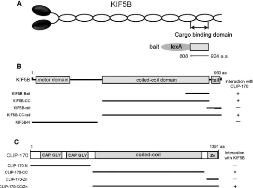
To identify proteins that interact with KIF5B, a bait con- struct encoding a fusion protein containing the cargo-bind- ing domain (amino acids 808-924) of mouse KIF5B was used for yeast two-hybrid screening (Fig. 1A). In screen of 7×10⁶ independent transformants, three positive clones were obtained. All positive clones were overlapped at the open reading frame (ORF) of CLIP-170. KIF5B is composed of a motor domain in N-terminal region, the central coiled-coil domain, and the C-terminal tail region [13]. Various frag- ments of KIF5B were constructed and tested for interaction with CLIP-170 using a yeast two-hybrid system. The result indicates that the region located within the C-terminal part of the coiled-coil domain of KIF5B is required for binding to CLIP-170 (Fig. 1B).
CLIP-170 was originally identified as a nucleotide-sensi- tive microtubule-binding protein in HeLa cells and is a mul- ti-domain protein comprised of a CAP-GLY domain (cytos- keleton-associated protein glycine-rich domain) in N-termi- nal region, a coiled-coil domain, and a Zinc knuckle domain in the tail region (Fig. 3C) [19]. To identify the domain of CLIP-170 required for the interaction with KIF5B, a series of deletion mutants of CLIP-170 was constructed and ana- lyzed for their interactions with KIF5B using the yeast two-hybrid assay. Results exhibited that only the coiled-coil domain of CLIP-170 interacted with KIF5B (Fig. 1C). This experiment demonstrated that the minimal binding domain was located in the coiled-coil domain of CLIP-170.
Subsequently, whether other KIF5s, KIF5A and KIF5C, KLC1, and KIF3A, a motor subunit of kinesin 2, interact with CLIP-170 was investigated. As shown in Fig. 2A and Fig.
2B, KIF3A and KLC1 did not interact with CLIP-170, but KIF5A and KIF5C bound to CLIP-170. GRIP1, known to in- teract with KIF5B [21], served as a positive control. This re- sult is not surprising because the cargo-binding domains of the three KIF5s share high identity in their amino acid se- quences (81-83% identity in the C-terminal region) [13]. To quantify the binding affinity of KIF5s to CLIP-170, the KIF5s or KLC1 expression plasmids were transformed into yeast and the β-galactosidase activity was measured in liquid cultures. The interaction of KIF5s with CLIP-170 yielded ap- proximately 520 units of β-galactosidase activity (Fig. 2C) and it suggests that KIF5s bound to CLIP-170 in protein level.
CLIP-170 interacts with KIF5s at protein level To further confirm the KIF5s and CLIP-170 interaction at
Fig. 1. Identification of the proteins interacted with KIF5B by yeast two-hybrid screening. (A) Schematic diagram of KIF5B. The cargo-binding domain of KIF5B used for the yeast two-hybrid screen. (B) Minimal CLIP-170 binding region in KIF5B. KIF5B has the motor domain, coiled-coil domain, and tail region, indicated in gray. The truncated forms of KIF5B were assessed in the yeast two-hybrid assay for interaction with CLIP-170. (C) KIF5B binding region in CLIP-170. CLIP-170 has the CAP-GLY domain, coiled-coil domain, Zinc knuckle (Zk) domain, indicated in gray. Different truncations of CLIP-170 were tested in the yeast two-hybrid assay for interaction with KIF5B.+, interaction -, no interaction; KIF5B, kinesin superfamily proteins 5B; CLIP-170, cytoplasmic linker protein 170; CAP-GLY, cytoskeleton-associated protein glycine-rich; aa, amino acids.
Fig. 2. Interaction of KIF5s with CLIP-170. (A, B) The tail region of each KIF5, KIF3A, and the full length KLC1 were tested for the interaction in the in the yeast two-hybrid assay. CLIP-170 specifically interacted with KIF5s but not with KIF3A or KLC1.
GRIP1 served as a positive control for interaction. (C) The strength of interactions between CLIP-170 and KIF5s or KLC1 was examined quantitatively using β-galactosidase activity in yeast two-hybrid reporter assay. KIF5: kinesin superfamily proteins 5, KLC1: kinesin light chain 1, CLIP-170: cytoplasmic linker protein 170, GRIP1: Glutamate receptor interacting protein 1.
A
B
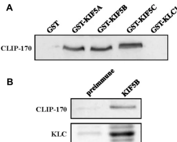
Fig. 3. Association of KIF5B with CLIP-170 in the pull-down and co-immunoprecipitation. (A) Proteins in the mouse brain lysate were allowed to bind to GST alone, GST-KIF5A, GST-KIF5B, GST-KIF5C, or GST-KLC1 fusion proteins.
The elution fractions were resolved by SDS-PAGE and immunoblotting was performed using an antibody to CLIP-170. (B) Mouse brain lysates were immunopreci- pitated with an anti-KIF5B antibody or preimmune se- rum, and then the precipitates were immunoblotted with anti-CLIP-170 or KLC antibodies. KIF5: kinesin super- family proteins 5, KLC1: kinesin light chain 1, CLIP-170:
cytoplasmic linker protein 170, GST: glutathione S-trans- ferase.
the protein level, the GST pull-down assays were performed.
Recombinant GST-KIF5s or GST-KLC1 fusion proteins were expressed in E. coli. After purification, the GST fusion pro- teins were allowed to interact with mouse brain lysates.
Immunoblot analyses revealed that CLIP-170 interacted with GST-KIF5s, but not with GST or GST-KLC1 (Fig. 3A). This result indicates that CLIP-170 associates with KIF5s at pro- tein level.
In order to address whether KIF5B interacts endogenously with CLIP-170 in mouse brain, immunoprecipitation analy- ses were performed. Lysates from mouse brain were in- cubated with an anti-KIF5B antibody. Protein G-agarose beads precipitated the immuno-complexes, which were then subsequently separated by SDS-PAGE and immunoblotted with anti-CLIP-170 and anti-KLC antibodies. As shown in Fig. 3B, antibody against KIF5B efficiently precipitated CLIP- 170 as well as KLC. Taken together, this result indicates that CLIP-170 interacts with KIF5B in brain.
Discussion
In this study, we show that KIF5B interacts with CLIP-170.
Using the cargo-binding domain of KIF5B as bait, we identi-
fied CLIP-170 in a yeast two-hybrid assay. The coiled-coil domain of CLIP-170 interacted with the cargo-binding do- main of KIF5B. Furthermore, KIF5s interacted with CLIP-170 at protein level and CLIP-170 was co-immunoprecipitated with KIF5B. Taking all of these results together, we hereby propose that kinesin 1 and CLIP-170 interaction may have a role in intracellular transport of microtubule plus-end tracking proteins (+TIPs).
Microtubule changes rapidly in response to the intra- cellular environment and microtubule dynamics are most ac- tive at the plus-end [5]. +TIPs are candidates to control the microtubules and relay their signals during cell homeostasis [5]. +TIPs can be divided into three categories: end-binding proteins (EBs), which are involved in recruiting binding partners such as CLIP-170, EB-dependent +TIPs, and EB-in- dependent +TIPs [23]. CLIP-170, the first identified +TIP, was initially characterized as a cytoplasmic linker between endosomes and microtubules [18]. In this study, we have shown that the cargo-binding domain of KIF5s interacts with the coiled-coil domain of CLIP-170.
What would be the role of the interaction between KIF5s and CLIP-170? CLIP-170 may be a scaffolding protein that links kinesin 1 and +TIPs. This binding is important for the growing microtubule plus-end localization of +TIPs.
Previous studies from yeast system suggested that yeast ki- nesin motor protein Kip2 forms complex with yeast CLIP-170 homolog Bik1 and transport along to the plus end of microtubules [1, 3, 6]. The Bik1-Kip2 complex dissociates upon arrival at plus ends [14]. In addition, the localization of CLIP-170 by kinesin is needed in microtubule stabilization [4]. Many different types of cargoes moved by kinesin 1 have been identified, including mitochondria and protein com- plex [9]. In some cases, these cargoes bind to soluble adaptor proteins/scaffolding proteins that mediate the attachment to kinesin 1 [9, 20]. Thus, CLIP-170 may serve as a scaffolding protein that can link kinesin 1 and +TIPs. Kinesin 1-medi- ated transport of CLIP-170 and/or CLIP-170-including +TIPs complex by the direct interaction with KIF5s may play a role in microtubule dynamics such as stability, assembly, and bundling.
Acknowledgment
This research was supported by Basic Science Research Program though the National Research Foundation of Korea (NRF) funded by the Ministry of Education, Science and
Technology (NRF-2015R1D1A1A01056820).
References
1. Boscheron, C., Caudron, F., Loeillet, S., Peloso, C., Mugnier, M., Kurzawa, L., Nicolas, A., Denarier, E., Aubry, L. and Andrieux, A. 2016. A role for the yeast CLIP170 ortholog, the plus-end-tracking protein Bik1, and the Rho1 GTPase in Snc1 trafficking. J. Cell Sci. 129, 3332-3341.
2. Brady, S. T. 1985. A novel brain ATPase with properties expected for the fast axonal transport motor. Nature 317, 73-75.
3. Browning, H., Hackney, D. D. and Nurse, P. 2003. Targeted movement of cell end factors in fission yeast. Nat. Cell Biol.
5, 812-818.
4. Busch, K. E., Hayles, J., Nurse, P. and Brunner, D. 2004.
Tea2p kinesin is involved in spatial microtubule organ- ization by transporting tip1p on microtubules. Dev. Cell 6, 831-843.
5. Carvalho, P., Tirnauer, J. S. and Pellman, D. 2003. Surfing on microtubule ends. Trends Cell Biol. 13, 229-237.
6. Caudron, F., Andrieux, A., Job, D. and Boscheron, C. 2008.
A new role for kinesin-directed transport of Bik1p (CLIP- 170) in Saccharomyces cerevisiae. J. Cell Sci. 121, 1506-1513.
7. Hirokawa, N., Noda, Y., Tanaka, Y. and Niwa, S. 2009.
Kinesin superfamily motor proteins and intracellular transport. Nat. Rev. Mol. Cell Biol. 10, 682-696.
8. Hirokawa, N. 1998. Kinesin and dynein superfamily pro- teins and the mechanism of organelle transport. Science 279, 519-526.
9. Hirokawa, N., Niwa, S. and Tanaka, Y. 2010. Molecular mo- tors in neurons: transport mechanisms and roles in brain function, development, and disease. Neuron 68, 610-638.
10. Jang, W. H. and Seog, D. H. 2013. Kinesin superfamily-asso- ciated protein 3 (KAP3) mediates the interaction between Kinesin-II motor subunits and HS-1-associated protein X-1 (HAX-1) through direct binding. J. Life Sci. 23, 978-983.
11. Kamal, A. and Goldstein, L. S. 2000. Connecting vesicle transport to the cytoskeleton. Curr. Opin. Cell Biol. 12, 503- 508.
12. Kanai, Y., Dohmae, N. and Hirokawa, N. 2004. Kinesin
transports RNA: isolation and characterization of an RNA- transporting granule. Neuron 43, 513-525.
13. Kanai, Y., Okada, Y., Tanaka, Y., Harada, A., Terada, S. and Hirokawa, N. 2000. KIF5C, a novel neuronal kinesin en- riched in motor neurons. J. Neurosci. 20, 6374-6384.
14. Maekawa, H. and Schiebel, E. 2004. CLIP-170 family mem- bers: a motor-driven ride to microtubule plus ends. Dev. Cell 6, 746-748.
15. Muresan, Z. and Muresan, V. 2005. Coordinated transport of phosphorylated amyloid-beta precursor protein and c-Jun NH2-terminal kinase-interacting protein-1. J. Cell Biol. 171, 615-625.
16. Nakajima, K., Yin, X., Takei, Y., Seog, D. H., Homma, N.
and Hirokawa, N. 2012. Molecular motor KIF5A is essential for GABA(A) receptor transport, and KIF5A deletion causes epilepsy. Neuron 76, 945-961.
17. Nirschl, J. J., Magiera, M. M., Lazarus, J. E., Janke, C. and Holzbaur, E. L. 2016. α-Tubulin tyrosination and CLIP-170 phosphorylation regulate the initiation of dynein-driven transport in neurons. Cell Rep. 14, 2637-2652.
18. Pierre, P., Scheel, J., Rickard, J. E. and Kreis, T. E. 1992.
CLIP-170 links endocytic vesicles to microtubules. Cell 70, 887-900.
19. Rickard, J. E. and Kreis, T. E. 1990. Identification of a novel nucleotide-sensitive microtubule-binding protein in HeLa cells. J. Cell Biol. 110, 1623-1633.
20. Seog, D. H., Lee, D. H. and Lee, S. K. 2004. Molecular motor proteins of the kinesin superfamily proteins (KIFs): struc- ture, cargo and disease. J. Kor. Med. Sci. 19, 1-7.
21. Setou, M., Seog, D. H., Tanaka, Y., Kanai, Y., Takei, Y., Kawagishi, M. and Hirokawa, N. 2002. Glutamate-receptor- interactingprotein GRIP1 directly steers kinesin to dendrites.
Nature 417, 83-87.
22. Tanaka, Y., Kanai, Y., Okada, Y., Nonaka, S., Takeda, S., Harada, A. and Hirokawa, N. 1998. Targeted disruption of mouse conventional kinesin heavy chain, kif5B, results in abnormal perinuclear clustering of mitochondria. Cell 93, 1147-1158.
23. Willige, V. D., Hoogenraad, C. C. and Akhmanova, A. 2016.
Microtubule plus-end tracking proteins in neuronal devel- opment. Cell Mol. Life Sci. 73, 2053-2077.
초록:미세소관의 plus end dynamics를 조절하는 CLIP-170과 kinesin 1의 KIF5s를 통한 결합
장원희1․정영주1․이원희2․김무성2․김상진3․엄상화4․석대현1*
(1인제대학교 의과대학 생화학교실, 2인제대학교 의과대학 신경외과학교실, 3인제대학교 의과대학 신경과학교실,
4인제대학교 의과대학 예방의학교실)
미세소관을 따라 이동하는 모터단백질들은 세포내 물질수송에 필수적인 역할을 한다. Kinesin 1은 세포내에서 미세소관을 따라 움직이는 모터단백질로서 다양한 소포, mRNA, 그리고 단백질의 세포내 수송에 관여한다.
Kinesin 1은 2개의 장쇄단위체(KHCs, 또는 KIF5s)와 2개의 경쇄단위체(KLCs)로 구성되어 있다. KIF5s는 N-말단 에 모터도메인을 가지고 있고 C-말단의 운반체 결합도메인을 통해 다양한 운반체와 결합한다. 본 연구에서 KIF5B 와 결합하는 단백질을 분리하기 위하여 효모 two-hybrid 탐색을 수행한 결과 미세소관의 plus end 결합단백질인 cytoplasmic linker protein 170 (CLIP-170)을 분리하였다. CLIP-170의 coiled-coil 도메인은 KIF5B의 운반체 결합 도메인과 결합하였다. 또한 CLIP-170은 KIF5A와 KIF5C와도 결합하였다. 그리고 glutathione S-transferase (GST) pull-down을 통해 KIF5s와 CLIP-170이 단백질수준에서 결합함을 확인하였다. 생쥐 뇌파쇄액을 KIF5B 항체로 면 역침강한 결과 CLIP-170이 같이 침강함을 확인하였다. 이러한 결과들은 kinesin 1이 세포내에서 CLIP-170을 운반 함을 시사한다.