Copyright ⓒ 2013, The Microbiological Society of Korea
Construction, Expression, and Purification of N-Terminal Variants of Lumazine Protein from Photobacterium leiognathi
Kyoung-Suk Kang, So-Young Kim, Ji-Sun Choi, Young-Doo Kim, Robert Pokoo, Ki-Seok Nam, and Chan Yong Lee*
Department of Biochemistry, Chungnam National University, Daejeon 305-764, Republic of Korea
발광세균 Photobacterium leiognathi의 돌연변이 아미노-말단 루마진 단백질들의 제조, 발현 및 정제
강경숙․김소영․최지선․김영두․로버트 포쿠․남기석․이찬용*
충남대학교 생화학과
(Received March 18, 2013 / Accepted June 24, 2013)
Lumazine protein is a fluorescent protein isolated from the bioluminescent bacteria of Photobacterium species. To generate minimal size of lumazine protein with possessing fluorescent characteristic, the gene coding for the wild type N-terminal domain of lumazine protein (N-LumP 118) containing amino acids up to 118 from Photobacterium leiognathi was produced. In addition, the genes coding for the variant proteins of N-LumP 118, replaced with one tryptophan amino acid (N-LumP 118 V41W, S48W, T50W, D64W, and A66W), were also constructed by Polymerase Chain Reaction and Site Directed Mutagenesis. These proteins were expressed in Escherichia coli by transformation with recombinant plasmids and purified by 6X-His tagging system. Spectroscopic studies have show that the purified proteins are capable of binding to the fluorescent ligand 6,7-dimethyl-8-ribityllumazine, resulted in showing of fluorescent characteristic with the minimal size of protein. From these studies, the mutant proteins containing single tryptophan amino acid residue, possessing its own intrinsic flouophore character at the different position, will be able to the use as a probe for further studies to deduce their three dimensional structure and the binding modes.
Keywords: Photobacterium, bioluminescence, lumazine protein, site directed mutagenesis
*For correspondence. E-mail: [email protected]; Tel.: +82-42-821-5482;
Fax: +82-42-822-7548
Bioluminescence refers to the emission of light without external source in specific environments (Herring, 1987;
Campbell, 1989). The luminescent bacteria emits the blue-green light during the reduced riboflavin phosphate (FMNH2) and long chain fatty-aldehyde are oxidized as the reaction scheme below (Hastings and Nealson, 1977; Meighen, 1988, 1991).
FMNH2 + RCHO + O2 → FMN + H2O + RCOOH + light
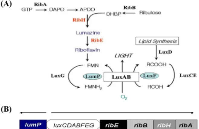
It was revealed that several genes involved in the riboflavin (vitamin B2) biosynthesis were present at the downstream of lux operon in Photobacterium species (Fig. 1A) (Lee et al., 1994;
Meighen, 1994). The riboflavin biosynthesis in bioluminescent bacteria is essential as it is the precursor of flavin substrate for the light emitting reaction. In addition, the fact that the gene (ribE) codes for the riboflavin synthase is interested in the aspect of molecular genetics as it shows 30% amino acid homology with lumazine protein (O’Kane et al., 1991; Lee et al., 2007) and the lumP gene coding the lumazine protein is transcribed in opposite direction with luxC gene (Lee and Meighen, 2000) (Fig. 1B).
Lumazine protein consisting of 185 amino acids is a fluorescent protein and was found in Photobacterium phosphoreum at the second half of 1970 (Gast and Lee, 1978; Small et al., 1980).
The strong fluorescent protein forms non-covalent bond interacted with 6,7-dimethyl-8-ribityllumazine (O’Kane et al., 1985; O’Kane and Lee, 1985) (It was hereafter referred to as lumazine) known to be a precursor for direct biosynthesis of
(A)
(B)
Fig. 1. (A) The genes involved in bacterial bioluminescence reaction.
The functions of the gene products are as follows: luxABCDE genes coding for lux proteins (luciferase and fatty acid reductase complex) involved in bioluminescence reaction, lumP for lumazine protein, ribA for GTP cyclohydrolase II, ribB for dihydroxy-butanone 4-phosphate synthase, ribH for lumazine synthase, and ribE for riboflavin synthase, and luxF for non-fluorescent flavoprotein. (B) Gene organization of lux operon region of P. leiognathi. Arrows indicate the direction of transcription.
(A)
(B)
Fig. 2. (A) Internal amino acid sequences between N-terminal half and C-terminal half of lumazine protein from P. leiognathi.
Identical amino acids are marked in box and similar amino acids shown in grey letters. The amino acids from the number of 97 to 118 containing N101 and I102 are underlined. (B) Model of binding site topology of lumazine protein of 20 kDa with 6,7-dimethyl-8-ribityllumazine obtained from author’s previous paper (Illarionov et al., 2007).
vitamin B2 (Koka and Lee, 1979; Fischer and Bacher, 2005).
This protein is considered to be generated through riboflavin gene duplication and is under the superfamily of riboflavin synthase (O’Kane et al., 1991). The lumazine protein has a monomeric structure showing the intramolecular sequence similarity between N-terminal and C-terminal half domain (Fig.
2A). It was reported that the protein binds to one molecule of lumazine in N-terminal half (Illarionov et al., 2007; Chatwell et al., 2008). In this study, to generate a minimal size of fluorescent lumazine protein and to provide a basis for the future binding study, the wild type and the mutant genes for the half of the N-terminal region of lumazine protein from Photobacterium leiognathi, were designed by PCR (Polymerase Chain Reaction) and site directed mutagenesis and expressed to be purified.
Studies have shown that asparagine 101 and isoleucine 102, located beyond N-terminal region, are involved in binding of the lumazine ligand in additions to the amino acids such as serine 48, threonine 50, and alanine 66 at the binding sites in N-terminal half of lumazine protein (Illarionov et al., 2007;
Chatwell et al., 2008) (Figs. 2A and 2B). Therefore, the gene coding for the N-terminal lumazine protein (N-LumP 118) extending to the region of these two amino acids of asparagine 101 and isoleucine 102 was synthesized using pPhl36 (Illarionov et al., 1994) as a template in PCR. In order to amplify the gene coding for the 118th position of amino acids in lumazine protein, designed forward primer (5′-CTTTAACA AGGATCCGTAATGTTTAG-3′) and reverse primer (5′-GGG ATATCAATCACTGCAGATTAACCTGCGTC-3′) from Solgent
Co. were used. BamHI and PstI restriction sites on these primers were indicated by underlined. PCR conditions were as followed: pre-denaturation 95℃ 5 min, denaturation 95℃ 30 sec, annealing 60℃ 30 sec, extension 72℃ 1 min 20 sec, final extension 72℃ 7 min denaturation, annealing, extension. This program set-up was run 24 cycles repeatedly. Generation of mutants such as N-LumP 118 V41W, S48W, T50W, D64W, and A66W were produced from the recombinant plasmid containing the gene for the wild type lumazine protein by point mutagenesis in the PCR reaction (Fig. 3). The primers used in mutation were manufactured by Solgent and Finnzymes cooperations as listed in Table 1.
Confirmation of mutants was detected by restriction digestions using SnaBI and EcoRV restriction enzyme whose restriction sites were located on the primers (Table 1). Initial analysis of the agarose gel electrophoresis gave a clue that the mutation had occurred by new generated additional restriction sites. These mutant genes were cut with BamHI and PstI restriction enzymes and ligated with pQE30 vector that had also been cut with the above restriction enzymes (Fig. 3). The DNA was sent to Solgent Co. in order to confirm the sequence of the mutants of interest by automatic base-sequence analyzer.
The recombinant plasmids, containing pQE30 vector inserted with the gene coding for N-LumP 118 of wild and mutant types, were inserted into E. coli XL-1 blue by transformation using the heat shock method and the resultant mixture was on Luria Bertani (LB) plates containing 100 μg/ml of ampicillin. Single colony out of the numerous colonies obtained was also further cultured in LB (tryptone 10 g, yeast-
Fig. 3. Nucleotide sequences and their corresponding translated amino acid sequences of N-LumP 118 wt. The nucleotide sequence and its translated amino acid sequences (MRGSHHHHHHGSV) of 6X-His tagging system are included. The amino acid sequences replaced with tryptophan were marked in underlined-bold.
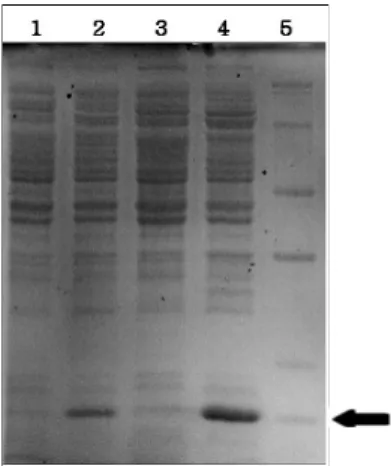
Fig. 4. SDS-PAGE analysis of N-terminal lumazine protein (N-LumP 118) obtained from different growing conditions. Lanes: 1, N-LumP 118 D64W no IPTG in LB media; 2, N-LumP 118 D64W with IPTG induction at 30℃ overnight in LB; 3, N-LumP 118 D64W no IPTG in TB media; 4, N-LumP 118 D64W with IPTG induction at 30℃ overnight in TB media; 5, Protein size marker (14, 20, 24, 29, 36, 45 kDa, respectively).
V41W
5′-AGTATCTACATCAACAAGTTGAAACATACCCTC-3′
S48W
5′-ATGGCCTAAAATACGTACAACTGTCAACCAACAACCATTTAC-3′
T50W
5′-CGCCTAAAATACGTACAACCCACAATGAACAACC-3′
D64W
5′- GATATCAAAGTACACCATATCGCCTAAAATACG-3 A66W
5′ GTTGTCGTACCCAACCATTGATCGATATCAAAGTA-3′
Table 1. Oligonucleotides used as primer for site directed mutagesesis of the gene for N-terminal domain of lumazine protein from P. leiognathi.
Restriction sites of EcoRV (GATATC) and SnaBI (TACGTA) are underlined.
extract 5 g, NaCl 10 g/L) media containing similar antibiotics as above.
The recombinant plasmid DNA was then transformed into M15 competent cell for protein expression. After heat-shock, they were streaked and incubated on LB plates with kanamycin (25 μg/ml) and ampicillin (100 μg/ml). The absorbance at a
wavelength of 600 nm of the cultured media was measured during the culturing of the cell containing the recombinant plasmid. IPTG inducer of known concentration was added at the OD value of 0.6. The expression levels of the protein were analyzed by loading the samples on SDS-PAGE electrophoresis.
The composition of the media for protein expression are as follows: LB and TB (Terrific Broth) (tryptone 12 g, yeast-extract 24 g, 4 ml glycerol, 2.31 g KH2PO4, 12.54 g K2HPO4/L). These media were used interchangeably to identify the best media for protein expression.
A high yeast-extract containing media TB provided the better expression than LB for protein expression with stable pH by using sodium phosphate in the experiment (Fig. 4).
Probably, it is due to the fact that TB media is suitable condition for growing in E. coli M15 host cell. The grown cell media was centrifuged for a known period of time at a specific gravity to separate the supernatant from the pellets (precipitate).
The precipitate was dissolved in lysis buffer solution containing 50 mM NaH2PO4, 300 mM NaCl, and 10 mM imidazole and this was followed with sonication.
After cell sonication, it was observed that proteins expressed by N-LumP 118 wt and S48W genes were found on the supernatant in step of cell lysis. In contrast, N-LumP 118 V41W, D64W, and A66W were found on pellet in step of cell lysis (data not shown). Hence, in order to obtain the protein from the precipitate, it was dissolved in a buffer solution containing 100 mM NaH2PO4, 10 mM Tris-Cl, and 8 M urea at pH 8.0 according to the manual (Palmer and Wingfield, 1995).
After all the pellets had evenly dissolved, the resultant solution was mixed with Ni-NTA resin from Qiagen in the column. The
Fig. 5. SDS-PAGE analysis of wt and mutants N-terminal lumazine protein (N-LumP 118) after purification. Lanes: 1, N-LumP 118 wt;
2, N-LumP 118 V41W; 3, N-LumP 118 S48W; 4, N-LumP 118 T50W; 5, N-LumP 118 D64W[1]; 6, N-LumP 118 D64W[2]; 7, N-LumP 118 A66W; 8, Protein size marker (15, 20, 30, 40, 50, 70, and 100 kDa, respectively). The following results were recorded:
N-LumP 118 wt protein, 334 μM; V41W, 704 μM; S48W, 222 μM;
T50W, 567 μM; D64W [1], 121 μM; D64W [2], 81 μM; A66W 100 μM, respectively.
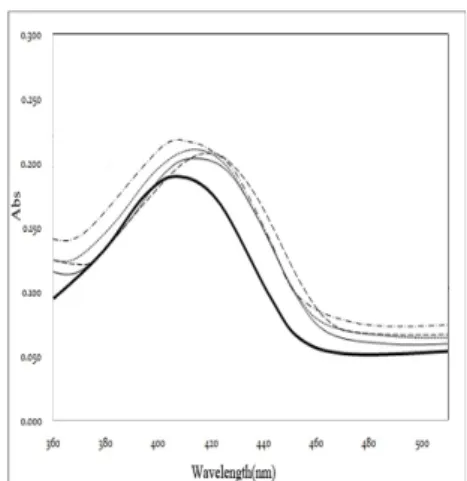
Fig. 6. Absorbance spectra of 6,7-dimethyl-8-ribityllumazine, free or in complex with variants of N-terminal domain of lumazine protein. Thick solid line, free lumazine (30 µM); Thin solid line, N-LumP 118 wt; Dotted line, N-LumP 118 V41W; Dashed line, N-LumP 118 S48W; Dash-dotted line, N-LumP 118 T50W. All protein concentrations were adjusted to 28 µM.
purified protein was eluted with imidazole of concentration 20 mM and 50 mM, respectively. In each step of protein elution, the eluent was analyzed for the presence of protein by SDS-PAGE analysis.
From the procedure above, the proteins were separated and purified by using buffer solutions at different pH conditions.
Undesirable proteins from the sonicated sample were washed off using a buffer of pH 6.3 whiles elution of protein of interest was done at pH 5.9 and pH 4.5. It was observed that some of the protein of interest was present in each of the above mentioned steps. After confirming the separated and purified proteins on SDS-PAGE (Fig. 5), they were emptied into a dialysis bag containing 50 mM NaH2PO4 5 L buffer and dialysed at 4℃ overnight. The proteins obtained after the dialysis step, were further concentrated to 1 ml and stored at 4℃.
The quantification of proteins was tested by using QubitTM from Invitrogen. To determine the protein concentration precisely, the absorption and literatures(Illarionov et al., 1994;
Lee et al., 2007) value of molar extinction coefficient (ε280
=5,800 M-1cm-1) were also counted. To remove the lumazine ligand bound to the proteins during purifications, the solutions containing 100 mM phosphate, pH 7.0, and the N-LumP proteins were dialyzed against 100 mM phosphate pH 7.0, containing 4 mM dithiothreitol and 6 M urea. For refolding, the apoprotein was dialyzed overnight against 100 mM phosphate, pH 7.0 containing 0.3 mM dithiothreitol.
As the analysis of spectroscopic properties of wild type and mutant of N-LumP, the possibility of binding of N-terminal
lumazine protein (N-LumP 118) to lumazine ligand was investigated by measuring the absorbance from the protein and ligand. The protein and its ligand lumazine were mixed together at a concentration ratio of 1:5, after which the resultant mixture was evenly shaken and dialyzed in a dialysis bag at 4℃
for 8 h. The dialysis bag contained 500 ml of 50 mM NaH2PO4. The absorbance spectra of the proteins were analyzed at a wavelength of range spanning 250 nm to 650 nm using Agilent 8453 Spectrophotometer.
As shown in Fig. 6, the following solutions 27 μM N-LumP 118 wt and N-LumP S48W, T50W, and A66W incubated with lumazine recorded a maximum peak around at 410 nm. These results indicate that wild type and mutants N-terminal lumazine proteins bind to the lumazine ligand. Comparing to the absorbance peak of free lumazine ligand, the λmax (maximum absorption) of lumazine (407 nm) generally shifted over a longer wavelength upon binding of ligands to N-LumP (409-415 nm). Similarly, the accurate ratio of binding was determined based on the value of maximum absorption and literaturevalue of molar extinction coefficient of lumazine ligand (ε408=10,700 M-1cm-1). The value of 28 μM of lumazine ligand was obtained from the calculation, suggesting that the molar ratio of binding of N-LumP protein to lumazine was 1:1.
Fluorescence intensities are also measured using FP-750 series of Fluorescence Spectrometer (JASCO). The emission spectra of the fluorescent ligand bound proteins at 407 nm of fixed excitation were shown to maximum fluorescence intensity at 490 nm region (data not shown). The following results of relative fluorescence intensities were recorded: free lumazine 1.0; N-LumP 118 wt, 1.10; N-LumP 118 V41W,
(A) (B)
(C) (D)
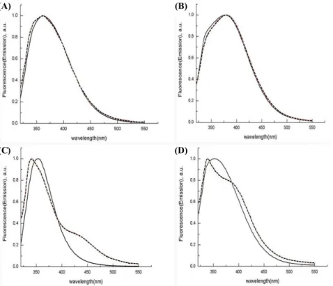
Fig. 7. Fluorescence emission spectra of wt and mutants N-LumP 118 (solid line) and N-LumP in the presence of 1 M urea (dashed line).
Excitation at 295 nm. (A) N-LumP 118 wt, (B) N-LumP 118 S48W, (C) N-LumP 118 T50W, (D) N-LumP 118 A66W. The concentration of each protein is 5 μM, respectively.
0.75; N-LumP 118 S48W, 0.90; N-LumP 118 T50W, 0.90;
N-LumP 118 A66W, 0.75. Considering to the previous result that the fluorescence intensity was quenched to 10% by binding N-terminal domain of riboflavin synthase (Lee et al., 2007), it can be suggested that all mutants N-LumP still possess the capability of fluorescent character. The observations, slight decreasing fluorescence intensities of N-LumP tryptophan mutants comparing to wild type, are in agreement with the fact that the artificial introduced aromatic acid residue (tryptophan) are close to protein-bound fluorophore. From these results, fluorescence quenching could be due to resonance transfer between the lumazine ligand and aromatic side chain.
The mutant proteins contain only one tryptophan amino acid residue. Therefore, it was also tested emission spectra arose from the excitation of the fluorescent tryptophan amino acids by the fixed at 295 nm (Fig. 7). Each tryptophan containing mutant proteins (N-LumP 118 V41W, S48W, and T50W) shows different pattern due to the specific environment around each tryptophan amino acid residue. The fluorescence emission peak of each purified protein were also changed quite differently by adding 1 M urea, indicated that the environmental conditions leading to urea denaturation are peculiar among these proteins (Fig. 7). From these results, it can be raised the possibility that these proteins can be used as a probe for the
detection of conformational change by its fluorescent character.
In summary, generating minimal size of lumazine protein with possessing fluorescent characteristic, wild type N-terminal half of lumazine protein as well as variant proteins mutated at putative binding sites of amino acids with tryptophan were constructed by PCR and site directed mutagenesis. The proteins were purified by 6X-His tagging affinity system to test the binding properties of the lumazine ligand as well as the fluorescent characteristics. These proteins will be helpful for further studies to deduce the three dimensional structure such as distance and orientations between protein and lumazine ligand as well as the binding modes of N-terminal domain of lumazine protein by using fluorescence character of tryptophan amino acid and of fluorescent ligand.
적 요
루마진 단백질은 발광 세균인 Photobacterium 종에서 추출된 형광성 단백질이다. 형광성을 지닌 최소 크기의 Photobacterium leiognathi 야생형 아미노-말단 도메인 루마진 단백질(N-terminal domain of lumazine protein 118 wt)과 여러 영역에 tryptophan을 생성시킨 돌연변이 단백질들(N-LumP 118 V41W, S48W, T50W, D64W, A66W)을 코드하는 유전자들을 위치 지정 돌연변이(Site Directed Mutagenesis)와 중합효소 연쇄 반응(Polymerase Chain
Reaction)을 통해 제조하였다. 위의 유전자들이 포함된 재조합 플라스미드를 대장균에 형질 전환시켜 과발현시키는 최적의 조 건을 찾았으며, 발현된 야생형 및 돌연변이 아미노-말단 영역 루 마진 단백질을 6X-His tag system을 이용하여 정제 하였다. 흡 광 및 형광 분광광도계를 이용한 실험 결과 이들 단백질들은 리 간드인 6,7-dimethyl-8-ribityllumazine과 결합하여 형광성을 보 유함을 보였다. 따라서 이들은 형광성을 지니게 되는 최소 크기 의 루마진 단백질일 뿐만 아니라 형광성을 지닌 아미노산인 tryptophan이 여러 위치에 유일하게 존재함으로써 배향성 및 거 리 등의 단백질의 구조 및 결합에 관한 심도 있는 연구에 탐침자 로써 유용하게 활용 될 수 있을 것이다.
Acknowledgements
This work was partly supported by the Exchanging Program (NRF 617-2010-C00001) and by Basic Science Research Program (NRF-2010-0022701) through the National Research Foundation of Korea (NRF) funded by the Minister of Education, Science and Technology. The authors also thank to Prof. Markus Fischer for his providing the space and equipment during authors visiting studies at University of Hamburg.
References
Campbell, A.K. 1989. Living light: biochemistry, function, and biomedical applications. Essays Biochem. 24, 41–76.
Chatwell, L., Illarionova, V., Illarionov, B., Eisenreich, W., Huber, R., Skerra, A., Bacher, A., and Fischer, M. 2008. Structure of lumazine protein, an optical transponder of luminescent bacteria. J. Mol. Biol.
382, 44–55.
Fischer, M. and Bacher, A. 2005. Biosynthesis of flavocoenzymes. Nat.
Prod. Rep. 22, 324–350.
Gast, R. and Lee, J. 1978. Isolation of in vivo emitter in bacterial bioluminescence. Proc. Natl. Acad. Sci. USA 75, 833–837.
Hastings, J.W. and Nealson, K.H. 1977. Bacterial bioluminescence.
Annu. Rev. Microbiol. 31, 549–545.
Herring, P.J. 1987. Systematic distribution of bioluminescence in living organisms. J. Biolumin. Chemilumin. 1, 147–163.
Illarionov, B., Eisenreich, W., Wirth, M., Lee,C.Y., Woo, Y.E., Bacher, A., and Fischer, M. 2007. Lumazine proteins from Photobacteria
: localization of the single ligand binding site to the N-terminal domain. Biol. Chem. 388, 1313–1323.
Illarionov, B., Illarionova, V., Lee, J., van Dongen, W., and Vervoort, J.
1994. Expression and properties of the recombination lumazine (riboflavin) protein from Photobacterium leiognathi. Biochim.
Biophys. Acta 1201, 251–258.
Koka, P. and Lee, J. 1979. Separation and structure of prosthetic group of the blue fluorescent protein from the bioluminescent bacterium Photobacterium phosphoreum. Proc. Natl. Acad Sci. USA 76, 3068–
3072.
Lee, C.Y., Illarionov, B., Woo, Y.E., Kemter, K., Kim, R.R., Eberhardt, S., Cushman, M., Eisenreich, W., Fischer, M., and Bacher, A. 2007.
Ligand binding properties of the N-terminal domain of riboflavin synthase from Escherichia coli. J. Biochem. Mol. Biol. 40, 239–246.
Lee, C.Y. and Meighen, E.A. 2000. The expression and DNA sequence of the gene coding for the lux specific fatty acyl-CoA reductase from Photobacterium phosphoreum. J. Microbiol. 38, 80–87.
Lee, C.Y., O'Kane, D.J., and Meighen, E.A. 1994. Riboflavin synthesis genes are linked with the lux operon of Photobacterium phosphoreum. J. Bacteriol. 176, 2100–2104.
Meighen, E.A. 1988. Enzymes and genes from the lux operons of bioluminescent bacteria. Annu. Rev. Microbiol. 42, 151–179.
Meighen, E.A. 1991. Molecular biology of bacterial bioluminescence.
Microbiol. Rev. 55, 123–142.
Meighen, E.A. 1994. Genetics of bacterial of bioluminescence. Annu.
Rev. Genet. 28, 117–139.
O'Kane, D.J., Karle, A.J., and Lee, J. 1985. Purification of lumazine protein from Photobacterium leiognathi and Photobacterium phosphoreum: bioluminescence properties. Biochemistry 24, 1454–
1455.
O'Kane, D.J. and Lee, J. 1985. Chemical characterization of lumazine protein from Photobacterium leiognathi: Comparison with lumazine protein from Photobacterium phosphoreum.
Biochemistry 24, 1467–1475.
O'Kane, D.J., Woodward, B., Lee, J., and Prasher, D.C. 1991. Borrowed proteins in bacterial bioluminescence. Proc. Natl. Acad. Sci. USA 88, 1100–1104.
Palmer, I. and Wingfield, P.T. 1995. Preparation and extraction of insoluble (inclusion-body) proteins from Escherichia coli. pp. 6.3.1.
–6.3.15. In Coligan, J.E., Dunn, B.M., Ploegh, H.L., Speicher, D.W., and Wingfield, P.T. (eds.). Current protocols in protein science, Vol.
1, John Wiley and Sons, New York, USA.
Small, E.D., Koka, P., and Lee, J. 1980. Lmazine protein from the bioluminescent bacterium Photobacterium phosphoreum. Purification and characterization. J. Biol. Chem. 255, 8804–8810.