loid Cells 1 Protein의 진단적 유용성
이화여자대학교 의과대학 내과학교실 심윤수, 이진화, 천은미, 장중현
Diagnostic Utility of Pleural Fluid Soluble Triggering Receptor Expressed on Myeloid Cells 1 Protein in Patients with Exudative Pleural Effusion
Yun Su Sim, M.D., Jin Hwa Lee, M.D., Eun Mi Chun, M.D, Jung Hyun Chang, M.D.
Department of Internal Medicine, Ewha Womans University College of Medicine, Seoul, Korea
Background: Triggering receptor expressed on myeloid cells 1 protein (TREM-1) is a cell surface molecule expressed on neutrophils and monocytes, and it plays an important role in myeloid cell-activated inflammatory response. The aim of this study was to investigate the diagnostic efficiency of soluble (s) TREM-1 in the patients who had pleural effusion from various causes.
Methods: Forty-five patients with exudative pleural effusion were included in this study. The level of sTREM-1 was measured in both the serum and pleural fluids by immunoblot assay with using human-sTREM-1 antibody.
Results: The pleural fluid sTREM-1 was significantly different in the three groups of exudative pleural effusion (p=0.011). Particularly, the patients with parapneumonic effusion were found to have significantly higher pleural fluid levels of sTREM-1 than patients with tuberculous (p<0.05) and malignant effusion, respectively (p<0.05). However, the serum sTREM-1 did not show a significant difference in the three groups. In order to evaluate the diagnostic utility of pleural fluid sTREM-1, the receiver operating characteristic (ROC) curve was constructed and the area under the curve (AUC) was 0.818 (p=0.001). Using a cutoff value of 103.5 pg/mL for the pleural fluid sTREM-1, the sensitivity and specificity were 73% and 81%, respectively, for differentiating parapneumonic effusion from tuberculous or malignant effusions.
Conclusion: Pleural fluid sTREM-1 can be an additional marker for making the differential diagnosis of pleural effusion. (Tuberc Respir Dis 2007; 62: 499-505)
Key words: Triggering receptor expressed on myeloid cells 1 protein, Pleural effusion, Exudates.
Address for correspondence: Jin Hwa Lee, M.D.
Department of Internal Medicine, Ewha Womans University, College of Medicine, 911-1 Mok-dong Yancheon-gu, Seoul 158-710, Korea
Phone: 82-2-2650-6007, Fax: 82-2-2655-2076 E-mail: jinhwalee@ewha.ac.kr
Received: Mar. 26. 2007 Accepted: May. 28. 2007
서 론
흉수의 원인질환을 진단하기 위해서는 임상양상과 영상의학과 촬영 결과와 함께 흉수 분석 결과가 중요 하다. 흉수천자로 얻은 흉수의 생화학적 분석 결과를 토대로 우선 여출성과 삼출성 흉수를 구분한다. 삼출 성 흉수는 흉막의 염증이나 감염, 손상 또는 림프관의 폐색으로 발생하며, 폐렴, 악성신생물, 결핵, 폐경색
등이 주요 원인질환으로 감별진단이 용이하지 않은 경우가 종종 있다1,2.
Triggering receptor expressed on myeloid cells-1(TREM-1)은 최근 발견된 세포표면수용체의 일종으로, 주로 과립백혈구, 단핵구, 대식세포 표면에 존재하는 30 kDa의 당단백질로 이루어진 면역글로블 린이며, 활성화된 대식세포에서 떨어져 나와 체액에 서 수용성 상태로 발견된다3-5. TREM-1은 결합단백 질인 DAP12의 도움으로 활성화되며 Toll-like receptor 의 신호전달에 상승작용을 일으켜 interleukin-10, tumor necrosis factor-α 등 여러 염증전달물질을 증 폭시키는 역할을 한다. TREM-1은 주로 세포외 세균 이나 진균의 세포표면 구성성분에 의해 발현이 증가 되지만, 결핵균은 TREM-1의 발현을 증가시키지 않 는 것으로 알려져 있다3-6. 또한, 건선, 궤양성대장염,
혈관염 등의 비감염성 염증질환이나 결핵이나 이물질 로 비롯된 육아종성 병변에 의한 TREM-1의 발현은 미미한 것으로 보고되고 있다3,4,7.
최근에 기계환기 중인 환자의 기관지폐포세척액에 서 측정한 soluble (s) TREM-1의 농도가 기계환기관 련폐렴의 강력한 독립적 예측인자가 되고8 혈청 sTREM-1이 패혈증 환자의 임상 경과와 관련이 있다 고 보고된 바 있다9.
저자들은 삼출성흉수를 가진 환자의 혈청과 흉수에 서 sTREM-1을 측정하여, sTREM-1이 흉수의 원인 질환을 감별하는데 유용한 지표로 사용될 수 있는지 알아보고자 하였다.
대상 및 방법 1. 대 상
2003년 3월부터 2006년 12월까지 이화여자대학교 의과대학부속 목동병원 호흡기내과에 입원한 삼출성 흉수 환자 45명을 대상으로 하였다. 삼출성흉수는 Light의 진단기준에 맞추어 혈청에 대한 흉수의 단백 질의 비가 0.5를 초과하거나 혈청에 대한 흉수의 lactic acid dehydrogenase(LD)의 비가 0.6을 초과하 거나 흉수의 LD가 혈청 LD의 정상 상한가의 2/3을 초과하는 경우로 정의하였다1. 악성흉수는 흉수의 세 포학적 검사나 흉막 생검에서 악성세포가 증명된 경 우로 정의하였고, 결핵성흉수는 흉수의 항산균 도말 또는 결핵균 배양이 양성이거나 흉막 생검 결과 치즈 양괴사를 동반한 육아종이 발견되거나 림프구 우세 흉수이면서 흉수 adenosine deaminase(ADA)가 40 U/L 이상으로 상승하여 있으면서 항결핵제 투약 후 호전된 경우로 정의하였다. 부폐렴성흉수는 폐렴에 동반되거나 흉수 그람염색 도말에서 균을 확인하였거 나 흉수 배양 결과 균이 동정된 경우이면서 항생제 투 약 후 임상 경과가 호전된 경우로 정의하였다. 45명의 삼출성흉수 환자 중에 2명에서는 흉수의 원인이 정확 히 밝혀지지 않았고, 2명은 악성 흉수가 의심되었지만 조직학적 진단이 되지 않아 제외하였다. 41명의 환자
중에 결핵성흉수 환자는 18명, 부폐렴성흉수 환자는 15명, 악성흉수 환자는 8명이었다.
2. 방 법
모든 환자에게 치료 전에 같은 날 혈액과 흉수를 채취하여, 각각 원심분리 후 상청액을 sTREM-1을 측정하기 전까지 -80℃에서 냉동 보관하였다. sTREM-1 은 상품화된 human sTREM-1 ELISA kit (Quantine, R&D system, Minneapolis, MN, USA)를 이용하여 공급자의 권고에 따라 human TREM-1 항체를 사용 하여 면역점적법(immunoblot assay)으로 측정하였 다. sTREM-1의 측정은 각 검체의 질환을 모르는 상 태에서 시행되었다.
모든 흉수에서 포도당, 단백질, LD, ADA, cari- noembryonic antigen(CEA)을 측정하였고, 적혈구와 백혈구 수 및 백혈구 중 림프구와 중성구 비율을 측정 하였으며, 그람염색, 항산균염색, 및 배양, 그리고 세 포학적 검사를 시행하였다. 임상양상, 방사선촬영 및 흉수 분석으로 진단이 어려운 일부 환자에서 흉막 생 검을 시행하였다. 또한 모든 환자의 혈액에서 혈액학 적 지표로 백혈구 수, 적혈구침강속도(erythrocyte sedimentation rate, ESR), 혈청 C-reactive protein (CRP), LD, 단백질, 알부민, CEA를 측정하였다.
3. 통계분석
모든 자료는 평균과 표준편차 또는 빈도로 표시하 였고, 통계분석은 SPSS 12.0(SPSS Inc, Chicago, IL, USA) 프로그램을 이용하였다. 세 질환군 사이에 평 균을 비교하기 위하여 일원배치분산분석(one-way ANOVA)과 post-hoc test of Scheffe를 적용하였고, 범주형 변수를 비교하기 위하여 카이제곱검정을 이용 하였다. 흉수 sTREM-1을 이용한 부폐렴성흉수의 진 단율을 평가하기 위하여, receiver-operating charac- teristic(ROC) 곡선을 구한 후, 감수성과 특이성을 최 대로 할 수 있는 cutoff 값을 정하였다. p<0.05인 경우 를 통계적으로 유의한 것으로 해석하였다.
Tuberculous PE (n=18)
Parapneumonic PE (n=15)
Malignant PE (n=8)
Male 9 (13%) 13 (87%) 4 (50%)
Age* 42±19
†49±14
†68±10
Associated diseases Diabetes mellitus Hypertension
2 (11%) 2 (11%)
1 (7%) 2 (13%)
2 (25%) 3 (37%) Smoking
Smoker Pack-years
10 (56%) 32±29
10 (77%) 28±15
5 (73%) 43±25 Data are mean±SD or number (frequency).
PE: pleural effusion.
*p<0.01 by one-way ANOVA, †p<0.05 vs. malignant PE by multiple comparison using post-hoc test of Scheffe.
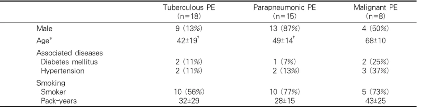
Table 1. Clinical characteristics in patients with pleural effusion
Tuberculous PE (n=18)
Parapneumonic PE (n=15)
Malignant PE (n=8) WBC (x1000/mm
3)
CRP (mg/dL)
*ESR (mm/hr) Protein (g/dL) Albumin (g/dL) LD (IU/L)
†CEA (ng/mL)
*6.7±2.2 9.1±8.9
‡32±16 6,4±0.6 3.4±0.4 467±100 2.5±1.5
§10.7±5.7 20.2±15.7
54±39 6.6±0.4 3.1±0.5 378±148
§2.2±1.1
§9.7±8.0 6.5±7.5 43±28 6.0±1.0 3.4±0.5 710±291 28.5±35.3 Data are mean±SD.
PE: pleural effusion; WBC: white blood cell count: CRP: C-reactive protein: ESR: erythrocyte sedimentation rate:
LD: lactic acid dehydrogenase: CEA, carcinoembryonic antigen.
*p<0.05, †p<0.01 by one-way ANOVA, ‡p<0.05 vs. parapneumonic PE, §p<0.05 vs. malignant PE by multiple comparison using post-hoc test of Scheffe.
Table 2. Laboratory characteristics in patients with pleural effusion
결 과흉수의 원인 질환에 따라 나누어 비교하였을 때, 평 균 나이가 유의한 차이를 보였는데(p=0.002), 악성흉 수 환자가 결핵성흉수와(p<0.05) 부폐렴성흉수 환자 보다 유의하게 나이가 많았다(p<0.05). 성별, 동반질 환 및 흡연력에는 각 흉수 질환 간에 유의한 차이를 보이지 않았다(Table 1).
혈액검사결과 CRP가 세 질환군 사이에 유의한 차 이를 보였는데(p=0.016), 부폐렴성흉수 환자에서 혈청 CRP가 결핵성흉수보다 통계학적으로 유의하게 높았 다(p<0.05). 혈청 LD(p=0.002)와 CEA도 세 질환군 사 이에 유의한 차이가 있었는데(p=0.011), 악성흉수 환 자의 LD가 부폐렴성흉수 환자보다 유의하게 높았고 (p<0.05), 악성흉수 환자의 혈청 CEA가 결핵성흉수
와(p<0.05) 부폐렴성흉수 환자보다 유의하게 높았다 (p<0.05)(Table 2).
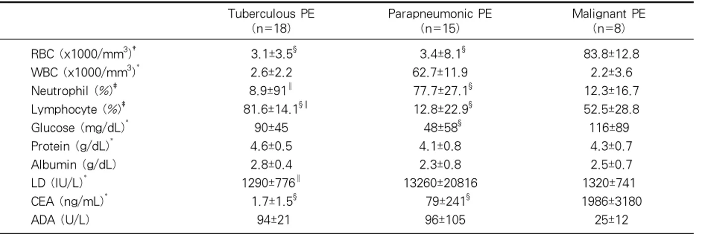
흉수 분석 결과 적혈구 수와(p=0.003) 백혈구 수가 세 질환군 사이에 유의한 차이를 보였는데(p=0.049), 특히 악성흉수의 적혈구 수가 결핵성흉수와(p<0.05) 부폐렴성흉수보다 유의하게 많았다(p<0.05). 흉수의 백혈구 중 중성구와(p<0.001) 림프구가 차지하는 비 율이 세 질환 사이에 유의한 차이를 보였는데 (p<0.001), 부폐렴성흉수의 중성구 비율이 결핵성흉 수와(p<0.05) 악성흉수보다 유의하게 높았고(p<0.05), 림프구 비율은 결핵성흉수, 악성흉수, 부폐렴성흉수 순으로 유의하게 낮아졌다(p<0.05). 흉수의 포도당도 세 군 사이에 유의한 차이가 있었으며(p=0.03), 부폐 렴성흉수의 포도당 농도가 악성흉수보다 유의하게 낮 았다(p<0.05). 흉수 단백질과(p=0.035) LD도 세 군 사
Tuberculous PE (n=18)
Parapneumonic PE (n=15)
Malignant PE (n=8) RBC (x1000/mm
3)
†WBC (x1000/mm
3)
*Neutrophil (%)
‡Lymphocyte (%)
‡Glucose (mg/dL)
*Protein (g/dL)
*Albumin (g/dL) LD (IU/L)
*CEA (ng/mL)
*ADA (U/L)
3.1±3.5
§2.6±2.2 8.9±91
║81.6±14.1
§║90±45 4.6±0.5 2.8±0.4 1290±776
║1.7±1.5
§94±21
3.4±8.1
§62.7±11.9 77.7±27.1
§12.8±22.9
§48±58
§4.1±0.8 2.3±0.8 13260±20816
79±241
§96±105
83.8±12.8 2.2±3.6 12.3±16.7 52.5±28.8 116±89 4.3±0.7 2.5±0.7 1320±741 1986±3180
25±12 Data are mean±SD.
PE: pleural effusion: RBC: red blood cell count: WBC: white blood cell count: LD: lactic acid dehydrogenase:
CEA: carcinoembryonic antigen ADA, adenosine deaminase.
*p<0.05, †p<0.01, ‡p<0.001 by one-way ANOVA, §p<0.05 vs. malignant PE, ║p<0.05 vs. parapneumonic PE by multiple comparison using post-hoc test of Scheffe.
Table 3. Pleural fluid analysis in patients with pleural effusion
Tuberculous Parapneumonic Malignant
pleural fluid sTREM-1
1200
1000
800
600
400
200
0
p<0.05 p<0.05
Tuberculous Parapneumonic Malignant
pleural fluid sTREM-1
1200
1000
800
600
400
200
0
p<0.05 p<0.05
Figure 1. sTREM-1 in the pleural fluid of patients with tuberculous (n=18), parapneumonic (n=15), and malignant pleural effusions (n=8). Horizontal bars represent mean values.
Tuberculous PE (n=18) Parapneumonic PE (n=15) Malignant PE (n=8)
Serum sTREM-1 (pg/mL) 57±150 19±6 20±7
Pleural fluid sTREM-1 (pg/mL)
*117±138
†329±325 72±72
†Data are mean±SD.
PE: pleural effusion.
*p<0.05 by one-way ANOVA, †p<0.05 vs. parapneumonic PE by multiple comparison using post-hoc test of Scheffe.
Table 4. Soluble triggering receptor expressed on myeloid cells-1 (sTREM-1) in serum and pleural fluid of patients with pleural effusion
이에 유의한 차이를 보였으며(p=0.025), 부폐렴성흉수 의 LD가 결핵성흉수보다 유의하게 높았다(p<0.05).
흉수의 CEA도 세 군 사이에 유의한 차이를 보였는데 (p=0.012), 악성흉수의 CEA가 결핵성흉수와(p<0.05) 부폐렴성흉수와 비교하여 유의하게 높았다(p<0.05).
흉수 ADA는 세 군 사이에 유의한 차이가 없었다 (Table 3).
혈청 sTREM-1은 세 질환군에서 유의한 차이가 없 었으나, 흉수 sTREM-1은 통계학적으로 유의한 차이 를 보였다(p=0.011). 특히 부폐렴성흉수의 sTREM-1 이 결핵성흉수와(p<0.05) 악성흉수보다 유의하게 높 았다(p<0.05)(Table 4, Figure 1).
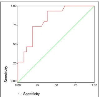
부폐렴성흉수를 진단하는 데 있어서 흉수 sTREM-1 의 유용성을 평가하는 ROC 곡선의 곡선밑면적(area under the curve, AUC)은 0.818이었다(p=0.001)
1 - Specificity
1.00 .75
.50 .25
0.00
Se n s itivity
1.00
.75
.50
.25
0.00
Figure 2. ROC curve for pleural fluid sTREM-1. The area under curve of pleural fluid sTREM-1 was 0.818(p=0.001).
(Figure 2). 흉수 sTREM-1의 cutoff 값을 103.5 pg/mL로 하였을 때, 부폐렴성흉수의 진단에 대한 민 감도가 73%, 특이도가 81%이었다.
고 찰
흉수는 흔하게 부딪치는 임상양상이나 원인질환의 감별이 어려운 경우가 있다. 흉수의 생화학적, 세포학 적 분석 결과가 감별진단에 도움을 주지만, 어느 한가 지 자료만으로 확실한 진단을 내리기는 어렵다. 흉수 의 감별진단에는 임상특징과 방사선촬영 결과 및 흉 수 분석 결과에 대한 종합적인 판단이 필요하다. 간혹 진단을 내리지 못하는 경우, 침습적 검사로 흉막 생검 을 추가로 시행할 수 있으나, 맹검적 흉막 생검은 추 가적 진단율이 낮다고 알려져 있다10. 결국 정확한 진 단을 위해서는 흉강경을 이용한 흉막 생검을 하여야 하나11, 검사에 따른 합병증과 위험이 증가한다. 따라 서, 비침습적인 진단방법으로서 흉수에 대한 새로운 생화학적 표지물질이 계속 개발되고 있다.
본 연구에서는 삼출성흉수를 일으키는 대표적인 질 환인 결핵, 폐렴, 악성종양 환자를 대상으로 혈청과 흉수에서 sTREM-1을 측정한 결과, 부폐렴성흉수의
sTREM-1이 결핵성흉수나 악성흉수보다 유의하게 높았다. 이는 지금까지 흉수 sTREM-1을 측정하여 보고한 유일한 연구인 Liu 등12의 결과와 일치하는 것 으로, Liu 등은 감염성흉수의 sTREM-1이 여출액이 나 악성흉수보다 높고, 다시 감염성흉수를 부폐렴성 흉수와 결핵성흉수로 나누어 비교하였을 때 부폐렴성 흉수의 sTREM-1이 결핵성흉수보다 높다고 보고하 였다. 흉수의 sTREM-1이 결핵성흉수에서 비교적 낮 게 측정되는 이유는, 결핵성흉수가 흉막에 존재하는 결핵균 항원에 의한 지연성과민반응으로 생성되며 림 프구가 주로 작용하는 염증 반응이기 때문으로 생각
된다13-15. 결핵성흉수의 실제 결핵균 배양률은 30%
이하로 알려져 있다13,14. TREM-1은 단핵구, 중성구, 대식세포 표면에 존재하며, 흔히 세포 외에 존재하는 세균이나 진균에 의해 발현되므로, 세포 내에 존재하 는 결핵균에 의해 주로 림프구의 증식을 초래하는 결 핵에서는 TREM-1의 발현이 작을 수 밖에 없고, 비 감염성질환인 여출액과 악성흉수에서도 TREM-1의 증가가 미미할 것이다3-7.
흉수 sTREM-1이 세 가지 흉수질환 사이에 통계적 으로 유의한 차이를 보이는 반면, 혈청 sTREM-1은 서로 유의한 차이가 없었다. Richeldi 등16이 결핵, 간 질성폐렴, 지역사회획득폐렴 환자를 대상으로 한 연 구에서도, 지역사회획득폐렴 환자의 기관지폐포세척 술액에서 sTREM-1의 농도가 결핵이나 간질성폐렴 환자보다 높았지만, 혈청 sTREM-1은 유의한 차이를 보이지 않았다. 이는 세균 감염에 의한 TREM-1의 발현이 주로 감염부위에 상주하는 대식세포와 감염부 위로 소집된 중성구에 의해 이루어지기 때문인 것으 로 생각된다. 한편, 패혈증 환자를 대상으로 한 다른 연구에서는, 비감염성쇼크 환자나 대조군에 비해 패 혈증 환자에서 측정한 혈청 sTREM-1이 유의하게 높 은 수치를 보였다9,17-19. 결국 감염이 국소적으로 조절 되는 상태에서는 혈청 sTREM-1이 증가하지 않지만, 전신적인 염증반응을 일으키게 되는 패혈증에서는 혈 청 sTREM-1이 증가하는 것이다.
본 연구에서 평균 연령은 악성흉수 환자에서 가장 높았는데, 흉수 환자에서 연령이 높은 경우 악성질환 의 가능성을 염두에 두어야 한다는 것은 잘 알려져 있
다. 또한, bloody 흉수인 경우 악성흉수일 가능성이 높다는 것도 잘 알려진 사실인데, 본 연구의 흉수 분 석 결과에서도 악성흉수에서 적혈구 수가 가장 높았 다. 부폐렴성흉수에서 백혈구 중 중성구 비율이 유의 하게 높은데 반해, 림프구 비율은 결핵성흉수와 악성 흉수에서 더 높았는데, 이는 결핵성흉수와 악성흉수 에서 대체로 림프구가 50% 이상이라는 보고와 일치
한다20,21. 흉수의 림프구나 중성구의 비율은 감별진단
의 폭을 줄이는 데 도움이 되지만, 원인질환으로서 결 핵 또는 폐렴을 특이적으로 진단하는 기준은 되지 못 한다. 그 외 흉수의 포도당, 단백질, LD, 그리고 CEA 도 흉수의 원인 질환에 따라 차이를 보였는데, 흉수 포도당이 60 mg/dL 이하일 경우 합병증이 동반된 부 폐렴성흉수일 가능성이 높다고 알려져 있다22. 흉수 LD는 흉수의 염증정도에 비례하고11, 흉수 CEA는 악 성흉수의 진단에 대해 82%의 민감도와 94%의 특이 도를 가진다고 보고된 바 있다23. 흉수 ADA는 40 U/L 이상에서 결핵성흉수의 가능성이 높다고 잘 알려져 있지만24, 본 연구에서는 일부 부폐렴성흉수에서도 높 은 농도로 측정되어 두 가지 질환에서 평균값이 통계 적으로 유의한 차이가 없었으며, ROC 곡선을 통한 분 석 결과 결핵성흉수의 진단에 민감도는 매우 높으나 특이도가 좀 낮았다.
본 연구는 대상 환자 수가 작고 원인질환이 세 가 지로 국한되어 다양한 원인질환 별로 흉수의 sTREM-1을 분석하지 못하였다는 제한점이 있다. 그 러나, 부폐렴성흉수를 진단하는데 흉수 sTREM-1의 민감도와 특이도가 비교적 높아 흉수의 원인질환을 감별하는 데 추가적인 자료를 제공할 수 있을 것으로 기대된다. 특히 임상양상이 잘 구별되지 않으면서 흉 수의 림프구와 호중구의 비율이 비슷하고 흉수의 ADA가 높은 경우, 추가로 흉수 sTREM-1을 측정해 본다면, 결핵성흉수와 부폐렴성흉수를 감별하는 데 도움이 될 것이다. 앞으로 더 많은, 다양한 흉수 환자 를 대상으로 sTREM-1을 측정해 본다면, 흉수의 감 별진단에 대한 sTREM-1의 유용성을 더 정확히 평가 할 수 있겠다.
요 약
배 경: TREM-1은 중성구, 단핵구, 대식세포 표면 에 존재하는 세포표면수용체로, 세균에 의해 그 발현 이 증가하여 여러 염증전달물질을 증폭시키는 역할을 한다. 저자들은 삼출성흉수를 가진 환자의 혈청과 흉 수에서 soluble (s) TREM-1을 측정하여 흉수의 원인 진단에 대한 유용성을 알아보고자 하였다.
방 법: 2003년 3월부터 2006년 12월까지 삼출성흉 수로 입원한 환자 45명을 대상으로 하여, 혈청과 흉수 에서 human sTREM-1 항체를 사용하여 면역점적법 (immunoblot assay)으로 sTREM-1을 측정하였다.
원인질환에 따라 결핵성, 부폐렴성, 악성흉수로 나누 어 비교하였다.
결 과: 혈청 sTREM-1은 원인질환 별로 유의한 차 이를 보이지 않았으나, 흉수 sTREM-1은 원인질환 별로 유의한 차이를 보였으며(p=0.011), 특히 부폐렴 성흉수의 sTREM-1이 결핵성흉수와(p<0.05) 악성흉 수보다 유의하게 높았다(p<0.05). 부폐렴성흉수를 진 단하는 데 흉수 sTREM-1의 유용성을 평가하고자 ROC 곡선을 그린 결과 곡선밑면적은 0.818이고 (p=0.001), 흉수 sTREM-1의 cutoff 값을 103.5 pg/mL로 하였을 때 민감도가 73%, 특이도가 81%이 었다.
결 론: 흉수의 sTREM-1은 삼출성흉수 중 부폐렴 성흉수를 진단하는 유용한 지표로 판단된다.
감사의 글
이 연구는 2002학년도 이화여자대학교 교내연구비 지원에 의한 연구임.
참 고 문 헌