Impact of Clinicopathologic Factors on Subclinical Central Lymph Node Metastasis in Papillary Thyroid Microcarcinoma
Bo-Yeon Kim,
1Chan-Hee Jung,
1Jae-Wook Kim,
2Seung-Won Lee,
2Chul-Hee Kim,
1Sung-Koo Kang,
1and Ji-Oh Mok
11Division of Endocrinology & Metabolism, Department of Internal Medicine and
2Department of Otorhinolaryngology-Head and Neck Surgery, Soonchunhyang University School of Medicine, Bucheon, Korea.
Received: August 24, 2011 Revised: November 22, 2011 Accepted: November 28, 2011 Corresponding author: Dr. Ji-Oh Mok, Division of Endocrinology & Metabolism, Department of Internal Medicine,
Soonchunhyang University Bucheon Hospital, 170 Jomaru-ro, Wonmi-gu,
Bucheon 420-767, Korea.
Tel: 82-32-621-5156, Fax: 82-32-621-5018 E-mail: byby815@schmc.ac.kr
∙ The authors have no financial conflicts of interest.
© Copyright:
Yonsei University College of Medicine 2012 This is an Open Access article distributed under the terms of the Creative Commons Attribution Non- Commercial License (http://creativecommons.org/
licenses/by-nc/3.0) which permits unrestricted non- commercial use, distribution, and reproduction in any medium, provided the original work is properly cited.
Purpose: We evaluated whether the clinicopathological factors of papillary thy- roid microcarcinoma (PTMC), especially tumoe size, are associated with subcini- cal central lymph node metastasis. Materials and Methods: A total of 160 pa- tients diagnosed with PTMC who underwent total thyroidectomy with bilateral central lymph node dissection were enrolled in this study. All patients were clini- cally lymph node negative PTMC. Patients were divided into 2 groups according to the size of tumor (≤5 mm vs. >5 mm). Clinicopathologic risk factors for sub- clinical central lymph node metastasis were analyzed. Results: Subclinical central lymph node metastasis was detected in 61 (38.1%). Patients with tumors ≤5 mm had a lower frequency of extrathyroidal extension, multifocality and subclinical central lymph node metastasis. On multivariate analysis, only male and tumor size
>5 mm were independent predictors of subclinical central lymph node metastasis;
age, multifocality, bilaterality, extrathyroidal extension, lymphvascular invasion and lymphocytic thyroiditis were not. Conclusion: In this study, male and tumor size >5 mm were two independent predictive factors for subclinical central lymph node metastasis in PTMC. These are easier factors to assess before surgery than other factors when planning the central lymph node dissection. However, further long-term follow-up studies are needed to confirm the prognostic significance of subclinical central lymph node metastasis in PTMC.
Key Words: Papillary thyroid microcarcinoma, tumor size, subclinical central lymph node metastasis
INTRODUCTION
In 2008, according to the National Cancer Registry data of Korea, thyroid cancer was reported as the most prevalent cancer in women.1 In particular, papillary thy- roid microcarcinoma (PTMC) is rapidly increasing.
Although PTMC generally has a highly favorable prognosis, the long term re- currence rate of PTMC has been reported as up to 10%.2 The majority of PTMC recurrence are locoregional, in the thyroid bed and in the neck lymph node.3 Cen- tral lymph node metastasis is an important risk factor of recurrence, and often is
eral dissection of bilateral paratracheal, pretracheal and prelaryngeal lymph nodes. Diagnosis of PTMC was recon- firmed by the surgical pathology for all patients. PTMC was defined as tumor 10 mm or less along its greatest di- mension, in accordance with the histologic classification of thyroid tumors by the World Health Organization.11 This study was approved by the Institutional Review Board of Soonchunhyang University Bucheon Hospital.
We carried out retrospective chart review of all patients treated at our institution for total thyroidectomy with cen- tral lymph node dissection. Various clinicopathologic prog- nostic factors described in literature such as age, gender, tu- mor size, extrathyroidal extension, multifocality, bilaterality and central lymph node dissection were reviewed. Lym- phovascular invasion and lymphocytic thyroiditis were also recorded. Patients were classified into two groups; those with PTMC tumor size ≤5 mm and those with PTMC >5 mm as done in previous studies. The extent of bilateral cen- tral neck dissection was similar between those 2 groups. In cases with the presence of more than two malignancies, multifocality was defined as multiple malignancies in one lobe and bilaterality was defined as multiple malignancies in both lobes of thyroid.
Statistical analysis
Data are presented as the mean±SD. SPSS 14.0 for Win- dows® software package (SPSS Inc., Chicago, IL, USA) was used for statistical analysis. Student t-test was used to compare the clinicopathologic factors between the tumor size ≤5 mm group and >5 mm group.
Univariate analysis was used for the assessment of the factors associated with subclinical central lymph node dis- section. Statistically significant results obtained at univariate analysis were submitted to multivariate logistic regression.
A p-value of <0.05 was considered statistically significant.
RESULTS
Of 160 patients, 19 (9.1%) were men and 141 (91.9%) were women. The mean age at initial treatment was 47 years (range, 20 to 78 years). The largest dimensions of primary tumors ranged from 2 to 10 mm, with a mean of 6.8 mm.
Subclinical central lymph node dissection was detected in 61 (38.1%) of 160 patients, with clinically node-negative PTMC. Multifocal lesions were found in 54 (33.8%) and bilateral lesions in 37 (23.1%) patients. Extrathyroidal and not detected clinically. Prevalence of the subclinical central
lymph node metastasis has been reported to be as great as 30-65% in PTMC; however, the role of routine central lymph node dissection in the treatment of PTMC remains debated.4,5 Routine prophylactic central lymph node dissec- tion may be overtreatment in many patients with PTMC.
Therefore, identification of predictive factors associated with subclinical central lymph node metastasis may help tai- lor appropriate surgical strategies for patients with PTMC.
In addition, endoscopic thyroidectomy is rapidly increas- ing in Korea for preoperatively diagnosed low-risk PTMC due to the attractive advantage of good cosmetic results in Korea.6,7 However, endoscopic thyroidectomy has some lim- itations such as a difficulty in achieving sufficient lymphade- nectomy and a narrower operative space.6,7 Therefore, know- ing about the absence or presence of subclinical central lymph node metastasis can help physicans choose an ap- propriate method and extent of surgery.
Recently, tumors ≤5 mm along their great dimension in PTMC are increasing as a result of earlier diagnosis by high resolution ultrasonographic examination and skillful fine needle aspiration biopsy as part of the routine health check- ups.8 Results of studies on whether the tumor size is an in- dependent predictive factor for the presence of subclinical central lymph node metastasis or not are not consistent.9,10
We thought, therefore, that if there was any difference found in subclinical central lymph node metastasis on the basis of clinicopathologic features, especially the tumor size of PTMC, we could administer more aggressive treatment to the group of patients who have more aggressive PTMC.
Therefore, the aim of our study was to find out which factors are associated with subclinical central lymph node metastasis of PTMC, and also to test whether the diameter of the carci- noma is a significant prognostic factor for the lymph node metastasis.
MATERIALS AND METHODS
A total of 160 patients diagnosed with PTMC who under- went total thyroidectomy with bilateral central lymph node metastasis between January 2006 and July 2010 were en- rolled in this study. Of the 160 patients, 31 (19.4%) patients underwent endoscopic surgery. Based on findings from pre- operative ultrasonography and fine needle aspiration biop- sy, all patients were clinically lymph node negative PTMC.
Bilteral central lymph node dissection was defined as bilat-
with PTMC >5 mm. Patients with tumors ≤5 mm had a lower frequency of extrathyroidal extension than the pa- tients with tumors >5 mm (31.7% vs. 58.0%, p=0.003).
Also, patients with tumors ≤5 mm had a lower frequency of multifocality than patients with tumors >5 mm (22% vs.
37.8%, p=0.046). All patients with lymphovascular inva- sion had tumors >5 mm. However, two groups did not have significant differences in age, bilaterality, number of re- moved lymph node, lymphocytic thyroiditis and proportion of endoscopic thyroidectomy.
Association between clinicopathologic factors and subclinical central lymph node metastasis
The frequency of central lymph node metastasis was great- lymphovascular invasion of primary tumors were detected
in 82 (51.3%), and 4 (2.5%) patients, respectively. Lym- phocytic thyroiditis was detected in 66 (40.7%). Proportion of endoscopic thyroidectomy was 19.4% (31 to 160) (Table 1). In 61 patients with subclinical central lymph node me- tastasis, the mean number of metastatic lymph nodes was 2.6±2.9 (data not shown).
Clinicopathologic features of patients with PTMC ≤5 mm and those with PTMC >5 mm
The size of the thyroid cancer was 5 mm or less in 41 pa- tients (25.6%), and larger than 5 mm in 119 patients (74.4%).
Table 2 compares the differences in clinicopathologic fea- tures between patients with PTMC ≤5 mm and the patients Table 1. Patients Demographics and Clinical Characteristics
Characteristics Values
No. of patients 160
Sex (M/F) (%) 19/141 (9.1/91.9)
Age (yrs), mean±SD (range) 47.3±11.8 (20-78)
Tumor size (main) (cm), mean±SD (range) 0.68±0.21 (0.2-1.0)
Multifocality, n (%) 54 (33.8)
Bilaterality, n (%) 37 (23.1)
Extrathyroidal extension, n (%) 82 (51.3)
Central lymph node metastasis, n (%) 61 (38.1)
Lymphovascular invasion, n (%) 4 (2.5)
Lymphocytic thyroditis, n (%) 66 (40.7)
Endoscopic surgery, n (%) 31 (19.4)
T1/T3, n (%) 78/82 (48.7/51.3)
N0/N1a/N1b, n (%) 99/57/4 (61.9/35.6/2.5)
M0/M1, n 0/0
Table 2. Clinical Characteristics at Presentation, Classified Into Two Subgroups According to the Tumor Size
Characteristics Tumor size
p value
≤5 mm >5 mm
No. of patients (%) 41 (25.6) 119 (74.4)
Sex (M/F) (%) 5/36 (12.2/87.8) 14/105 (11.8/88.2) 0.568
Age (yrs), mean±SD (range) 46.2±12.0 (20-78) 47.7±11.7 (22-76) 0.472
Tumor size (main) (cm), mean±SD (range) 0.40±0.10 (0.2-0.5) 0.78±0.13 (0.6-1.0) <0.001
Multifocality, n (%) 9 (22) 45 (37.8) 0.046
Bilaterality, n (%) 6 (14.6) 31 (26.1) 0.098
Extrathyroidal extension, n (%) 13 (31.7) 69 (58.0) 0.003
Central lymph node metastasis, n (%) 6 (14.6) 55 (46.2) <0.001
Lymphovascular invasion, n (%) 0 (0) 4 (3.4) 0.302
Lymphocytic thyoiditis, n (%) 16 (39.0) 49 (41.5) 0.464
Removed lymph node, n (%) 7.59±7.79 (2-34) 7.32±4.99 (2-23) 0.801
Endoscopic surgery, n (%) 12 (29.3) 19 (16.0) 0.071
T1/T3, n (%) 28/13 (68.3/31.7) 50/69 (42.0/58.0) 0.003
N0/N1a/N1b, n (%) 35/6/0 (85.4/14.6/0) 64/51/4 (53.8/42.9/3.4) 0.001
p values, chi-squire for group comparison of categorical variables, and Student t-test for mean comparison.
DISCUSSION
Results from this study showed that male gender and tumor size >5 mm were independent predictors of subclinical cen- tral lymph node metastasis. Age, multifocality, bilaterality, extrathyroidal extension, lymphovascular invasion and lym- phocytic thyroiditis were not predictive of subclinical cen- tral lymph node metastasis.
Despite good overall prognosis of PTMC, recurrence of the disease after initial surgical cure remains a troublesome problem.9,12,13 According to the study by Hay, et al.,14 the re- currence within cervical lymph nodes was more than 80%.
They noted “nodes beget nodes”. Central lymph node metas- er in male patients (p=0.005), and in patients with tumor
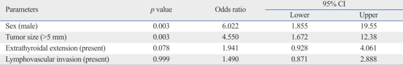
size >5 mm (p<0.001). The presence of extrathyroidal ex- tension and lymphovascular invasion were statistically re- lated to subclinical central lymph node metastasis (p=0.021 and p=0.02, respectively). However, age, bilaterality, multi- focality of tumor and lymphocytic thyroiditis were not sig- nificantly related to subclinical central lymph node metasta- sis (Table 3). Logistic regression analysis identified the most influential variables associated with subclinical central lymph node metastasis. On multivariate analysis, only male gender (odds ratio, 6.022; 95% CI 1.86-19.55) and tumor size >5 mm (odds ratio, 4.550; 95% CI 1.67-12.38) were independent predictors of subclinical central lymph node metastasis (Table 4).
Table 3. Clinicopathologic Parameters and Central Lymph Node Metastasis
Parameters Central lymph node metastasis
number (%) p value
Age (yrs) 0.507
<45 27 of 72 (37.5)
≥45 34 of 88 (38.6)
Sex 0.005
Male 13 of 19 (68.4)
Female 48 of 141 (34.0)
Tumor size (main) (mm) <0.001
≤5 6 of 41 (14.6)
>5 55 of 119 (46.2)
Multifocality 0.375
Solitary 39 of 106 (36.8)
Multifocal 22 of 54 (40.7)
Bilaterality 0.436
Unilateral 46 of 123 (37.4)
Bilateral 15 of 37 (40.5)
Extrathyroidal extension 0.021
Absent 23 of 78 (29.5)
Present 38 of 82 (46.3)
Lymphovascular invasion 0.020
Absent 57 of 156 (36.5)
Present 4 of 4 (100)
Lymphocytic thyoiditis 0.557
Absent 36 of 156 (38.3)
Present 25 of 66 (38.7)
p values, chi-squire for group comparison of categorical variables, and Student t-test for mean comparison.
Table 4. Multivariate Analysis for Risk Factors of Central Lymph Node Metastasis
Parameters p value Odds ratio 95% CI
Lower Upper
Sex (male) 0.003 6.022 1.855 19.55
Tumor size (>5 mm) 0.003 4.550 1.672 12.38
Extrathyroidal extension (present) 0.078 1.941 0.928 4.061
Lymphovascular invasion (present) 0.999 1.490 0.871 2.888
ever, studies by So, et al.16 and Roh, et al.5 reported that tu- mor size was not an independent predictor of subclinical central lymph node metastasis in multivariate analysis.
Recently, several authors subdivided tumor size using various cutoff values. Recently, Lee, et al.21 divided the pa- tients into two groups using cutoff values of 5-9 mm; ones with tumors less than the cutoff value and the other with tu- mors at or more than the cutoff value. They reported that PTMC aggressiveness is present mainly in patients with PTMC >7 mm and a tumor size >7 mm was an indepen- dent factor associated with central lymph node metastasis.
In the study by Pakdaman, et al.,17 a threshold of ≥4 mm was found to be more significant than one of 5 mm for con- ferring increased risk for extrathyroidal spread. However, they did not study differences in lymph node metastasis ac- cording to cutoff of sizes of 5 mm vs. 4 mm. Up to now, many studies have analyzed the aggressiveness of PTMC based on a size of 5 mm. The debate as to the clinical impli- cations of PTMC especially <5 mm in size, and the neces- sity for central lymph node dissection will continue.
We found no relation between tumor multifocaltiy, bilat- erality and central lymph node metastasis.4,16,22 In the study by Hay, et al.14 multifocality increased the risk of later nod- al recurrence, with 11% of multifocal tumors exhibiting re- currence, compared with only 4% of unifocal tumors. On the other hand, Roh, et al.,5 Lee, et al.18 and Lee, et al.23 re- ported that there was no relation between tumor multifocal- ity and central lymph node metastasis.
In the present study, there was no association between age and subclinical central lymph node metastasis, in con- sistent with some earlier studies.5,16,24
Our finding of male gender as an unfavorable prognostic factor coincides with previous studies identifying the role of gender as an important demographic variable in risk stratification for patients with papillary thyroid carcinoma (PTC).18,25,26 Previous studies found inconsistent results re- garding the association between gender and lymph node metastasis, and Roh, et al.5 reported that there was no asso- ciation between male gender and central lymph node me- tastasis.
Extrathyroidal extension of thyroid carcinoma was asso- ciated with poor prognosis. Extrathyroidal extension was found in 51.3% of patients in our study, and these results are very similar to other studies.16 Most cases of extrathy- roidal extension in our study showed minimal invasion of perithyroidal soft tissue or strap muscle. Not only gross in- vasion into extrathyroidal structures but also microscopic tasis is believed to be related to poor prognosis because they
occurs spontaneously or precede the occurrence of distant metastases.15 Despite the absence of palpable neck nodes, PTMC has a considerable rate of lymph node metastasis to the central compartment.3-5 Our study showed that subclini- cal central lymph node metastasis was detected in 38.1% of cases. Previous studies showed results similar to those of our study.5,16 In contrast, the studies of Pakdaman, et al.17 and Lee, et al.18 showed lower frequencies than our study.
The indications of central lymph node dissection are an- other major issue in PTMC management and debate goes on regarding the optimal treatment for this disease. Routine pro- phylactic central lymph node dissection for PTMC has been in debated because prophylactic central lymph node dissec- tion seems to have little prognostic benefit.3,4 However, cen- tral lymph node metastasis is an important risk factor of re- currence, and often is not detected clinically. Thus, it is reasonable to perform elective central lymph node dissection after predicting the presence of subclinical central lymph node metastasis. If we can predict central lymph node metas- tasis through assessing clinical factors, it would be a useful information to help us decide on the extent of surgery.
Several studies have described clinicopathological fac- tors associated with subclinical central lymph node metas- tasis in patients with PTMC, but results from those studies were inconsistent. In particular, it is considered important whether or not tumor size is predictor of subclinical central lymph node metastasis. Although assessment of tumor size depends on a large number of uncontrolled variables, high resolution ultrasonography by skilled physicians give good images and can be used to assess the size of tumor. Because we can know approximate tumor size by high resolution ul- trasonography and can plan the extent of surgery ahead at preoperation. The present study showed that subclinical central lymph node metastasis is significantly associated with tumor size in univariate analysis and tumor size is an independent predictor of subclinical central lymph node me- tastasis in multivariate analysis. Kasai and Sakamoto19 re- ported that patients with a tumor size >5 mm had central lymph node metastasis more frequently than with the pa- tients with a tumor size ≤5 mm. Also, several authors have demonstrated that PTMC tumors >5 mm are more signifi- cantly associated with central lymph node metastasis than with those of less than 5 mm.4,18,20 In addition, our study showed that a PTMC >5 mm related to signs of tumor ag- gressiveness (multifocality, extrathyroidal extension) in agreement with other studies. In contrast to our study, how-
population was a cohort of patients cared for in a single cen- ter. Therefore, it is unlikely that this cohort of small provides reliable statements, and a much larger number of subjects in multicenter will be needed to generalize this results. In con- clusion, in the present study, male gender and tumor size greater than 5 mm were two independent predictive factors for subclinical central lymph node metastasis in PTMC.
These are factors to more easily assess before surgery than other factors when planning the central lymph node dissec- tion. However, these data should be interpreted with caution because of retrospective analysis and lack of data for the prognostic significance.
REFERENCES
1. Jung KW, Park S, Kong HJ, Won YJ, Lee JY, Park EC, et al. Can- cer statistics in Korea: incidence, mortality, survival, and preva- lence in 2008. Cancer Res Treat 2011;43:1-11.
2. Giordano D, Gradoni P, Oretti G, Molina E, Ferri T. Treatment and prognostic factors of papillary thyroid microcarcinoma. Clin Otolaryngol 2010;35:118-24.
3. Ito Y, Tomoda C, Uruno T, Takamura Y, Miya A, Kobayashi K, et al. Clinical significance of metastasis to the central compartment from papillary microcarcinoma of the thyroid. World J Surg 2006;
30:91-9.
4. Wada N, Duh QY, Sugino K, Iwasaki H, Kameyama K, Mimura T, et al. Lymph node metastasis from 259 papillary thyroid microcar- cinomas: frequency, pattern of occurrence and recurrence, and op- timal strategy for neck dissection. Ann Surg 2003;237:399-407.
5. Roh JL, Kim JM, Park CI. Lateral cervical lymph node metastases from papillary thyroid carcinoma: pattern of nodal metastases and optimal strategy for neck dissection. Ann Surg Oncol 2008;15:
1177-82.
6. Koh YW, Park JH, Kim JW, Lee SW, Choi EC. Endoscopic hemithyroidectomy with prophylactic ipsilateral central neck dis- section via an unilateral axillo-breast approach without gas insuf- flation for unilateral micropapillary thyroid carcinoma: prelimi- nary report. Surg Endosc 2010;24:188-97.
7. Chung YS, Choe JH, Kang KH, Kim SW, Chung KW, Park KS, et al. Endoscopic thyroidectomy for thyroid malignancies: com- parison with conventional open thyroidectomy. World J Surg 2007;31:2302-6.
8. Pelizzo MR, Boschin IM, Toniato A, Pagetta C, Piotto A, Ber- nante P, et al. Natural history, diagnosis, treatment and outcome of papillary thyroid microcarcinoma (PTMC): a mono-institutional 12-year experience. Nucl Med Commun 2004;25:547-52.
9. Mazzaferri EL, Jhiang SM. Long-term impact of initial surgical and medical therapy on papillary and follicular thyroid cancer. Am J Med 1994;97:418-28.
10. Hay ID, Grant CS, Taylor WF, McConahey WM. Ipsilateral lo- bectomy versus bilateral lobar resection in papillary thyroid carci- noma: a retrospective analysis of surgical outcome using a novel prognostic scoring system. Surgery 1987;102:1088-95.
11. Hedinger C, Williams ED, Sobin LH. The WHO histological clas- sification of thyroid tumors: a commentary on the second edition.
extension outside of thyroid capsule is included in the diag- nosis of extrathyroidal extension in this study as well as in many other Korean studies.16 Although extrathyroidal ex- tension is well known as a prognostic factor in differentiat- ed thyroid carcinoma, minimal extrathyroidal extension has not been associated with recurrence in a few studies.27 In this study, extrathyroidal extension was statistically signifi- cantly associated with tumor size and subclinical central lymph node metastasis, but extrathyroidal extension was not an independent predictive factor for subclinical central lymph node metastasis in multivariate analysis. Moon, et al.28 showed that minimal extrathyroidal extension was sig- nificantly associated with tumor size and central lymph node metastasis.
It is known that the prevalence of lymphocytic thyroiditis is higher in patients with PTC.29 The effect of coexistant lymphocytic thyroiditis on prognosis in PTC patients re- mains controversial and only a few studies have been per- formed in PTMC patients.30 Kim, et al.31 noted that 15% of patients with PTC of mean tumor size 2.2 cm had lympho- cytic thyroiditis, and lymphocytic thyroiditis was associated with smaller size of the primary tumor at presentation.
However, there were no differences in the prevalence of central lymph node metastasis according to the presence of lymphocytic thyroiditis. In a PTMC study, So, et al.16 re- ported that 24.9% of 551 patients with PTMC had lympho- cytic thyroiditis and there was no association between lym- phocytic thyroiditis and lymph node metastasis, and Kim, et al.32 showed that the presence of lymphocytic thyroiditis did not influence the frequency of lymph node metastasis in all PTMC cases. In our study, the prevalence of lymphocyt- ic thyroiditis was higher than in other studies, and there was no association between lymphocytic thyroiditis and central lymph node metastasis.
Lymphovascular invasion was not associated with central lymph node metastasis in our study. Kim, et al.33 reported that lymphovascular invasion was associated not with cen- tral lymph node metastasis but with lateral cervical lymph node metastasis in patients with PTC. Our results are con- sistent with other studies for patients with PTMC.5,16,33
Our study has several limitations that must be taken into account. First, since this study was a retrospective analysis, the prognostic significance of subclinical lymph node me- tastasis such as the relation to tumor recurrence has not been fully investigated. The long-term follow-up studies are needed to confirm the prognostic significance of subclinical central lymph node metastasis in PTMC. Second, our study
microcarcinoma. Laryngoscope 2008;118:659-62.
24. Lim YC, Choi EC, Yoon YH, Kim EH, Koo BS. Central lymph node metastases in unilateral papillary thyroid microcarcinoma.
Br J Surg 2009;96:253-7.
25. Shaha AR, Shah JP, Loree TR. Risk group stratification and prog- nostic factors in papillary carcinoma of thyroid. Ann Surg Oncol 1996;3:534-8.
26. Besic N, Pilko G, Petric R, Hocevar M, Zgajnar J. Papillary thy- roid microcarcinoma: prognostic factors and treatment. J Surg Oncol 2008;97:221-5.
27. Ito Y, Tomoda C, Uruno T, Takamura Y, Miya A, Kobayashi K, et al. Minimal extrathyroid extension does not affect the relapse-free survival of patients with papillary thyroid carcinoma measuring 4 cm or less over the age of 45 years. Surg Today 2006;36:12-8.
28. Moon HJ, Kim EK, Chung WY, Yoon JH, Kwak JY. Minimal ex- trathyroidal extension in patients with papillary thyroid microcar- cinoma: is it a real prognostic factor? Ann Surg Oncol 2011;18:
1916-23.
29. Matsubayashi S, Kawai K, Matsumoto Y, Mukuta T, Morita T, Hi- rai K, et al. The correlation between papillary thyroid carcinoma and lymphocytic infiltration in the thyroid gland. J Clin Endocri- nol Metab 1995;80:3421-4.
30. Loh KC, Greenspan FS, Dong F, Miller TR, Yeo PP. Influence of lymphocytic thyroiditis on the prognostic outcome of patients with papillary thyroid carcinoma. J Clin Endocrinol Metab 1999;84:
458-63.
31. Kim EY, Kim WG, Kim WB, Kim TY, Kim JM, Ryu JS, et al.
Coexistence of chronic lymphocytic thyroiditis is associated with lower recurrence rates in patients with papillary thyroid carcino- ma. Clin Endocrinol (Oxf) 2009;71:581-6.
32. Kim HS, Choi YJ, Yun JS. Features of papillary thyroid microcar- cinoma in the presence and absence of lymphocytic thyroiditis.
Endocr Pathol 2010;21:149-53.
33. Kim JM, Kim TY, Kim WB, Gong G, Kim SC, Hong SJ, et al.
Lymphovascular invasion is associated with lateral cervical lymph node metastasis in papillary thyroid carcinoma. Laryngoscope 2006;116:2081-5.
Cancer 1989;63:908-11.
12. Appetecchia M, Scarcello G, Pucci E, Procaccini A. Outcome af- ter treatment of papillary thyroid microcarcinoma. J Exp Clin Cancer Res 2002;21:159-64.
13. Hay ID, Grant CS, van Heerden JA, Goellner JR, Ebersold JR, Bergstralh EJ. Papillary thyroid microcarcinoma: a study of 535 cases observed in a 50-year period. Surgery 1992;112:1139-46.
14. Hay ID, Hutchinson ME, Gonzalez-Losada T, McIver B, Reinalda ME, Grant CS, et al. Papillary thyroid microcarcinoma: a study of 900 cases observed in a 60-year period. Surgery 2008;144:980-7.
15. Harwood J, Clark OH, Dunphy JE. Significance of lymph node metastasis in differentiated thyroid cancer. Am J Surg 1978;136:
107-12.
16. So YK, Son YI, Hong SD, Seo MY, Baek CH, Jeong HS, et al.
Subclinical lymph node metastasis in papillary thyroid microcar- cinoma: a study of 551 resections. Surgery 2010;148:526-31.
17. Pakdaman MN, Rochon L, Gologan O, Tamilia M, Garfield N, Hier MP, et al. Incidence and histopathological behavior of papil- lary microcarcinomas: study of 429 cases. Otolaryngol Head Neck Surg 2008;139:718-22.
18. Lee NS, Bae JS, Jeong SR, Jung CK, Lim DJ, Park WC, et al.
Risk factors of lymph node metastasis in papillary thyroid micro- carcinoma. J Korean Surg Soc 2010;78:82-6.
19. Kasai N, Sakamoto A. New subgrouping of small thyroid carcino- mas. Cancer 1987;60:1767-70.
20. Roti E, Rossi R, Trasforini G, Bertelli F, Ambrosio MR, Busutti L, et al. Clinical and histological characteristics of papillary thyroid microcarcinoma: results of a retrospective study in 243 patients. J Clin Endocrinol Metab 2006;91:2171-8.
21. Lee KJ, Cho YJ, Kim SJ, Lee SC, Kim JG, Ahn CJ, et al. Analysis of the clinicopathologic features of papillary thyroid microcarci- noma based on 7-mm tumor size. World J Surg 2011;35:318-23.
22. Chow SM, Law SC, Chan JK, Au SK, Yau S, Lau WH. Papillary microcarcinoma of the thyroid-Prognostic significance of lymph node metastasis and multifocality. Cancer 2003;98:31-40.
23. Lee SH, Lee SS, Jin SM, Kim JH, Rho YS. Predictive factors for central compartment lymph node metastasis in thyroid papillary