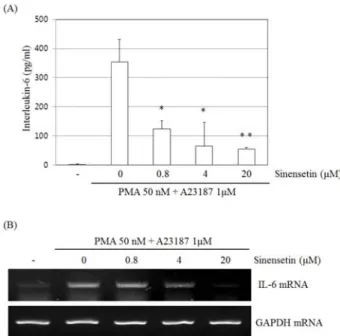
Sinensetin Inhibits Interleukin-6 in Human Mast Cell - 1 Via Signal Transducers and Activators of the Transcription 3 (STAT3) and Nuclear Factor Kappa B (NF-κB) Pathways
4
0
0
전체 글
(2)
(3)
(4)
수치
관련 문서
