Two rhesus monkeys in the Korea National Primate Research Center were found to suffer from acute gastrointestinal dilation
5
0
0
전체 글
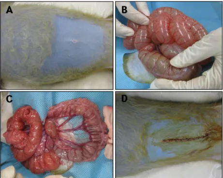
(2) 218. Kyoung-Min Kim et al.. Figure 1. Gastric dilation in a laboratory rhesus monkey. A) Excessive gastric dilation was observed. B) Severe congestion was found in the lung. Bar=2 cm.. Figure 2. Gastrointestinal dilation in a laboratory rhesus monkey. (A) Abdominal distension by acute gastrointestinal dilation was observed. (B) The extended small intestine was found to extrude from the abdominal cavity. (C) The gas in the gastrointestinal tract was removed. (D) After surgery, the abdomen returned to its normal state.. suffer from acute GI dilation. They were housed and maintained in the specific pathogen-free facility at the Korea National Primate Research Center, according to Korea Research Institute of Bioscience and Biotechnology (KRIBB) Institutional Animal Care and Use Committee Guidelines (Approval No. KRIBB-ACE-11010). The monkeys were kept in indoor individual cages and were fed commercial monkey chow (LabDiet, Harlan Laboratories, Inc., USA) supplemented daily with fruits and supplied water ad libitum. One of the monkeys showed severe gastric bloating after general anesthesia with 2% isoflurane (IFRAN LIQ, Hana Pharm. Co., Korea) for MR imaging for other neuroscientific purposes, where after it died suddenly (Figure 1A). Lab Anim Res | September, 2012 | Vol. 28, No. 3. During necropsy, severe congestion of the lung was observed (Figure 1B). No pathologic changes were detected in the other organs. These results suggest that the cause of sudden death may have been the severely dilated stomach, which strongly compressed the lung, resulting in subsequent depressed respiration. Interestingly, although feed restriction was performed for >20 hours before general anesthesia, much undigested and watery food was contained in the stomach of the monkey. The GI contents were collected for microbiologic examination. The other monkey was accidently found to have a dilated abdomen (Figure 2A) and has showed a little anorexia and depression for 1 day before finding the abdominal dilatation. No experiments or treatments were.
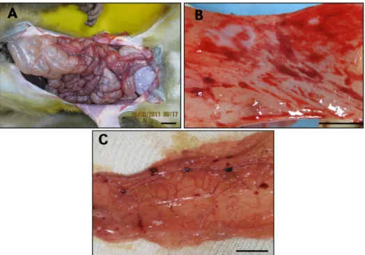
(3) Acute gastrointestinal dilation in laboratory monkeys. 219. Figure 3. Postmortem examination of a rhesus monkey with gastrointestinal dilation. (A) Excessive large intestinal dilation was found. Bar=2 cm. (B) Severe hemorrhagic lesions were detected in the small intestine. Bar=1 cm. (C) Focal hemorrhagic lesions were found in the large intestine. Bar=1 cm.. conducted before the symptom was found. The monkey showed signs of respiratory distress caused by GI dilation, lay prone without moving, and showed severe symptoms of salivation due to breathing difficulty. Surgical treatment was performed immediately using the following procedure: atropine sulfate (Jeil Pharm. Co., Korea) was administered intramuscularly (0.04 mg/kg) to prevent salivation, and the monkey was anesthetized with intramuscular injection of ketamine HCl (Yuhan Co., Korea). When the abdominal wall was opened, the small intestine was found to extrude from the abdominal cavity because of the increase in abdominal pressure caused by GI dilation (Fig. 2B). After laparotomy, removal of the gas contained in the GI tract was performed with a trocar, and then the GI contents were collected for microbiologic examination. Immediately thereafter, saline containing an antibiotic (cefazolin, 50 mg/kg, Chong Kun Dang Pharm. Co., Korea) and an antifoaming agent (Bloten Liquid, 20 mL, Vetco Pharma, India) was flushed through the system to prevent secondary infection and gas formation. As a results, gas that had accumulated abnormally in the GI tract was removed and the GI tract was restored to normal (Figure 2C and D). As post-operative management, cefazolin (40 mg/kg) and metoclopramide HCl (Meckool, 5 mg/ kg, Jeil Pharm. Co.) were injected intramuscularly twice. a day, and the monkey was treated intravenously with fluid therapy consisting of 5% dextrose (Daihan Pharm. Co., Korea) for 3 days. The activity status of the monkey improved gradually, but the monkey was found dead on the 8th day after surgery. During necropsy, excessive dilation of the large intestine was observed (Figure 3A). Severe congestion was detected in the small intestine Figure 3B) and the large intestine (Figure 3C). Tissues were fixed in 10% neutral-buffered formalin and then embedded in paraffin for histologic assessment. Tissue sections (5 µm) were stained with hematoxylin and eosin. Histopathologically, erythrocytes were found to fill the alveoli and alveolar capillaries of the lungs (Figure 4A). In the stomach, epithelial cells were found to be sloughed from the mucosal layer, and erythrocytes were found to fill the blood vessels in the submucosal and mucosal layer (Figure 4B). In the small intestine and large intestine, epithelial cells were also found to be sloughed from the mucosal layer, and inflammatory cells were found to have infiltrated the submucosa (only in the large intestine) and mucosa (Figure 4C and D). The organism receiving the most attention as the primary cause of acute gastric dilatation is Clostridium perfringens, and this gas-producing, anaerobic bacillus has been isolated from the gastric contents of monkeys with acute gastric dilatation [3]. Other bacteria isolated Lab Anim Res | September, 2012 | Vol. 28, No. 3.
(4) 220. Kyoung-Min Kim et al.. Figure 4. Histologic findings in a rhesus monkey with gastrointestinal dilation. (A) Erythrocytes were found to fill the alveoli and alveolar capillaries. Lung, hematoxylin and eosin (H&E) stain (×100). Bar=100 µm. (B) Epithelial cells were found to be sloughed from the mucosal layer, and erythrocytes were found to fill the blood vessels in the submucosal and mucosal layer of the stomach. Stomach, H&E stain (×100). Bar =100 µm. (C) Epithelial cells were found to be sloughed from the mucosal layer, and inflammatory cells were found to have infiltrated the mucosa of the small intestine. Small intestine, H&E stain (×200). Bar=200 µm. (D) Epithelial cells were found to be sloughed from the mucosal layer, and inflammatory cells were found to have infiltrated the mucosa and submucosa of the large intestine. Large intestine, H&E stain (×200). Bar=200 µm.. from the gastric contents of monkeys with acute gastric dilatation have included Lactobacillus spp., alpha Streptococcus, Enterobacter cloacae, and Escherichia coli [4,5]. In our monkeys, these pathogenic bacteria were not detected in the GI contents, and only Staphylococcus haemolyticus and Enterococcus faecalis were isolated. S. haemolyticus is a member of the coagulase-negative staphylococci [6] that also colonizes prosimians, monkeys, and domestic animals [7]. S. haemolyticus infections can be localized or systemic and are often associated with the insertion of medical devices [8-10]. S. haemolyticus is a difficult pathogen to treat because it has a highly antibiotic-resistant phenotype and the ability to form biofilms [11]. E. faecalis can cause life-threatening infections in humans and has a naturally high level of antibiotic resistance, which contributes to its pathogenicity [12]. It also can cause endocarditis and bacteremia and other infections in humans [13]. However, S. haemolyticus and E. faecalis were not the cause of the observed GI dilation, because neither of them forms gas in the GI tract. Nevertheless, these bacteria may have been the cause of death in our monkeys, because they have high levels of antibiotic resistance and can induce Lab Anim Res | September, 2012 | Vol. 28, No. 3. systemic bacteremia during antibiotic treatment. Many reports have described acute gastric dilation in humans after anesthesia [14,15]. Further, the quantity and composition of monkey diets, especially a commercial biscuit type diet, are important factors in the pathogenesis of bloating. Water being supplied ad libitum may also cause bloating [3,16]. In our monkeys, general anesthesia with 2% isoflurane was performed before the occurrence of bloating, a commercial biscuit type diet was supplied to the monkeys, and water also was supplied ad libitum in our center. Thus, these results suggest that the cause of the acute GI dilation observed in our monkeys was not infection by gas-forming bacteria, but rather multiple factors such as diet, anesthesia, and excessive water consumption.. Acknowledgments This research was supported by a grant from the KRIBB Research Initiative Program (KGM4241231) and by the National Research Foundation of Korea funded by the Ministry of Education, Science and Technology, Republic of Korea (NBC5001113)..
(5) Acute gastrointestinal dilation in laboratory monkeys. References 1. Pond CL, Newcomer CE, Anver MR. Acute gastric dilatation in nonhuman primates: review and case studies. Vet Pathol Suppl 1982; 7: 126-133. 2. Min BR, Pinchak WE, Anderson RC, Hume ME. In vitro bacterial growth and in vivo ruminal microbiota populations associated with bloat in steers grazing wheat forage. J Anim Sci 2006; 84(10): 2873-2882. 3. Soave OA. Observations on acute gastric dilatation in nonhuman primates. Lab Anim Sci 1978; 28(3): 331-334. 4. Elwell MR, DePaoli A. Gastric dilatation and volvulus in a squirrel monkey. J Am Vet Med Assoc 1978; 173(9): 1235-1236. 5. Smith AW, Casey HW, LaCroix JT, Johnson DK. Acute bloat syndrome (gastric dilatation) in Macaca mulatta. J Am Vet Med Assoc 1969; 155(7): 1241-1244. 6. Weinstein MP, Mirrett S, Van Pelt L, McKinnon M, Zimmer BL, Kloos W, Reller LB. Clinical importance of identifying coagulasenegative staphylococci isolated from blood cultures: evaluation of MicroScan Rapid and Dried Overnight Gram-Positive panels versus a conventional reference method. J Clin Microbiol 1998; 36(7): 2089-2092. 7. Fischetti A, Novick RP, Ferretti JJ, Portnoy DA, Rood JI, Lina G, Etienne J, Vandenesch F. Biology and pathogenicity of staphylococci other than Staphylococcus aureus and Staphylococcus epidermidis. In: Gram-positive pathogens, ASM Press, Washington, DC, 2000;. 221. pp 450-462. 8. Falcone M, Giannella M, Raponi G, Mancini C, Venditti M. Teicoplanin use and emergence of Staphylococcus haemolyticus: is there a link? Clin Microbiol Infect 2006; 12(1): 96-97. 9. Poyart C, Quesne G, Boumaila C, Trieu-Cuot P. Rapid and accurate species-level identification of coagulase-negative staphylococci by using the sodA gene as a target. J Clin Microbiol 2001; 39(12): 4296-4301. 10. Viale P, Stefani S. Vascular catheter-associated infections: a microbiological and therapeutic update. J Chemother 2006; 18(3): 235-249. 11. de Allori MC, Jure MA, Romero C, de Castillo ME. Antimicrobial resistance and production of biofilms in clinical isolates of coagulase-negative Staphylococcus strains. Biol Pharm Bull 2006; 29(8): 1592-1596. 12. Ryan KJ, Ray CG. Sherris Medical Microbiology, 4th ed, McGraw Hill, 2004; pp 294-295. 13. Murray BE. The life and times of the Enterococcus. Clin Microbiol Rev 1990; 3(1): 46-65. 14. Byrne JJ, Cahill JM. Acute gastric dilation. Am J Surg 1961; 101: 301-309. 15. Danhof IE. The clinical gas syndromes: a pathophysiologic approach. Ann N Y Acad Sci 1968; 150(1): 127-140. 16. Newton WM, Beamer PD, Rhoades HE. Acute bloat syndrome in stumptailed macaques (Macaca arctoides): a report of four cases. Lab Anim Sci 1971; 21(2): 193-196.. Lab Anim Res | September, 2012 | Vol. 28, No. 3.
(6)
수치

관련 문서