INTRODUCTION
In clinical trials for acute ischemic stroke, imaging modalities such as computed tomography (CT) or magnetic resonance imaging (MRI) may play critical roles by ruling out acute hemorrhage, allowing detection of the infarct
Neuroimaging in Randomized, Multi-Center Clinical Trials of Endovascular Treatment for Acute Ischemic Stroke:
A Systematic Review
Chong Hyun Suh, MD
1, Seung Chai Jung, MD, PhD
1, Byungjun Kim, MD, PhD
2, Se Jin Cho, MD
1, Dong-Cheol Woo, PhD
3, Woo Yong Oh, MA
4, Jong Gu Lee, PhD
4, Kyung Won Kim, MD, PhD
1, 51Department of Radiology and Research Institute of Radiology, University of Ulsan College of Medicine, Asan Medical Center, Seoul, Korea;
2Department of Radiology, Korea University Anam Hospital, Korea University College of Medicine, Seoul, Korea; 3Bioimaging Center, Biomedical Research Center, Asan Institute for Life Sciences, Asan Medical Center, Seoul, Korea; 4Clinical Research Division, National Institute of Food and Drug Safety Evaluation, Ministry of Food and Drug Safety, Cheongju, Korea; 5Asan Image Metrics, Clinical Trial Center, Asan Institute for Life Sciences, Asan Medical Center, Seoul, Korea
Appropriate use and analysis of neuroimaging techniques is an inevitable aspect of clinical trials for patients with acute ischemic stroke. Neuroimaging examinations were recently used to define the core eligibility criteria and outcomes in acute ischemic stroke research. Recent clinical trials for endovascular treatment in acute ischemic stroke have also demonstrated the efficacy or safety of endovascular treatment using various imaging modalities as well as clinical indices. Furthermore, independent imaging reviews and imaging core laboratory assessments are essential to manage and analyze imaging data in order to enhance the reliability of the outcomes. Therefore, we systematically reviewed the use of neuroimaging in recent randomized clinical trials for endovascular treatment of acute ischemic stroke in order to provide a thorough summary, which would serve as a resource guiding the use of appropriate imaging protocols and analyses in future clinical trials for acute ischemic stroke. This review will help researchers select appropriate imaging biomarkers among the various imaging protocols available and apply the selected type of imaging examination for each study in accordance with the academic purpose.
Keywords: Acute ischemic stroke; Endovascular treatment; Clinical trials; Neuroimaging; Systematic review
Received June 4, 2019; accepted after revision October 1, 2019.
This study was supported by a grant from the Ministry of Food and Drug Safety in 2018 (No. 18182MFDS402).
Corresponding author: Seung Chai Jung, MD, PhD, Department of Radiology and Research Institute of Radiology, University of Ulsan College of Medicine, Asan Medical Center, 88 Olympic-ro 43-gil, Songpa-gu, Seoul 05505, Korea.
• Tel: (822) 3010-3949 • Fax: (822) 476-4719
• E-mail: [email protected]
This is an Open Access article distributed under the terms of the Creative Commons Attribution Non-Commercial License (https://creativecommons.org/licenses/by-nc/4.0) which permits unrestricted non-commercial use, distribution, and reproduction in any medium, provided the original work is properly cited.
core or vessel occlusion, evaluating collateral circulation, and assessing the penumbra (1, 2). In 1995, the European Cooperative Acute Stroke Study (ECASS) and The National Institute of Neurological Disorders and Stroke rt-PA stroke study group used noncontrast-enhanced CT (NECT) findings as the eligibility criteria and safety outcomes for detection of intracranial hemorrhage (3, 4). In seven recent successful clinical trials for determining the efficacy of endovascular treatment (endovascular mechanical thrombectomy) in acute ischemic stroke (5-11), neuroimaging was used not only for selecting eligible patients for the trials but also for evaluating one of the primary or secondary outcomes.
The recommendations from the American Heart Association/
American Stroke Association (AHA/ASA) 2018 guidelines for the early management of patients with acute ischemic stroke reconfirmed the role of imaging modalities in diagnosing acute ischemic stroke and guiding treatment options (2).
Korean J Radiol 2020;21(1):42-57 https://doi.org/10.3348/kjr.2019.0354
Appropriate use and analysis of imaging techniques is essential for successfully conducting clinical trials for acute ischemic stroke. Imaging-based outcomes such as the infarct core volume, including infarct growth, revascularization, recanalization, and reperfusion as well as hemorrhage, are gaining popularity as primary or secondary outcomes in clinical trials for acute ischemic stroke (5-20).
In addition, independent imaging reviews and imaging core laboratory assessments are essential for conducting clinical trials with reliable imaging-based outcomes.
Recent clinical trials for acute ischemic stroke applied various imaging modalities to each study appropriately by using NECT, CT angiography (CTA, single-phase or multiple- phase), CT perfusion (CTP), diffusion-weighted imaging (DWI), MR angiography (MRA), T2*-weighted gradient echo (GRE), MR perfusion (MRP), or fluid attenuated inversion recovery (FLAIR).
Therefore, we systematically reviewed the use of neuroimaging in recent, randomized clinical trials for the efficacy of endovascular treatment for acute ischemic stroke so as to provide a thorough summary that could serve as a resource guiding the use of appropriate imaging protocols and analyses in future clinical trials for acute ischemic stroke. This review will help researchers select appropriate imaging biomarkers among the various imaging protocols available and apply the selected imaging biomarkers to each study in accordance with the purpose.
MATERIALS AND METHODS
This study was performed and reported in accordance with the Preferred Reporting Items for Systematic Reviews and Meta-Analyses guidelines (21). The following research questions were established: What are the roles, appropriate protocols, and reviewing systems for neuroimaging
assessments in randomized, multi-center clinical trials of endovascular treatment for acute ischemic stroke?
Literature Search
A systematic, computerized search of Ovid-MEDLINE and EMBASE was performed to identify published randomized trials of endovascular treatment for acute ischemic stroke. The search terms were as follows: ((stroke)) AND ((thrombectomy)) AND ((randomized) OR (randomly)). The databases were searched for articles published on or before May 22, 2018. This systematic, computerized search was limited to clinical trials. The bibliographies of the identified
articles were screened manually to expand the literature search. Our search was not limited by sample size, type of design, endpoint selection, or blinding. There were also no limits related to the language of publication, the trial’s geographic origin, or sponsorship.
Eligibility Criteria
Randomized trials of endovascular treatment for acute ischemic stroke were included. Conference abstracts without published full texts were not considered to be eligible for this study. Review articles, non-randomized trials, protocols, observational studies, and case reports were also excluded.
Subgroup analyses or sequential studies of original randomized trials were excluded.
Data Extraction
A standardized, predesigned data extraction form was used. The data were independently extracted from each eligible randomized trial by two authors. Publication year, sample size, eligibility criteria, imaging-based outcomes or radiologic outcomes, imaging core laboratory or imaging core lab, imaging modalities for initial work-up or follow-up imaging, imaging protocols for vessel occlusion or perfusion, use of the Alberta Stroke Program Early CT (ASPECT) score, the definition of the ischemic core, the definition of the penumbra, analysis software, reviewing systems, and standardization were reviewed. The data retrieved from all included trials were cross-checked, and a third author was consulted for consensus in cases of unclear definitions.
RESULTS
Literature Search
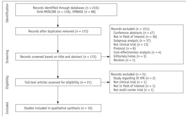
The systematic literature search identified 216 articles.
Full-text reviews of 21 potentially eligible articles were conducted and five studies were excluded due to the following reasons: two studies assessed intravenous tissue plasminogen activators (IV tPAs) (22, 23), one study was not a randomized trial (24), one study was not in the field of interest (25), and one study was a single-center study (26). Finally, a total of 16 randomized, multi-center clinical trials published between 2012 and 2018 were identified for our study (Fig. 1) (5-20). The eligible trials covered a total of 4080 patients.
Characteristics of the Included Studies
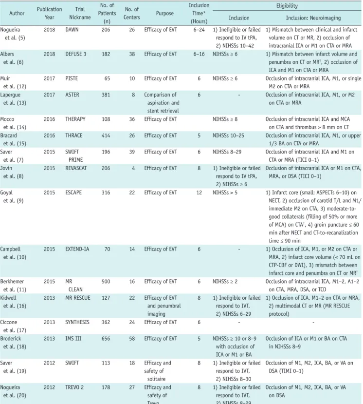
The characteristics of the included trials are described in
Table 1. The size of the study populations ranged from 65 to 656 patients, and the number of participating centers ranged from 4 to 58. The purpose of the clinical trials was to demonstrate the efficacy or safety of endovascular treatment in comparison with conventional medical treatment. The inclusion criteria for the time after symptom or randomization onset ranged from 5 to 24 hours, which indicates the eligible time to perform endovascular treatment. In the THERAPY trial, the time was 3–4.5 hours based on the IV tPA (14). Other trials applied the following inclusion times on the basis of the endovascular treatment:
6 hours in eight trials (7, 10-13, 15, 17, 18), within 8 hours in four trials (8, 16, 19, 20), and within 12 hours in one trial (9). Two trials (2%) published in 2018 included patients 6–16 hours (6) and 6–24 hours (5) after symptoms onset. The techniques used for endovascular treatments varied from intra-arterial thrombolysis using urokinase, suction or aspiration using a penumbra or catheter, or stent retriever. Four clinical trials used a single device for endovascular treatment, including the Solitaire (7-10) and Trevo retrieval stent (5). Three clinical trials compared the efficacy and safety of the devices as follows: comparison between aspiration and stent retriever treatment (13);
between Solitaire stent and Merci device (19); and between Trevo and Merci device (20). Only 10 of the 16 trials
demonstrated the efficacy of endovascular treatments (5-11, 15, 19, 20).
Eligibility and Neuroimaging Criteria for Acute Ischemic Stroke Trials
The eligibility and neuroimaging criteria used in the included trials are described in Tables 1 and 2. All trials except the SYNTHESIS trial included patients with arterial occlusion that was assessed on vessel imaging.
Among the 15 trials, 10 included only anterior circulation occlusion (internal carotid artery [ICA] and the middle cerebral artery [MCA]) (5-10, 12-14, 16), 1 included only anterior circulation occlusion (anterior cerebral artery as well as ICA or MCA) (11), and 4 included posterior circulation (basilar artery or vertebral artery) as well as anterior circulation (15, 18-20). Although most trials used CTA or MRA for vessel imaging, two trials used only CTA (7, 14), two trials used only digital subtraction angiography (DSA) (19, 20), and one trial included transcranial Doppler ultrasonography as well as CTA, MRA, or DSA (11). Neuroimaging played a critical role in the exclusion criteria in the trials for detecting intracranial hemorrhage, significant mass effect with midline shift, intracranial tumors except small meningiomas, extensive infarct core volumes, and pre-existing proximal arterial
Records identified through databases (n = 216):
Ovid-MEDLINE (n = 118), EMBASE (n = 98)
Records after duplicates removed (n = 172) Records excluded (n = 151):
Conference abstracts (n = 47) Not in field of interest (n = 38) Subgroup analysis (n = 37) Not clinical trial (n = 13) Protocol (n = 8)
Cost-effectiveness analysis (n = 4) Editorials/notes (n = 3)
Reviews (n = 1)
Records excluded (n = 5):
Study regarding IV tPA (n = 2) Not clinical trial (n = 1) Not in field of interest (n = 1) Not multi-center trial (n = 1) Records screened based on title and abstract (n = 172)
Full-text articles assessed for eligibility (n = 21)
Studies included in qualitative synthesis (n = 16)
IdentificationScreeningEligibilityIncluded
Fig. 1. Study selection protocol. IV tPA = intravenous tissue plasminogen activator
Table 1. Characteristics of Included Clinical Trials Author Publication
Year
Trial Nickname
No. of Patients
(n)
No. of
Centers Purpose
Inclusion Time*
(Hours)
Eligibility
Inclusion Inclusion: Neuroimaging Nogueira
et al. (5)
2018 DAWN 206 26 Efficacy of EVT 6–24 1) Ineligible or failed respond to IV tPA, 2) NIHSSs 10–42
1) Mismatch between clinical and infarct volume on CT or MR, 2) occlusion of intracranial ICA or M1 on CTA or MRA Albers
et al. (6)
2018 DEFUSE 3 182 38 Efficacy of EVT 6–16 NIHSSs ≥ 6 1) Mismatch between infarct volume and penumbra on CT or MR†, 2) occlusion of ICA and M1 on CTA or MRA
Muir et al. (12)
2017 PISTE 65 10 Efficacy of EVT 6 NIHSSs ≥ 6 Occlusion of intracranial ICA, M1, or single M2 on CTA or MRA
Lapergue et al. (13)
2017 ASTER 381 8 Comparison of
aspiration and stent retrieval
6 - Occlusion of intracranial ICA, M1, or M2 on CTA or MRA
Mocco et al. (14)
2016 THERAPY 108 36 Efficacy of EVT NIHSSs ≥ 8 Occlusion of intracranial ICA and MCA
on CTA and thrombus > 8 mm on CT Bracard
et al. (15)
2016 THRACE 414 26 Efficacy of EVT 5 NIHSSs 10–25 Occlusion of intracranial ICA, M1, or upper 1/3 BA on CTA or MRA
Saver et al. (7)
2015 SWIFT PRIME
196 39 Efficacy of EVT 6 NIHSSs 8–29 Occlusion of intracranial ICA and M1 on CTA or MRA (TICI 0–1)
Jovin et al. (8)
2015 REVASCAT 206 4 Efficacy of EVT 8 1) Ineligible or failed respond to IV tPA, 2) NIHSSs ≥ 6
Occlusion of intracranial ICA or M1 on CTA, MRA, or DSA (TICI 0–1)
Goyal et al. (9)
2015 ESCAPE 316 22 Efficacy of EVT 12 NIHSSs > 5 1) Infarct core (small: ASPECTs 6–10) on NECT, 2) occlusion of carotid T/L and M1/
immediate M2 on CTA, 3) moderate-to- good collaterals (filling of 50% or more of MCA) on CTA‡, 4) groin puncture ≤ 60 min after NECT and CT-to-recanalization time ≤ 90 min
Campbell et al. (10)
2015 EXTEND-IA 70 14 Efficacy of EVT 6 - 1) Occlusion of ICA, M1, or M2 on CTA or
MRA, 2) infarct core volume (< 70 mL on CTP-CBF or DWI), 3) mismatch between infarct core and penumbra on CT or MR† Berkhemer
et al. (11)
2015 MR
CLEAN
500 16 Efficacy of EVT 6 NIHSSs ≥ 2 Occlusion of intracranial ICA, M1–2, A1–2 on CTA, MRA, DSA, or TCD
Kidwell et al. (16)
2013 MR RESCUE 127 22 Efficacy of EVT
and penumbral imaging
8 1) Ineligible or failed respond to IVT, 2) NIHSSs 6–29
1) Occlusion of ICA, M1–2 on CTA or MRA, 2) multimodal CT or MR (MR RESCUE protocol)
Ciccone et al. (17)
2013 SYNTHESIS 362 24 Efficacy of EVT 6 - -
Broderick et al. (18)
2013 IMS III 656 58 Efficacy of EVT 5 NIHSSs ≥ 10 or 8–9
with occlusion of ICA or M1 or BA
Occlusion of ICA or M1 or BA on CTA in NIHSSs 8–9
Saver et al. (19)
2012 SWIFT 113 18 Efficacy and
safety of solitaire
8 1) Ineligible or failed respond to IVT, 2) NIHSSs 8–30
Occlusion of M1, M2, ICA, BA, or VA on DSA (TIMI 0–1)
Nogueira et al. (20)
2012 TREVO 2 178 27 Efficacy and
safety of Trevo
8 1) Ineligible or failed respond to IVT, 2) NIHSSs 8–29
Occlusion of M1, M2, ICA, BA, or VA on DSA
*Inclusion time means eligible time to perform EVT, †Definition of mismatch were as follows: infarct core volume < 70 mL, penumbral to infarct core volume ≥ 1.8, absolute penumbral volume (Tmax > 6 s) ≥ 15 mL in DEFUSE 3 trial; infarct core volume > 50 mL, severe penumbral volume (Tmax ≥ 10 s) ≥ 100 mL, or penumbral to infarct core volume ≤ 1.8 and penumbral volume < 15 mL in SWIFT PRIME trial; infarct core volume < 70 mL on CTP-CBF or DWI, mismatch ratio > 1.2, and absolute mismatch volume > 10 mL (infarct core: CTP-CBF < 30% of normal tissue; penumbra: Tmax > 6 s on CTP or MRP) in EXTEND-IA trial, ‡Multiphase CTA was preferred. ASPECTs = Alberta Stroke Program Early CT score, BA = basilar artery, CBF = cerebral blood flow, CT = computed tomography, CTA = CT angiography, CTP = CT perfusion, DSA = digital subtraction angiography, DWI = diffusion-weighted imaging, EVT = endovascular treatment, ICA = internal carotid artery, IV tPA = intravenous tissue plasminogen activator, MCA = middle cerebral artery, MR = magnetic resonance, MRA = MR angiography, MRP = MR perfusion, NECT = noncontrast-enhanced CT, NIHSSs = National Institutes of Health Stroke Scale score, TCD
= transcranial Doppler, TICI = thrombolysis in cerebral infarction scale, TIMI = thrombolysis in myocardial ischemia, Tmax = time to maximum of residue function, VA = vertebral artery
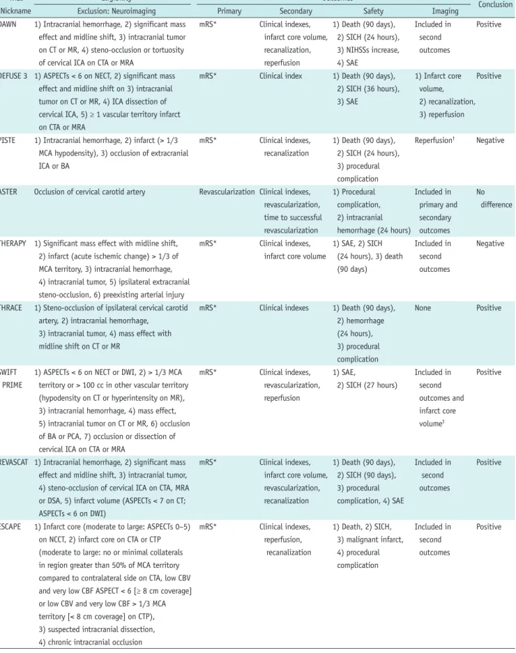
Table 2. Eligibility, Outcomes, Conclusion of Included Clinical Trials Trial
Nickname
Eligibility Outcomes
Conclusion
Exclusion: Neuroimaging Primary Secondary Safety Imaging
DAWN 1) Intracranial hemorrhage, 2) significant mass effect and midline shift, 3) intracranial tumor on CT or MR, 4) steno-occlusion or tortuosity of cervical ICA on CTA or MRA
mRS* Clinical indexes, infarct core volume, recanalization, reperfusion
1) Death (90 days), 2) SICH (24 hours), 3) NIHSSs increase, 4) SAE
Included in second outcomes
Positive
DEFUSE 3 1) ASPECTs < 6 on NECT, 2) significant mass effect and midline shift on 3) intracranial tumor on CT or MR, 4) ICA dissection of cervical ICA, 5) ≥ 1 vascular territory infarct on CTA or MRA
mRS* Clinical index 1) Death (90 days), 2) SICH (36 hours), 3) SAE
1) Infarct core volume, 2) recanalization, 3) reperfusion
Positive
PISTE 1) Intracranial hemorrhage, 2) infarct (> 1/3 MCA hypodensity), 3) occlusion of extracranial ICA or BA
mRS* Clinical indexes, recanalization
1) Death (90 days), 2) SICH (24 hours), 3) procedural complication
Reperfusion† Negative
ASTER Occlusion of cervical carotid artery Revascularization Clinical indexes, revascularization, time to successful revascularization
1) Procedural complication, 2) intracranial hemorrhage (24 hours)
Included in primary and secondary outcomes
No difference
THERAPY 1) Significant mass effect with midline shift, 2) infarct (acute ischemic change) > 1/3 of MCA territory, 3) intracranial hemorrhage, 4) intracranial tumor, 5) ipsilateral extracranial steno-occlusion, 6) preexisting arterial injury
mRS* Clinical indexes, infarct core volume
1) SAE, 2) SICH (24 hours), 3) death (90 days)
Included in second outcomes
Negative
THRACE 1) Steno-occlusion of ipsilateral cervical carotid artery, 2) intracranial hemorrhage,
3) intracranial tumor, 4) mass effect with midline shift on CT or MR
mRS* Clinical indexes 1) Death (90 days), 2) hemorrhage (24 hours), 3) procedural complication
None Positive
SWIFT PRIME
1) ASPECTs < 6 on NECT or DWI, 2) > 1/3 MCA territory or > 100 cc in other vascular territory (hypodensity on CT or hyperintensity on MR), 3) intracranial hemorrhage, 4) mass effect, 5) intracranial tumor on CT or MR, 6) occlusion of BA or PCA, 7) occlusion or dissection of cervical ICA on CTA or MRA
mRS* Clinical indexes, revascularization, reperfusion
1) SAE,
2) SICH (27 hours)
Included in second outcomes and infarct core volume‡
Positive
REVASCAT 1) Intracranial hemorrhage, 2) significant mass effect and midline shift, 3) intracranial tumor, 4) steno-occlusion of cervical ICA on CTA, MRA or DSA, 5) infarct volume (ASPECTs < 7 on CT;
ASPECTs < 6 on DWI)
mRS* Clinical indexes, infarct core volume, revascularization, recanalization
1) Death (90 days), 2) SICH (90 days), 3) procedural complication, 4) SAE
Included in second outcomes
Positive
ESCAPE 1) Infarct core (moderate to large: ASPECTs 0–5) on NCCT, 2) infarct core on CTA or CTP (moderate to large: no or minimal collaterals in region greater than 50% of MCA territory compared to contralateral side on CTA, low CBV and very low CBF ASPECT < 6 [≥ 8 cm coverage]
or low CBV and very low CBF > 1/3 MCA territory [< 8 cm coverage] on CTP), 3) suspected intracranial dissection, 4) chronic intracranial occlusion
mRS* Clinical indexes, reperfusion, recanalization
1) Death, 2) SICH, 3) malignant infarct, 4) procedural complication
Included in second outcomes
Positive
steno-occlusions, which represent ineligible conditions for undergoing endovascular treatment. In some trials, neuroimaging was used as a part of the core eligibility criteria for selection of patients with a small infarct or the presence of mismatch between the infarct core volume and the salvageable tissue volume (5-7, 10). This mismatch may have contributed to the extension of the therapeutic time window up to 16 and 24 hours from the
symptom onset for endovascular treatment in the DAWN and DEFUSE 3 trials. The DAWN trial enrolled patients who showed a mismatch between the severity of the clinical deficit and the infarct core volume as seen on DWI or CTP.
The mismatch was divided into three groups based on a patient age threshold of 80 years (5). The DEFUSE 3 trial included patients who had an initial infarct core volume
< 70 mL, a penumbral tissue volume to initial infarct core Table 2. Eligibility, Outcomes, Conclusion of Included Clinical Trials (Continued)
Trial Nickname
Eligibility Outcomes
Conclusion
Exclusion: Neuroimaging Primary Secondary Safety Imaging
EXTEND-IA 1) Infarct volume (hypodensity > 1/3 MCA territory) on NECT, 2) intracranial hemorrhage on CT or MR, 3) difficulty or inability to access to cerebral arteries (proximal stenosis, dissection)
Reperfusion, NIHSSs (3 days)
Clinical indexes, infarct core volume†, recanalization
1) Death, 2) SICH, 3) PH
Included in primary and secondary outcomes
Positive
MR CLEAN Intracranial hemorrhage on CT or MR mRS* Clinical indexes, infarct core volume, reperfusion, recanalization
1) Neurologic deterioration, 2) SICH, 3) procedural complication, 4) SAE (death)
Included in second outcomes
Positive
MR RESCUE 1) Intracranial hemorrhage, 2) cervical carotid steno-occlusion on CTA or MRA
mRS* Clinical indexes, infarct core volume, reperfusion, revascularization
1) Death (90 days), 2) ICH (7 days), 3) SAE
Included in second outcomes
Negative
SYNTHESIS 1) Intracranial hemorrhage, 2) intracranial tumor except small meningioma, 3) acute infarct (may be > 4.5 hours after onset)
mRS* Clinical indexes 1) Hemorrhage,
2) infarct, 3) death, 4) NIHSSs ≥ 4 increase, 5) extracerebral events at 7 days
None Negative
IMS III 1) Infarct (> 1/3 of MCA territory),
2) intracranial hemorrhage, 3) significant mass effect with midline shift, 4) intraparenchymal tumor, 5) baseline CTA without evidence of arterial occlusion
mRS* Clinical indexes, infarct core volume, reperfusion, recanalization
1) Death, 2) hemorrhage, 3) major complication due to nonintracerebral bleeding, 4) recurrent stroke, 5) device or procedural complication
Included in second outcomes
Negative
SWIFT 1) Infarct volume (> 1/3 MCA territory or >
100 cc of volume, 2) intracranial hemorrhage, 3) intracranial tumor or mass effect on CT or MR, 4) complete cervical carotid occlusion, carotid dissection on DSA
Recanalization Clinical indexes, time to successful recanalization
1) SICH, 2) death, 3) SAE
Included in primary outcomes
Positive
TREVO 2 1) Infarct volume (> 1/3 MCA territory or >
100 cc of volume), 2) intracranial hemorrhage, 3) significant mass effect with midline shift, 4) intracranial tumor on CT or MR, 5) cervical carotid steno-occlusion including excessive tortuosity
Reperfusion Clinical indexes, time to successful reperfusion, asymptomatic SICH
1) Death, 2) SICH, 3) SAE, 4) device or procedural complication
Included in primary outcomes
Positive
*mRS was evaluated at 90 days after symptom onset or randomization, †Tertiary outcomes, ‡Additional outcomes. CBV = cerebral blood volume, ICH = intracranial hemorrhage, mRS = modified Rankin scale, NCCT = noncontrast CT, PCA = posterior cerebral artery, PH = parenchymal hematoma, SAE = severe adverse event, SICH = symptomatic intracranial or intracerebral hemorrhage
volume ratio ≥ 1.8, and an absolute volume of penumbra ≥ 15 mL based on CTP, MRP, or DWI (6).
Penumbral Imaging
The DEFUSE 3, SWIFT PRIME, and EXTEND-IA trials used penumbral imaging data representing the mismatch between the volume of the ischemic core and salvageable tissue as eligibility criteria (6, 7, 10). The DEFUSE 3 and EXTEND- IA trials defined the penumbral volume as the time to a maximum of the residue function (Tmax) value > 6 seconds and the definition of the infarct core volume was relative to cerebral blood flow (CBF) < 30%, which was analyzed based on the software Rapid processing of Perfusion and Diffusion (RAPID, iSchemaView, Menlo Park, CA, USA) (6, 10). The SWIFT PRIME trial used penumbral imaging findings as the exclusion criteria (7). The DAWN trial used the mismatch between the severity of the clinical deficit and the infarct core volume, representing the infarct core volume on DWI or CTP-CBF to the National Institutes of Health Stroke Scale (NIHSS) score (5) (Table 1).
Outcomes in the Acute Ischemic Stroke Trials The outcomes and the neuroimaging data used in assessing the outcomes of the included trials are described in Table 2. Revascularization, reperfusion, or recanalization was adopted as the primary outcome in four trials (10, 13, 19, 20), while other trials used the modified Rankin scale (mRS) score at 90 days as the primary outcome.
Thirteen trials used imaging-based outcomes as secondary (n = 12) or tertiary (n = 1) outcomes as well as clinical indices such as the mRS score, the NIHSS score, Barthel index, and the EuroQol EQ-5D questionnaire score. Among the 13 trials, 7 and 12 adopted the infarct core volume and revascularization, reperfusion, or recanalization as secondary or tertiary outcomes, respectively. With respect to safety outcomes, neuroimaging was mainly used for detection of hemorrhage or malignant infarcts. The DEFUSE 3 trial presented imaging outcomes, including the infarct core volume, reperfusion, and recanalization separately from the primary or secondary outcomes (6). The PISTE trial separately presented reperfusion as a procedural outcome (12). The SWIFT PRIME trial presented infarct core volume as an additional outcome (7). The EXTEND-IA presented the infarct core volume (growth), reperfusion, and recanalization as tertiary outcomes (10).
Imaging-Based Outcomes for the Acute Ischemic Stroke Trial
Imaging-based outcomes and their definitions are described in Tables 3 and 4. The infarct core volume and infarct growth were assessed on CT or MR in the enrolled trials. The definition in each imaging modality does not appear to have been disclosed completely across the published articles, supplementary appendices, and protocols in some trials.
The THERAPY and IMS III trial evaluated the infarct core volume using the ASPECT score. All trials that adopted infarct core volume as an outcome used semi- or automated algorithms to segment the infarct core volume, except the THERAPY trial. RAPID was the most commonly used software (n = 4) and Olea (Olea Medical, La Ciotat, France) software was used as an alternative (n = 1). The REVASCAT trial used a semi-automated algorithm with a threshold-based region growing fashion, Quantomo (27). The MR RESCUE trial used a study-specific predictive model for assessing the initial infarct core volume, which was presented in the MR RESCUE protocol, and employed the hyperintensity in FLAIR and hypodensity in CT to determine the final infarct volume. The MR CLEAN trial used a semi-automated algorithm, with the intensity-based region growing algorithm centered on the seed point by an experienced radiologist, for determining the infarcted hypo-attenuated area on NECT (28).
Revascularization, reperfusion, or recanalization were interchangeably used for evaluation of the patency or perfusion in proximal arteries, distal vessels, or the downstream vascular territory. Revascularization was used in four trials as follows: DSA with the modified thrombolysis in cerebral infarction scale (mTICI, 2b–3) in three trials (7, 8, 13) and CTA or MRA with TICI (2a–3) (16). Reperfusion was used in 10 trials as follows: DSA with the mTICI (2b–3) or TICI (2b–3 or 2–3) scales in 7 trials (5-7, 11, 12, 18, 20) and CTP or MRP with the reduction percentage of perfusion lesion volumes in 4 trials (6, 7, 10, 16). Recanalization was used in nine trials as follows: CTA or MRA with various scoring systems in eight trials (5-12, 18) and DSA with the thrombolysis in myocardial ischemia (TIMI) (2–3) scale in one trial (19). The various scoring systems for recanalization included the modified arterial occlusive lesion scale (mAOL), TIMI, and the third international stroke trial CTA score (29) (Table 4). Recanalization was mainly evaluated between baseline and follow-up imaging assessments on CTA or MRA.
Among the eight trials, the ESCAPE trial involved follow- up imaging at 2–8 hours (9), and the other seven trials
involved follow-up imaging at 24 hours after the onset or randomization. The ESCAPE trial preferred to use multiphase CTA for quick determination of the collateral status: the first phase for a conventional arch to vertex CT-angiogram and the next two phases for sequential skull-base to vertex acquisitions obtained in the mid-venous and late venous phases (9).
The DEFUSE3, SWIFT PRIME, EXTEND-IA, and MR RESCUE trials evaluated reperfusion using CTP or MRP. The reperfusion was assessed with the percentage reduction in perfusion lesion volume, which was defined as Tmax >
6 seconds between baseline and follow-up imaging (6, 7, 10, 16). Three trials defined successful reperfusion as a reduction of perfusion lesion volume by 90% or more in the follow-up imaging examination in comparison with the baseline imaging examination (6, 7, 16).
Hemorrhagic transformation was assessed in most trials based on ECASS (n = 13), The Safe Implementation of Thrombolysis in Stroke-Monitoring Study (SITS-MOST, n = 4) or the Study specific definition (n = 1). The SYNTHESIS trial defined a hemorrhagic infarct as one or more hyperdensity areas due to presence of blood, with a speckled or mottled appearance and with indistinct margins, in the context of the area of low attenuation representing infarction or edema. The SYNTHESIS trial defined intracerebral hemorrhage as a very dense, homogeneous region of increased density with distinct margins with or without a mass effect, including all or the major part of the infarcted lesion, on CT. Symptomatic intracerebral hemorrhage in the SITS-MOST protocol was defined as local or remote parenchymal hemorrhage type 2 at 22–36 hours after treatment, which was characterized by neurological Table 3. Outcome Data for Infarct Core Volume and Hemorrhagic Transformation
Trial Nickname
Infarct Core Volume Hemorrhagic
Transformation Baseline 24 Hours 5–7 Days or
Discharge Definition Classification
DAWN DWI, CTP DWI, NECT - RAPID† (with semi-automated algorithm using manual lesion outlining; CTP-CBF, < 30% of contralateral normal tissue; DWI, based ADC)
Manually outlining hypodense lesion (NECT)
ECASS
DEFUSE 3 DWI, CTP MR (DWI), CT - RAPID ECASS
PISTE - - - - ECASS (PH 1, 2),
SITS-MOST
ASTER - - - - ECASS
THERAPY CT CT - ASPECTs ECASS
THRACE - - - - ECASS
SWIFT PRIME DWI, CTP DWI/FLAIR/MRP, NECT/CTP*
- RAPID (DWI [ADC], < 620 x 106 mm2; CTP-CBF, > 70% reduced region)
ECASS
REVASCAT DWI, NECT DWI, NECT - Quantomo ECASS, SITS-MOST
ESCAPE - - - - -
EXTEND-IA CTP DWI, NECT - RAPID (CTP-CBF, automated ischemic core volume < 30% of normal tissue), DWI or NECT (manually outlined)
SITS-MOST
MR CLEAN NECT, CTP - NECT Semi-automated algorithm for CT hypodensity ECASS
MR RESCUE DWI (MRP), CT
- FLAIR, CT Study-specific predictive model on baseline, hyperintensity (FLAIR), hypodensity (CT)
ECASS
SYNTHESIS - - - - Study specific
definitions
IMS III CT CT - ASPECTs, digital measurement ECASS
SWIFT - - - - ECASS
TREVO 2 - - - - ECASS, SITS-MOST
*At 27 hours, †RAPID, iSchemaView. ADC = apparent diffusion coefficient, ECASS = European cooperative acute stroke study (Hemorrhage was classified based on ECASS study), FLAIR = fluid attenuated inversion recovery, RAPID = Rapid processing of Perfusion and Diffusion, SITS-MOST = Safe Implementation of Thrombolysis in Stroke-Monitoring Study (Hemorrhage was classified based on SITS-MOST study)
deterioration indicated by an NIHSS score increase ≥ 4 compared to the baseline score, the lowest NIHSS score between baseline and 24 hours, or patient death (30).
Independent Image Review, Imaging Core Laboratory Assessments, and Standardization
Neuroimaging data in all trials were evaluated on the basis of an independent image review system using a centralized imaging core laboratory. The imaging core laboratory assessment was used to provide an unbiased, independent assessment of imaging-based outcomes, and images were sent directly from the site to the imaging core laboratory.
The imaging review in the imaging core laboratory was performed blinded to treatment information. The imaging
review in most clinical trials was performed by multiple reviewers. Five trials included only neuroradiologists, two trials included neuroradiologists and a neurologist or other experts, and one trial included only interventionists as reviewers. However, detailed information, such as the reviewers and review systems, standardization including imaging protocols, and quality control or assurance were not presented completely in some trials.
The DAWN trial recommended in the DAWN supplementary appendix that sites should be encouraged to use the same imaging modality at 24 hours as the one used at baseline.
The DEFUSE 3 trial presented detailed imaging protocols and parameters for CT and MR. They commented that the baseline and follow-up imaging assessments should be Table 4. Outcome Data for Revascularization, Reperfusion, or Recanalization
Trial Nickname
Revascularization Reperfusion Recanalization
Imaging Time
Interval Definition Imaging Time
Interval Definition Imaging Time
Interval Definition
DAWN - - - DSA Post-
procedure
mTICI (2b–3) CTA or
MRA
24 hours No, partial, or complete
DEFUSE 3 - - - 1) CTP or MRP,
2) DSA
1) 24 hours, 2) post- procedure
1) Reduction (> 90%) in perfusion lesion volume with Tmax > 6 s, 2) mTICI (2b–3)
CTA or MRA
24 hours Complete or not
PISTE - - - DSA Post-
procedure
mTICI (2b–3) CTA or
MRA
24 hours IST-3 CTA score
ASTER DSA Post-
procedure mTICI (2b–3)
- - - - - -
THERAPY - - - - - - - - -
THRACE - - - - - - - - -
SWIFT PRIME
DSA Post-
procedure mTICI (2b–3)
CTP or MRP 27 hours Reduction (≥ 90%) in perfusion lesion volume
- - -
REVASCAT DSA Post-
procedure mTICI (2b–3)
- - - CTA or
MRA
24 hours Patent or occluded
ESCAPE - - - DSA Post-
procedure
TICI (2b–3) CTA 2–8 hours mAOL (2–3)
EXTEND-IA - - - CTP or MRP 24 hours RAPID (reduction [%] in perfusion
lesion volume with Tmax > 6 s)
CTA or MRA
24 hours TIMI (2–3)
MR CLEAN - - - DSA Post-
procedure
mTICI (2b–3) CTA or
MRA
24 hours mAOL (2–3)
MR RESCUE CTA or MRA
7 days TICI (2a–3)
CTP or MRP 7 days Reduction (≥ 90%) in perfusion lesion volume with Tmax > 6 s
- - -
SYNTHESIS - - - - - - - - -
IMS III - - - DSA Post-
procedure
TICI (2–3) CTA >
MRA
24 hours Partial or complete recanalization
SWIFT - - - - - - DSA Post-
procedure
TIMI (2–3)
TREVO 2 - - - DSA Post-
procedure
TICI (2–3) - - -
IST-3 CTA score = third international stroke trial CTA score, mAOL = modified arterial occlusive lesion scale, mTICI = modified thrombolysis in cerebral infarction scale
performed with the DEFUSE 3 protocol, which was installed at all study sites. The THERAPY trial required thin section thickness (≤ 2.5 mm) for NECT (14). The SWIFT PRIME trial commented that the sponsor will collaborate with the participating centers to evaluate and optimize the quality of imaging and image transfer in the SWIFT PRIME protocol.
The ESCAPE trial acknowledged that imaging quality is critical in the trial even though the trial showed significant variation across imaging modalities (9). Therefore, they presented dedicated imaging protocols for NECT and CTA in the ESCAPE protocol. The EXTEND-IA trial declared that
the imaging protocol will follow international consensus guidelines (31) in the EXTEND-IA protocol. In the SWIFT protocol, the SWIFT trial commented that the same imaging modality should be performed at 24 hours as the one used at baseline (32). The DAWN and DEFUSE 3 trials revealed that MR is preferred to CT for follow-up imaging (Table 5).
DISCUSSION
We systematically reviewed 16 recent randomized clinical trials for establishing the efficacy or safety of endovascular
Table 5. Independent Image Review, Imaging Core Laboratory, Standardization, and Proportions of CT:MR Trial
Nickname
Independent Image Review and
Core Laboratory
Reviewers Standardization CT:MR*
DAWN Used - Same imaging modality is encouraged to
be used during follow-up
131:75 (63.6:36.4%)
DEFUSE 3 Used - Baseline and follow-up imaging should
be performed with DEFUSE 3 protocol, which is installed at all study sites
133:49 (73.1:26.9%)
PISTE Used 3 neuroradiologists - -
ASTER Used 2 + 1 - -
THERAPY Used 1 neuroradiologist Nonenhanced thin-section (≤ 2.5 mm) CT -
THRACE Used 4 neuroradiologists for CT and MR, 3 interventional neuroradiologists for DSA
- -
SWIFT PRIME Used 2 + 1 Sponsor will collaborate with participating
centers to evaluate and optimize quality of imaging and image transfer
189:15 (92.6:7.4%)
REVASCAT Used - - -
ESCAPE Used - NECT and CTA protocols were presented 13:54
(19.4:80.6%
at 24 hours) EXTEND-IA Used Neuroradiologist/stroke neurologist Imaging protocols will follow current
international consensus guidelines.
Standard CT and MR protocols were presented
-
MR CLEAN Used Two neuroradiologists - 24:94
(20:80%)
MR RESCUE Used - MR RESCUE protocols were presented -
SYNTHESIS Used - - -
IMS III Used 3 CT experts (including one neuroradiologist was mandatory)
- -
SWIFT Used 2 neurointerventionalists It is preferred that whether CT or MR is taken at baseline, same imaging modality should be obtained at follow-up
-
TREVO 2 Used - - -
*Data indicates numbers of patients and parentheses indicate proportions.
treatment for acute ischemic stroke in terms of the role, protocols, and reviewing system for neuroimaging. Among the 16 trials, 15 included neuroimaging data in the eligibility criteria and 14 used neuroimaging data as the main outcomes. Infarct core volume and revascularization, recanalization, or reperfusion were core imaging-based outcomes that were evaluated on the basis of independent image review systems with the imaging core laboratory.
The independent imaging core laboratory played a critical role in the acquisition, transfer, processing, and analysis of neuroimaging data, which contributed to standardization and enhanced the reliability of study outcomes.
The definition of infarct core volume varied across the clinical trials. RAPID, Quantomo, and study-specific algorithms used automated segmentation. Automated methods mainly segmented the infarct core based on the thresholding apparent diffusion coefficient or CTP-CBF value. The infarct core volume for CTP-CBF was defined as the region that is reduced by less than 30% of the normal tissue. Semi-automated algorithms consisted of manual interventions (outlining or placing seed) followed by automated intensity-based region growing. The ASPECT score was also adopted even though it may have limited reproducibility (33, 34).
Some clinical trials used similar assessment methods for revascularization, recanalization, and reperfusion, even though these three are often used interchangeably.
Revascularization reflects all treatment-related flow improvement, including local arterial recanalization and reperfusion of the downstream territory. Recanalization is required for antegrade tissue reperfusion but may not be necessary for reperfusion in distal regions (35, 36). Revascularization and reperfusion appear to be interchangeable terms while recanalization seems to focus on the restoration of proximal vessel patency. In this review, we attempted to reflect the terms and meaning that they represented in each trial.
Clinical trials in oncology and Alzheimer’s dementia have emphasized standardization of imaging protocols because imaging data were used as the main outcomes in these clinical trials (37-41). Consensus recommendations expressed concerns that minor differences in imaging scanners and parameters may result in significant changes in image contrast, leading to significant measurement discordance across centers and masking the effect of the disease (42). Although unlike tumor imaging, acute stroke imaging has an inherent limitation due to the need
for urgent management, groups leading stroke imaging research have postulated the need for standardized imaging techniques and imaging assessment, especially with respect to the final infarct volume, in order to enhance its reliability, even in acute stroke research (31, 43, 44).
Therefore, maintaining the balance between the urgent management pathway and standardization will be important in future clinical trials of acute stroke patients, and this may lead to a greater role for the imaging core laboratory.
The relatively larger salvageable tissue volume to the infarct core volume became more important with the extension of the therapeutic time window for endovascular treatment up to 24 hours. The DEFUSE 3 and DAWN trials demonstrated that appropriate selection of eligible patients for endovascular treatment using neuroimaging assessments for the infarct core and penumbral volume is a key point to extend the time window. However, the difference in the cutoff values to determine the infarct core or penumbral volume across the trials remains a limitation.
Although there are no published thresholds for the infarct core and penumbra, the following criteria have been generally used: a decrease in CBF of 30–50% (5, 7, 22, 45, 46); cerebral blood volume less than 2 to 2.5 g/100 mL relative to the normal cerebral hemisphere for the infarct core (45, 47, 48); a Tmax delay of more than 6 seconds for the penumbra for CTP and DWI lesions for the infarct core (6, 10, 16); and a Tmax delay of more than 6 seconds for the penumbra on MRP (49). However, if imaging protocols or modalities are not standardized or synchronized, reliability cannot be guaranteed regardless of the threshold or post- processing software used.
Most of the studies evaluated hemorrhagic transformation by using the ECASS criteria. However, detection of
hemorrhagic transformation can vary depending on imaging modalities such as CT and MRI (50, 51). In CT criteria adapted to MRI, the detailed criteria may differ for each MRI sequence (52). Therefore, comparisons between baseline and follow-up assessments or among enrolled patients that ignore the imaging modalities should be cautiously performed.
A thorough survey of the imaging modalities and protocols used at each site in a multi-center study could help improve and enlarge the number of imaging protocols in future clinical trials or studies. Investigators should endeavor to balance imaging protocols between those used in study designs and those used under realistic circumstances across all centers. The imaging modalities
and protocols must follow the actual clinical protocols.
Therefore, guidelines for patients with acute ischemic stroke were considered (2) while planning imaging protocols in clinical trials or studies.
The three protocols based on imaging modalities are CT- based protocols, MR-based protocols, and mixed CT- and MR- based protocols. Based on the results of previous clinical trials, NECT and angiography are essential to exclude acute hemorrhage and evaluate occluded large arteries in acute stroke patients, which are the principal eligibility criteria (5, 6). NECT is inevitably the first screening modality, although MR-based protocols are also used; thus, there may be no pure MR-based protocol. However, NECT is much less sensitive for the detection of acute infarct core volume than DWI (47).
Furthermore, in small population studies in which the infarct core volume is the main outcome, NECT may not be sufficient for the analysis of infarct core volume and for measuring precise changes in this volume. Therefore, CTP in CT-based protocols or DWI in mixed CT- and MR-based protocols could be helpful in future imaging protocols. However, NECT and/
or CTP for the detection of the infarct core in posterior circulation stroke still remains challenging (53, 54).
Angiography can be performed with CTA, MRA, or DSA.
CTA has the advantage of allowing a combination of NECT and CTP, with collateral imaging performed using multiphase CTA. Because CT images can be acquired quickly, the results of whole protocols with NECT, CTP, and CTA can be obtained in minutes (55). However, radiation exposure and ionized contrast media may be obstacles, and precise evaluation of the infarct core volume in comparison with that of DWI may be limited, even when automated or semi-automated post- processing software or specific thresholds for NECT or CTP are used. The radiation dose increases as the coverage area increases when multiphase CTA and CTP are employed. The contrast media can be infused in double doses when CTP and CTA are combined. If MRA is chosen for the evaluation of occluded vessels, its combination with MRI, especially DWI, can be a good strategy. Time-of-flight MRA (TOF-MRA) can evaluate occluded arteries without the need for contrast media, but the scan time is quite long (56). TOF-MRA can reduce the scan time using advanced techniques such as compressed sensing. Contrast-enhanced MRA images can be acquired within minutes and used to simultaneously cover the intracranial and neck arteries, including the aortic arch (57). DSA is mainly used for endovascular thrombectomy rather than for pure evaluation of steno-occlusion.
MR has the advantage of providing information from
multiparametric sequences and allows precise delineation of the infarct core using DWI without any radiation hazard.
Conversely, it has the disadvantage of a longer scan time and has lower accessibility than CT. However, institutional trials designed to enhance accessibility (58) and fast-scan MR sequences (59, 60) may encourage the use of MR in acute stroke patients.
The guidelines recommend (2) that imaging modalities should not delay treatment and also state that the risk of contrast-induced nephropathy is low, especially in patients without prior renal impairment, although both CT and MRI may be necessary for the evaluation of kidney function using contrast media.
CTP or MRP is not essential in acute stroke imaging based on the guidelines (2), but they can be added because the penumbra has been reported to be important when considering treatment options in many clinical trials (5, 6, 10). Therefore, the first imaging modality is generally NECT, followed by CTA or MRA. If MRA is chosen, the mixed CT and MR protocol becomes the main imaging protocol and can include other MR protocols, generally DWI with the possible addition of GRE, FLAIR, CTP, or MRP. Therefore, the CT-based protocol generally consists of NECT and CTA, and CTP can be added according to the study design. NECT, DWI, and MRA (or CTA) are key imaging sequences, to which MRP, GRE, and FLAIR can be added. However, the combination of imaging protocols can vary depending on the nature of the clinical trial or study. In addition, pure MR-based protocols could be used in more sophisticated study designs, and strict eligibility criteria can be used to overcome ethical issues.
NECT and GRE are the main sequences used for evaluation of hemorrhagic transformation, but GRE is much more sensitive than NECT for the detection of hemorrhage (49, 51). Therefore, in studies focusing on hemorrhagic transformation, GRE may be another imaging option.
In addition, the results obtained using GRE can differ considerably depending on magnet strength (1.5T vs. 3T) or sequences (conventional GRE vs. susceptibility weighted imaging). Therefore, standardized imaging protocols are quite important and enhance reliability when GRE is used to evaluate hemorrhage.
Our study had several limitations. In an effort to overcome the paucity of randomized trials of endovascular treatment for acute ischemic stroke, we conducted an exhaustive systematic search and included all published trials regarding acute ischemic stroke. Therefore, our study included a variety of different study populations
with different eligibility criteria and thereby included data from trials with variable sample sizes. This could have potentially increased the level of bias. Furthermore, although we thoroughly reviewed the published articles, supplementary appendices, protocols, published protocols, and clinicalTrials.gov data for the selected studies, detailed information might have been missed. Therefore, our results need to be interpreted with caution.
SUMMARY
When researchers plan to perform a study regarding acute ischemic stroke, appropriate eligibility criteria and outcomes should be selected based on clinical and imaging indexes. The eligibility criteria based on clinical indexes may vary depending on the purpose, hypothesis, or sample size of the study. In the case of imaging indexes, angiographic information obtained using CTA, MRA, or DSA and data for the infarct core volume and penumbra volume obtained using CT or MR are used as eligibility criteria, but the detailed standards also vary depending on the study.
Therefore, a thorough review of previous studies may be helpful for planning a study.
Among clinical indexes, the mRS score at 90 days is used as the primary outcome in most studies, followed by the NIHSS score, Barthel index, and the EuroQol EQ- 5D questionnaire score as secondary or tertiary clinical outcomes. Among imaging indexes, infarct core volume, revascularization (or reperfusion), and hemorrhagic transformation are used as outcomes in most studies, but the detailed criteria for infarct core volume and revascularization are not standardized. Therefore,
neuroimaging assessments should preferably be performed using the same imaging protocols and analyzed by an independent image review committee and imaging core laboratory. However, it may be unreasonable or unrealistic to mandate (or encourage) the use of the same imaging modality for comparisons of outcomes because the same imaging protocol can be difficult to employ in urgent circumstances. A dedicated imaging protocol would be the best option in any study or clinical trial. In particular, a dedicated or standardized imaging protocol could be important in a small population study with limited resources.
If a dedicated imaging protocol is difficult to achieve in all patients, the same imaging modality, irrespective of CT or MRI, should be recommended for imaging the same patient in the baseline and follow-up examinations to
facilitate intra-individual comparisons. Infarct core volume is more likely to be analyzed quantitatively based on the recent semi- or fully automated segmentation relative to the ASPECT score alone. Hemorrhagic transformation is evaluated in most of the studies traditionally based on ECASS criteria. Revascularization in DSA is more likely to be analyzed based on the scales such as mTICI, including the local arterial recanalization and reperfusion of the downstream territory, while CTA or MRA may be preferred to analyze the recanalization scores such as mAOL, focusing on antegrade tissue reperfusion.
In conclusion, neuroimaging became an essential component to demonstrate the hypothesis effectively and to enhance the reliability of study outcomes in recent clinical trials for endovascular treatment in acute ischemic stroke. The role of neuroimaging may increase and extend in applied areas to various research studies beyond clinical trials for endovascular treatment in acute ischemic stroke.
However, it is difficult for each researcher to select the appropriate imaging protocols and analytic methods among the diverse imaging biomarkers proposed in various studies in accordance with one’s purpose and to decide the degree and extent of standardization in the imaging acquisition, protocols, and analysis methods, even though the standardized protocols may be necessary to improve the reliability and quality of studies. This review may serve as a reference helping each researcher to appropriately use neuroimaging biomarkers for observational studies as well as clinical trials in acute ischemic stroke.
Conflicts of Interest
The authors have no potential conflicts of interest to disclose.
ORCID iDs Seung Chai Jung
https://orcid.org/0000-0001-5559-7973 Chong Hyun Suh
https://orcid.org/0000-0002-4737-0530 Byungjun Kim
https://orcid.org/0000-0001-9462-5885 Se Jin Cho
https://orcid.org/0000-0001-6450-7554 Dong-Cheol Woo
https://orcid.org/0000-0001-8202-015X Woo Yong Oh
https://orcid.org/0000-0001-7442-7128