57
Korean J Ophthalmol 2011;25(1):57-59 DOI: 10.3341/kjo.2011.25.1.57 pISSN: 1011-8942 eISSN: 2092-9382
Case Report
A Case of Ocular Benign Lymphoid Hyperplasia Treated with Bevacizumab Injection
Doo Hwan Oh, Yeoun Sook Chun, Jae Chan Kim
Department of Ophthalmology, Chung-Ang University College of Medicine, Seoul, Korea
We report the first case of ocular benign lymphoid hyperplasia (BLH) treated with subconjunctival injection of bev- acizumab (Avastin). A 27-year-old man presented to our clinic with conjunctival masses and limbal neovascularization.
An incisional biopsy yielded the diagnosis of BLH. The patient was subsequently given a subconjunctival injection of bevacizumab (1.25 mg / 0.1 mL). The patient did not experience recurrence or malignant metaplasia during the one-year follow-up period. In patients with conjunctival BLH, subconjunctival injection of bevacizumab can be a use- ful treatment option in patients unable to undergo a surgical procedure due to limbal neovascularization.
Key Words: Bevacizumab, Conjunctival benign lymphoid hyperplasia, Subconjunctival injection
ⓒ2011 The Korean Ophthalmological Society
This is an Open Access article distributed under the terms of the Creative Commons Attribution Non-Commercial License (http://creativecommons.org/licenses /by-nc/3.0/) which permits unrestricted non-commercial use, distribution, and reproduction in any medium, provided the original work is properly cited.
Received: January 12, 2010 Accepted: May 11, 2010
Corresponding Author: Jae Chan Kim, MD, PhD. Department of Ophthalmology, Chung-Ang University Yongsan Hospital, Chung-Ang University College of Medicine, #65-207 Hangangno 3-ga, Yongsan-gu, Seoul 140-757, Korea. Tel: 82-2-748-9838, Fax: 82-2-6381-9838, E-mail:
Ocular adnexal lymphoproliferative lesions are lympho- histologic masses that can appear in various locations, in- cluding the conjunctiva, orbit, eyelid, lacrimal duct, and lacrimal gland. They can be both primary or secondary and encompass a wide disease spectrum ranging from benign lymphoid hyperplasia (BLH) to malignant lymphoma.
Conjunctival lymphoproliferative lesions have the best prog- nosis among ocular lymphoproliferative lesions; the ma- jority are diagnosed as BLH. It has been reported that more than 90% of such lesions do not go on to develop into sys- temic lymphoma [1]. There is no definitive recommended treatment for conjunctival BLH, but reported treatments in- clude cryotherapy and the combination of surgical excision and oral steroid administration [2,3]. There have also been reports of using local radiotherapy to prevent malignant proliferation and systemic invasion [4]. However, cryotherapy or surgical excision carry the risk of cosmetic problems due to scar formation. Furthermore, localized radiotherapy, cry- otherapy, or surgical excision would be difficult to perform in lesions with limbal neovascularization. As such, we re- port the first case of conjunctival BLH treated with bev- acizumab injection.
Case Report
A 27-year-old man with a two year history of bilateral me- dial conjunctival masses was referred to our hospital. His chief complaint was injection of both eyes. His best corrected visual acuity was 20/20 and the intraocular pressures in both eyes were within normal limits. Color vision testing, auto- mated visual fields, fundus examination, and extraocular muscle function were normal bilaterally. There were no palpable masses or edema in the eyelids and there was no exophthalmos.
Slit lamp examination revealed a protruding hypervascular mass with combined medial limbal neovascularization on each medial conjunctival surface. Specifically, examination revealed a salmon colored, elevated (2 mm), moderately firm patch (6 mm × 5 mm) on the nasal conjunctiva of the right eye with neovascularization. A faint, salmon colored, ele- vated (1 mm) mass (5 mm × 4 mm) on the nasal conjunctiva of the left eye, with thinner new vessels than in the right eye, was also observed (Fig. 1A and 1B).
Incisional biopsy was performed on the mass in the right eye for definitive diagnosis. Pathological examination revealed benign lymphohistiocytic infiltrates (Fig. 2). The lymphoid reaction showed T cells (CD3+) and B cells (CD20+) with no evidence of atypical malignant cells. The patient sub- sequently underwent a complete physical exam, including serology (thyroid function tests) and radiology (chest radiog- raphy and abdominal unltrasonography), to rule out systemic disease. There were no significant findings.
Given the combination of BLH with hypervascular masses
and medial limbal neovascularization, bevacizumab (2.5 mg
Korean J Ophthalmol Vol.25, No.1, 2011
58
A B
C D
E
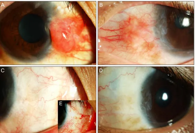
Fig. 1. (A) Slit lamp photograph of the patient’s right eye on initial presentation. Note the size, salmon color, and ele- vated appearance of the hypervascular lesion. (B) Slit lamp photograph of the patient’s left eye on initial presentation.
Note the size, faint salmon color, and mildly protruding mass. (C,D) Slit lamp photograph of both eyes 2 months follow- ing subconjunctival injection of bevacizumab. The lesions appear to be almost completely resolved with no obvious rem- nants seen on the sclera or conjunctiva. (E) Slit lamp photograph of the patient’s right eye after biopsy.
A B
Fig. 2. Haematoxylin and eosin staining. (A) ×40 magnification of the lesion biopsied in Fig. 1A. (B) ×200 magnifica- tion of the same lesion. Note the abundance of lymphocytes and the predominance of T cells (CD3+) and B cells (CD20+), a pattern typical of benign lymphoid hyperplasia.
/ 0.1 mL) (Avastin; Genentech, South San Francisco, CA, USA) was injected into both medial subconjunctival spaces. Two months after injection, both masses had almost completely disappeared and the accompanying neovascularization was
reduced (Fig. 1D and 1E). The patient did not experience re-
currence or any other complications during the one-year
follow-up period.
DH Oh, et al. Ocular Benign Lymphoma Treated with Bevacizumab