Clinical Features and Computed Tomography Characteristics of Non-Klebsiella pneumoniae Liver Abscesses in Elderly
(>65 Years) and Nonelderly Patients
Chih-Weim Hsiang,
1Chang-Hsien Liu,
1Hsiu-Lung Fan,
2Kai-Hsiung Ko,
1Chih-Yung Yu,
1Hong-Hau Wang,
1Wen-I Liao,
3Hsian-He Hsu,
1and Wei-Chou Chang
1Departments of 1Radiology and 3Emergency Medicine, Tri-Service General Hospital, National Defense Medical Center, Taipei, Taiwan;
2Division of General Surgery, Department of Surgery, Tri-Service General Hospital, National Defense Medical Center, Taipei, Taiwan, Republic of China.
Received: March 24, 1014 Revised: May 23, 2014 Accepted: May 28, 2014
Corresponding author: Dr. Wei-Chou Chang, Department of Radiology,
Tri-Service General Hospital, National Defense Medical Center, No. 325, Section 2, Cheng-Kung Road, Neihu 114, Taipei, Taiwan, Republic of China.
Tel: 886-2-87927244, Fax: 886-2-87927245 E-mail: [email protected]
∙ The authors have no financial conflicts of interest.
© Copyright:
Yonsei University College of Medicine 2015 This is an Open Access article distributed under the terms of the Creative Commons Attribution Non- Commercial License (http://creativecommons.org/
licenses/by-nc/3.0) which permits unrestricted non- commercial use, distribution, and reproduction in any medium, provided the original work is properly cited.
Purpose: To compare the clinical and computed tomography (CT) appearances of liver abscesses caused by non-Klebsiella pneumoniae bacterial pathogens in elderly and nonelderly patients. Materials and Methods: Eighty patients with confirmed non-Klebsiella pneumoniae liver abscesses (non-KPLAs) were enrolled and divid- ed into two age groups: elderly (age ≥65 years, n=42) and nonelderly (age <65 years, n=38). Diagnosis of non-KPLA was established by pus and/or blood culture.
We compared clinical presentations, outcomes, and CT characteristics of the two groups, and performed multivariate analysis for significant variables and receiver- operating-characteristic analysis to determine the cutoff value of abscess diameter for predicting non-KPLA. Results: Elderly patients with non-KPLA were associat- ed with a longer hospital stay (p<0.01). Regarding etiology, biliary sources had a strong association in the elderly group (p<0.01), and chronic liver diseases were re- lated to the nonelderly group (p<0.01). Non-KPLAs (52.5%) tended to show a large, multiloculated appearance in the elderly group and were associated with bile duct dilatation (p<0.01), compared with the nonelderly group. The abscess diameter (cutoff value, 5.2 cm; area under the curve, 0.78) between the two groups was pre- dicted. In multivariate analysis, underlying biliary tract disease [odds ratio (OR), 3.58, p<0.05], abscess diameter (OR, 2.40, p<0.05), and multiloculated abscess (OR, 1.19, p<0.01) independently predicted elderly patients with non-KPLA. Con- clusion: In the elderly patients with non-KPLA, a large, multiloculated abscess with a diameter greater than 5.2 cm was the predominant imaging feature.
Key Words: Liver abscess, Klebsiella pneumoniae, computed tomography, age
INTRODUCTION
A liver abscess is potentially life-threatening and has been reported worldwide in North America, Europe, Australia, and Asia. The reported incidence ranges from 8 to 22 cases per 100000 hospital admissions, while the fatality rate ranges from 7.8% to 28.6% due to differences in species of bacterial pathogens and the underly-
the written informed consent of patients because confidenti- ality was maintained. The study was also compliant with the Health Insurance Portability and Accountability Act.
We performed a retrospective computer search of our hos- pital database for a 7-year period (July 2003 to January 2009) using the International Classification of Diseases (9th Revision, code 572.0) to identify all patients at our institu- tion who had a diagnosis of pyogenic liver abscess other than that caused by K. pneumoniae (i.e., non-KPLA) during their hospitalization. Pyogenic liver abscess was defined as an abscess caused by at least one bacterial origin and with- out amebiasis or fungal sources. Furthermore, in this study, non-KPLA had to fulfill the following criteria: 1) presence of a focal lesion in the liver parenchyma on CT images; 2) pus drained from the abscess cavity through a diagnostic, therapeutic radiological, and/or surgical drainage proce- dure; 3) positive bacterial culture resulting from the abscess and/or blood cultures; and 4) K. pneumoniae not isolated from the cultures.
Based on our inclusion criteria, 80 patients with clinically confirmed non-KPLA were studied, including 44 males and 36 females, with a mean age of 60 years (range, 29‒95 years). In order to compare the clinical factors and imaging features of different ages, we divided our patients into two groups: elderly (age ≥65 years, n=42) and nonelderly (age
<65 years, n=38).
Microbiological etiology
The microbiology of the liver abscesses was defined as the organism recovered from the drained pus and/or blood cul- ture. Etiologic pathogens from blood cultures were consid- ered when no growth was seen in the pus culture from the liver abscess. Pus was taken for standard aerobic and anaer- obic cultures, and tested for antibiotic susceptibility, along with the blood samples. Cultures resulting from samples taken at other sites of the body, including sputum and cere- brospinal fluid, were cross-matched to confirm metastatic infection, upon clinical and/or radiological evidence. After initial workup of the pus and/or blood culture, broad-spec- trum antibiotics were given parenterally. The subsequent antibiotics were modified according to the results of the mi- crobiological cultures and antibiotic susceptibility tests.
Data collection
We collected data by reviewing the medical records of each patient for the following information: 1) demographic data;
2) coexisting medical conditions, including chronic obstruc- ing conditions of the infected patient, such as age, immune
status, alcoholism, diabetes, and chronic lung or liver diseas- es.1 Currently, due to their distinct clinical outcomes, the causative bacterial organisms of liver abscesses have usually been classified as either Klebsiella pneumoniae (K. pneu- moniae) or other groups of non-K. pneumoniae infections.
K. pneumoniae liver abscess (KPLA) is well documented in the literature, because of its tendency to occur in diabetic pa- tients, and it is highly associated with septic metastatic com- plications. With advances in diagnosis and treatment, the mortality rate of KPLA has decreased significantly in recent years. Unlike K. pneumoniae, non-K. pneumoniae patho- gens, including Escherichia coli, Enterococcus species, Staphylococcus species, Streptococcus species, and Bacte- roides species, are associated with a much higher mortality rate, even with an aggressive therapy, and a higher incidence of biliary disease than that of K. pneumoniae.2-5
Reviewing a few prior studies, Yang, et al.4-7 concluded that cases of non-KPLA tend to occur in geriatric popula- tions; however, this finding was controversial because other investigators found that there was no tendency for elderly patients to have non-KPLAs.2,3,6,8 Meanwhile, the risk fac- tors and outcomes of K. pneumoniae have been studied ex- tensively, although that has not been the case for non-K.
pneumoniae, which constitutes the greatest number of mor- talities. In fact, the importance of older patients with non- KPLA is growing because of changes in the age structure of populations.5-9 It should be assumed that patients with non-KPLA in relatively young and older age groups have different risk profiles and imaging features. A review of the literature revealed that no previous studies had explored how the clinical features and a suspicion of non-KPLA in elderly patients differ from those of relatively young indi- viduals. In addition, exactly how risk factors and imaging features differ between elderly and nonelderly groups with non-KPLA has not yet been established.
In order to improve knowledge of this entity, we evaluat- ed and compared the clinical manifestations, risk factors, outcomes, and computed tomography (CT) imaging fea- tures of non-KPLA patients in different age groups.
MATERIALS AND METHODS
Study population
This retrospective study was approved by the Institutional Review Board for human investigations and did not require
predefined enhancement threshold level of 120 Hounsfield units (HU) was set to trigger the data acquisition. The portal venous phase was scanned 40 s after initiation of the arterial phase CT scan. A scanning range from the hepatic dome of the lower lungs to the iliac crest was used. With CT, images were routinely acquired at a beam collimation of 0.5 mm and were viewed at a slice thickness of 5 mm.
CT interpretation
Two experienced abdominal radiologists retrospectively re- viewed the CT images and developed a consensus opinion.
They were aware that the patients had a liver abscess but were blinded to the patient’s clinical condition. During the analysis of the CT features, cases from the elderly and non- elderly group were randomly intermixed.
The CT characteristics of the liver abscesses were recorded in terms of the following parameters: 1) location (right, left, or both lobes of the liver); 2) margin definition (ill-defined or well-defined; ill-defined was used to describe cases where more than half of the margin was finely speculated); 3) num- ber (one, two, three, or more); 4) diameter (defined as the di- ameter of the largest abscess); 5) cystic appearance (defined as more than half the abscess cavity appearing liquefied, with an attenuation value of ≤20 HU on contrast-enhanced CT);
6) a multiloculated abscess (defined as an abscess with en- hancing internal septations); 7) thickening of the abscess wall (measurement of the maximum wall thickness, and thickened defined as thickness ≥2 mm); 8) enhancement of the rim (defined as more than half of the margin having a higher attenuation than the surrounding liver on contrast-en- hanced CT images); 9) the presence of gas density in the ab- scess; 10) bile duct dilatation (defined as a clear, hypodense tubular structure accompanying a contrast-opacified portal venous branch focally in the liver); 11) thrombophlebitis and/
or pylephlebitis (presence of filling defects in the hepatic vein, inferior vena cava and/or portal vein); 12) pneumobi- lia (presence of air density in the biliary tract).
Additionally, the radiologists also recorded the presence of any underlying biliary disease and coexisting lesions in other organs (pleural effusion, lung base, and abdominal or- gan metastatic infection).
Statistical analysis
Statistical analyses were performed with SPSS software (SPSS 18.0; SPSS Inc., Chicago, IL, USA), and differences were considered significant when p<0.05. A comparison of the clinical findings and CT features of the elderly and non- tive pulmonary disease, chronic kidney disease (defined as
estimated glomerular filtration rate <30 mL/min/1.73 m2), diabetes mellitus, biliary tract disease (defined as a stone in the bile duct, air in the biliary tree, bile duct obstruction, cholecystitis, or any previous hepatobiliary surgery), chronic liver disease (defined as alcoholism, chronic hepatitis B or C, and cirrhosis of the liver), and malignancy; 3) initial labo- ratory test data (including white blood cell count, hemoglo- bin, platelet count, C-reactive protein, total bilirubin, albu- min, aspartate aminotransferase, alanine aminotransferase, and glucose); 4) clinical symptoms (including fever or right upper quadrant abdominal pain); 5) the result of the drain- age procedure [successful percutaneous catheter drainage (PCD) defined as image-guided needle aspiration alone and successful PCD therapy; failed PCD defined as the patient’s clinical condition that required an additional surgical proce- dure, such as surgical drainage or resection. Catheter block- age or dislocation was not included in this group]; 6) mi- crobiological pathogen of the liver abscess; 7) number of days from the onset of symptoms to diagnosis; number of days that the fever subsided (body temperature of 37.5°C or less for 2 days), number of days that leukocytosis subsided (defined as a white blood count <11000 cells/per mm3) and duration of hospitalization (defined as the number of days in hospital after the drainage procedure was performed); 8) the occurrence of drainage-related complications, including catheter dislodgement, obstruction, kinking, pneumothorax, intra-abdominal hemorrhage and bile leakage; 9) the occur- rence of complications, including pleural effusion, meta- static infection, and septic shock during the same admis- sion; and 10) mortality related to the liver abscess and its complications.
CT protocol for the liver abscess
In this study, contrast-enhanced CT was performed in all pa- tients before treatment. Patients with contraindications to contrast-enhanced CT (pregnancy, acute kidney failure, or allergy to iodinated contrast agents) were excluded. The CT protocol for right upper quadrant abdominal pain at our in- stitution was planned as follows. Unenhanced CT scans are routinely performed without any oral administration. Intra- venous contrast (Omnipaque; GE Healthcare, Norway) was administered by a power injector using a contrast concentra- tion of 350 mg/mL, delivered at a rate of 2.5‒3.0 mL/s for a volume of 90 mL. The scan delay for the arterial phase im- ages was defined with bolus tracking, with a circular region of interest positioned at the level of the abdominal aorta; a
nificant difference in the proportions of each sex was ob- served between the groups.
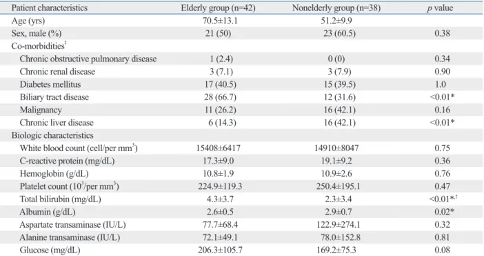
Underlying diseases of a biliary origin (identifiable on the CT images) were the most common in the elderly group, and there was a significantly higher prevalence thereof than in the nonelderly group (66.7% vs. 31.6%, p<0.01). Chron- ic liver diseases were obviously less frequent in the elderly group (14.3% vs. 42.1%, p<0.01). There were no differenc- es between groups regarding the incidence of chronic ob- structive pulmonary disease, chronic kidney disease, diabe- tes mellitus and malignancy. The findings of the laboratory tests showed higher serum total bilirubin in the elderly group than in the nonelderly group. This was considered to be rea- sonable considering that more biliary tract pathogens are associated with bile duct obstruction. There were no signifi- cant differences in the other laboratory tests at admission, except that higher total bilirubin and lower albumin levels were found in the elderly group than the nonelderly group.
Clinical characteristics of elderly and nonelderly patients with non-KPLA
There were no significant differences between the two groups with respect to symptoms including fever and right upper elderly groups was performed. For the two independent age
groups in our study, we used the Pearson’s chi-squared or Fisher’s exact test for categorical variables, and Student’s t- test for continuous variables that were expressed as mean±
standard deviation. If the variable was not normal distributed, we chose Mann-Whitney U test (Wilcoxon rank-sum test) to obtain the p value. Receiver-operating-characteristic (ROC) analysis was performed to determine the cutoff value for the optimal diameter between each study group. In addition, multivariable stepwise regression using binary logistic meth- od was performed for selecting the most significant vari- ables and obtaining their odds ratios (ORs).
RESULTS
Demographic and biologic characteristics of elderly and nonelderly patients with non-KPLA
Of the 80 patients with non-KPLA, 42 (52.5%) patients were in the elderly group, and 38 (47.5%) patients were in the nonelderly group (Table 1). The mean age was 70.5 years (range, 65‒95 years) in the elderly group and 51.2 years (range, 29‒64 years) in the nonelderly group. No sig-
Table 1. Demographic and Biologic Characteristics of Elderly and Nonelderly Patients with Non-KPLA
Patient characteristics Elderly group (n=42) Nonelderly group (n=38) p value
Age (yrs) 70.5±13.1 51.2±9.9
Sex, male (%) 21 (50) 23 (60.5) 0.38
Co-morbidities‡
Chronic obstructive pulmonary disease 1 (2.4) 0 (0) 0.34
Chronic renal disease 3 (7.1) 3 (7.9) 0.90
Diabetes mellitus 17 (40.5) 15 (39.5) 1.0
Biliary tract disease 28 (66.7) 12 (31.6) <0.01*
Malignancy 11 (26.2) 16 (42.1) 0.16
Chronic liver disease 6 (14.3) 16 (42.1) <0.01*
Biologic characteristics
White blood count (cell/per mm3) 15408±6417 14910±8047 0.75
C-reactive protein (mg/dL) 17.3±9.0 19.1±9.2 0.36
Hemoglobin (g/dL) 10.8±1.9 10.9±2.6 0.76
Platelet count (103/per mm3) 224.9±119.3 250.4±195.1 0.47
Total bilirubin (mg/dL) 4.3±3.7 2.3±3.4 <0.01*,†
Albumin (g/dL) 2.6±0.5 2.9±0.7 0.02*
Aspartate transaminase (IU/L) 77.7±68.4 122.9±274.1 0.32
Alanine transaminase (IU/L) 72.1±49.1 78.0±152.8 0.81
Glucose (mg/dL) 206.3±105.7 169.2±75.3 0.08
KPLA, Klebsiella pneumoniae liver abscess.
Continuous data are expressed as mean±standard deviations, and categorical data are expressed as numbers and percentages in parentheses (%).
*p<0.05.
†Since total bilirubin level is not normally distributed, Mann-Whitney U test was used for the p value. The median value of the total bilirubin level was 3.5 mg/dL in the elderly group and 1.2 mg/dL in the nonelderly group.
‡When patients fit into more than one category, they were counted in each category.
group, p=0.94) rarely occurred. Catheter problems were cor- rected by exchange or revision of the drainage tube. One pa- tient who experienced pneumothorax after the procedure in the nonelderly group underwent placement of a chest tube.
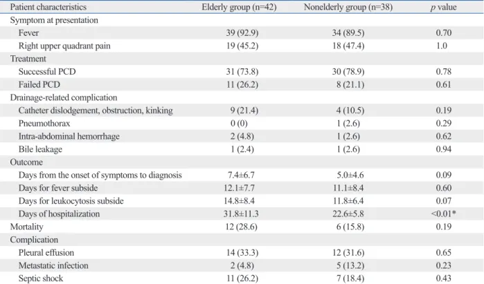
Hospital stay was longer in the elderly group (31.8±11.3 days) than the nonelderly group (17.6±5.9 days); this differ- ence was statistically significant (p<0.01). The median du- rations from the onset of symptoms to diagnosis, fever sub- sidence, and leukocytosis subsidence were higher in the elderly group; however, these differences did not reach statis- tical significance. Similar results were also found for the rates of mortality and associated complications of the two groups.
Microbiological isolates in pus and blood cultures in the elderly and nonelderly patients with non-KPLA
The species of organisms isolated in the two groups are summarized in Table 3. The overall positive growth rate of pus cultures was 77.1% (54/70), of which 70.4% (38/54) were monomicrobial. The overall positive growth rate of blood cultures was 67.1% (47/70), of which 85.1% (40/47) were monomicrobial. In both groups, the causative microor- ganism was predominantly Escherichia coli in both blood and pus cultures. Enterococcus species and coagulase-nega- quadrant abdominal pain (Table 2). All patients received
antibiotics and one or more drainage procedures. The rates of successful PCD in the elderly and nonelderly groups were 73.8% and 78.9%, respectively, which were not sig- nificantly different. The rates of failed PCD in the elderly and nonelderly groups were also not significantly different.
Specifically, in the elderly group, 11 patients experienced failed PCD therapy, with nine of these patients requiring additional surgical procedures after PCD and 2 patients re- ceiving a surgical drainage procedure initially.
According to our definition, drainage-related complications in our study occurred in 19, including 12 (28.5%) among the 42 elderly patients and seven (18.4%) among the 38 non- elderly patients. Catheter problems, including dislodgement, obstruction, and kinking, were the most common complica- tion in both elderly and nonelderly patient groups (n=9, or 21.4% in elderly group, and n=4, or 10.5% in nonelderly group, p=0.19). Other drainage-related complications, in- cluding pneumothorax (n=0, or 0% in elderly group, and n=1, or 2.6% in nonelderly group, p=0.29), intra-abdominal hemorrhage (n=2, or 4.8% in elderly group, and n=1, or 2.6% in nonelderly group, p=0.62), and bile leakage (n=1, or 2.4% in elderly group, and n=1, or 2.6% in nonelderly
Table 2. Clinical Characteristics of the Elderly and Nonelderly Patients with Non-KPLA
Patient characteristics Elderly group (n=42) Nonelderly group (n=38) p value
Symptom at presentation
Fever 39 (92.9) 34 (89.5) 0.70
Right upper quadrant pain 19 (45.2) 18 (47.4) 1.0
Treatment
Successful PCD 31 (73.8) 30 (78.9) 0.78
Failed PCD 11 (26.2) 8 (21.1) 0.61
Drainage-related complication
Catheter dislodgement, obstruction, kinking 9 (21.4) 4 (10.5) 0.19
Pneumothorax 0 (0) 1 (2.6) 0.29
Intra-abdominal hemorrhage 2 (4.8) 1 (2.6) 0.62
Bile leakage 1 (2.4) 1 (2.6) 0.94
Outcome
Days from the onset of symptoms to diagnosis 7.4±6.7 5.0±4.6 0.09
Days for fever subside 12.1±7.7 11.1±8.4 0.60
Days for leukocytosis subside 14.8±8.4 11.8±6.4 0.07
Days of hospitalization 31.8±11.3 22.6±5.8 <0.01*
Mortality 12 (28.6) 6 (15.8) 0.19
Complication
Pleural effusion 14 (33.3) 12 (31.6) 0.65
Metastatic infection 2 (4.8) 5 (13.2) 0.23
Septic shock 11 (26.2) 7 (18.4) 0.43
KPLA, Klebsiella pneumoniae liver abscess; PCD, percutaneous catheter drainage.
Continuous data are expressed as mean±standard deviations, and categorical data are expressed as numbers and percentages in parentheses (%).
*p<0.05.
Table 3. Microbiological Isolates in Pus and Blood Cultures in Elderly and Nonelderly Patients with Non-KPLA
Organism Elderly group (n=42) Nonelderly group (n=38)
Pus Blood Pus Blood
Positive growth 29 (69) 26 (62) 25 (65.8) 21 (55.3)
Monomicrobial growth 20 (47.6) 24 (57.1) 18 (47.4) 16 (42.1)
Gram-negative
Escherichia coli 12 12 10 9
Pseudomonas aeruginosa 2 2 2 1
Morganella morganii 2 3 0 0
Proteus species 2 0 1 1
Bacteroides species 2 2 2 1
Acinetobacter species 0 0 2 1
Citrobacter species 0 0 1 1
Serratia marcescens 0 0 1 1
Fusobacterium species 2 2 0 0
Gram-positive
Enterococcus species 6 4 4 5
Enterobacter species 2 1 2 1
Coagulase-negative staphylococci 5 1 5 5
Streptococcus species 2 1 1 0
Peptostreptococcus species 2 1 2 1
KPLA, Klebsiella pneumoniae liver abscess.
The categorical data are expressed as numbers and percentages in parentheses (%).
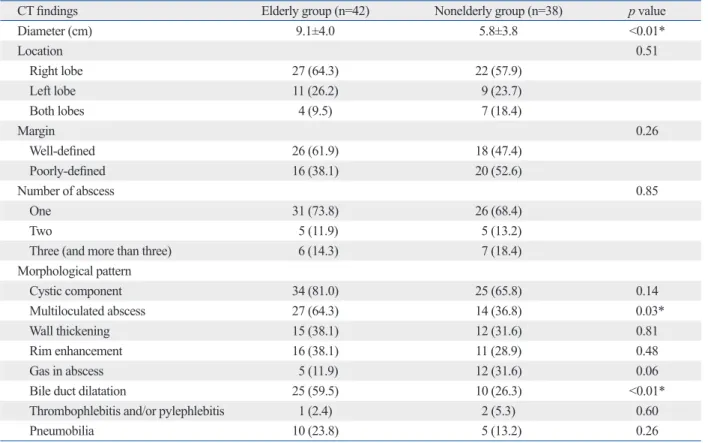
Table 4. CT Findings of Elderly and Nonelderly Patients with Non-KPLA
CT findings Elderly group (n=42) Nonelderly group (n=38) p value
Diameter (cm) 9.1±4.0 5.8±3.8 <0.01*
Location 0.51
Right lobe 27 (64.3) 22 (57.9)
Left lobe 11 (26.2) 9 (23.7)
Both lobes 4 (9.5) 7 (18.4)
Margin 0.26
Well-defined 26 (61.9) 18 (47.4)
Poorly-defined 16 (38.1) 20 (52.6)
Number of abscess 0.85
One 31 (73.8) 26 (68.4)
Two 5 (11.9) 5 (13.2)
Three (and more than three) 6 (14.3) 7 (18.4)
Morphological pattern
Cystic component 34 (81.0) 25 (65.8) 0.14
Multiloculated abscess 27 (64.3) 14 (36.8) 0.03*
Wall thickening 15 (38.1) 12 (31.6) 0.81
Rim enhancement 16 (38.1) 11 (28.9) 0.48
Gas in abscess 5 (11.9) 12 (31.6) 0.06
Bile duct dilatation 25 (59.5) 10 (26.3) <0.01*
Thrombophlebitis and/or pylephlebitis 1 (2.4) 2 (5.3) 0.60
Pneumobilia 10 (23.8) 5 (13.2) 0.26
KPLA, Klebsiella pneumoniae liver abscess.
The categorical data are expressed as numbers and percentages in parentheses (%).
*p<0.05.
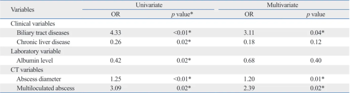
regression analyses of the selected significant variables are shown in Table 5. We chose five significant variables (clini- cal, laboratory, and imaging findings) and used a stepwise multivariate logistic regression model to identify the most significant predictors for elderly patients (>65 years) with non-KPLA. Of note, three variables (biliary tract disease, se- rum bilirubin level, and CT findings of biliary tract dilata- tion) were thought to be indicative of a similar entity; that is, highly associated with underlying biliary pathology. Since the three variables could affect each other, we decided to only include the clinical variable “biliary tract disease” as a predictor in the multivariate analysis. In the first step of mul- tivariable analyses, the ORs for each variable as being pre- dictive of elderly patients with non-KPLA were 3.11 for bili- ary tract disease, 2.39 for multiloculated abscess, 1.20 for abscess diameter, 0.68 for albumin level, and 0.18 for liver tive staphylococci were the second and third most common
pathogens, respectively. There was no significant difference in the distribution of microorganisms between groups.
CT characteristics of elderly and nonelderly patients with non-KPLA
The radiographic characteristics on CT exhibited several significant differences between the elderly and nonelderly groups (Table 4). Abscesses in the elderly group appeared significantly larger (9.1±4.0 mm vs. 5.8±3.8 mm, p<0.01) and more often had a multiloculated appearance (64.3% vs.
36.8%, p=0.03) than those in the nonelderly group (Fig. 1).
The rate of bile duct dilatation was significantly greater in the elderly group than in the nonelderly group (59.5% vs.
26.3%, p<0.01). There were no differences between the two groups with respect to the margin, number, and location of the abscesses, as well as the morphological pattern of the ab- scess cavity, including the cystic appearance, wall thicken- ing, rim enhancement, presence of dense gas, thrombophle- bitis, pylephlebitis, and pneumobilia.
ROC analysis (Fig. 2) was performed to determine the optimal value of abscess diameter in the elderly group for predicting non-KPLA in elderly patients. The ROC curve revealed that the cutoff value was 5.2 cm. Even with over- lapping appearances on CT, an abscess diameter greater than 5.2 cm was predictive of a liver abscess in the elderly group. The area under the ROC curve to predict the diame- ter of a liver abscess in the elderly group was 0.74.
Univariate and multivariate logistic regression analyses for predictors of elderly patients with non-KPLA The results of univariate and multivariate stepwise logistic
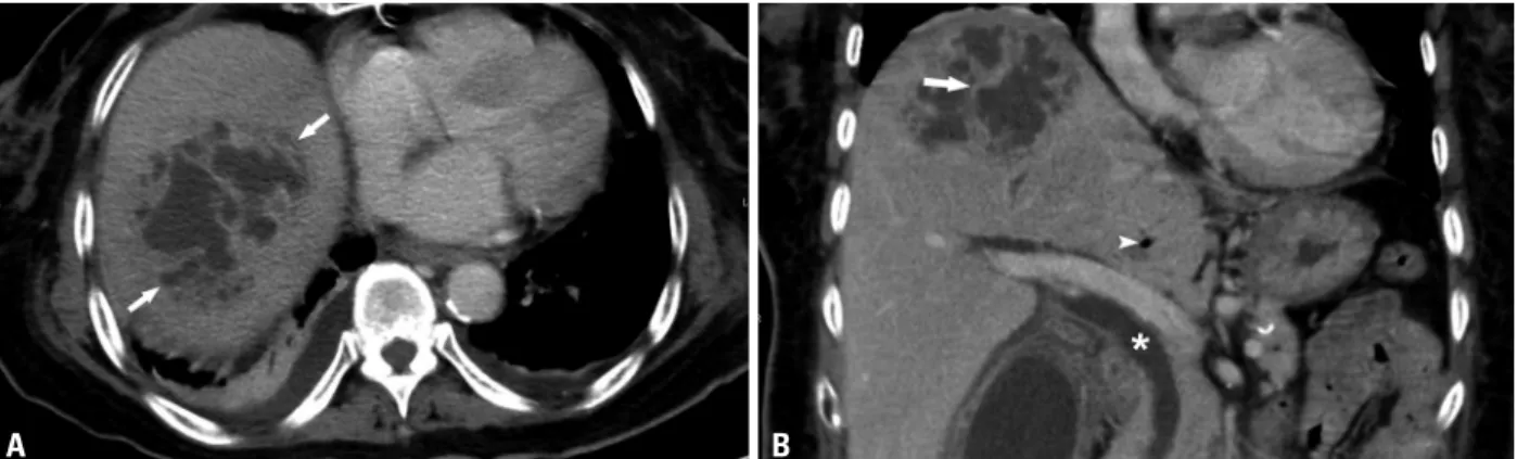
Fig. 1. A 72-year-old woman with non-KPLA (caused by Enterococcus) who presented with fever and right upper quadrant pain for 7 days. An axial, con- trast-enhanced CT image (A) shows a large (size: about 7.9 cm) abscess (arrows) in the dome. A coronal, contrast-enhanced reconstruction CT image (B) shows the abscess with a multiloculated appearance (arrow), which was associated with bile duct dilatation (star) and pneumobilia (arrowhead). Acute cholecystitis was found. This was the most common appearance of liver abscess in the elderly. Note also the mild bilateral pleural effusions and basal atel- ectasis. The patient received antibiotics and PCD drainage, and she was discharged after 25 days. KPLA, Klebsiella pneumoniae liver abscess; PCD, percu- taneous catheter drainage.
Fig. 2. Receiver-operating-characteristic (ROC) analysis of the minimal di- ameter for abscesses in the elderly group. AUC, the area under the ROC curve.
100-specificity
Sensitivity
0 20 40 60 80 100
0 20 60 40 80 100
Cut of value: 5.2 cm AUC: 0.748
A B
Size
rate. In our institution, an active drainage strategy is per- formed in patients with larger liver abscesses (diameter >5 cm) by implanting more than one drainage tube with a thick diameter. By our study, we have demonstrated that an ac- tive drainage strategy could provide a better clinical out- come than the previous published studies. Although there were significant differences in terms of larger abscess diam- eter, multiloculated abscesses, and hospital stay between el- derly and nonelderly patients, we believe that an active drain- age strategy could overcome drainage failures in certain cases with larger abscess diameter and reach a comparable success rate to that in the nonelderly patients.
Multiloculated abscesses were a distinctive imaging fea- ture of non-KPLA in the elderly patients. Multiloculated ab- scesses have been considered to be an important factor in determining the outcome of liver abscess treatment, from ei- ther a clinical or a radiological perspective.10,13,14 Liu, et al.15 showed that with an adequate drainage strategy, multilocu- lated abscesses show no differences in PCD failure rate and mortality rate, compared with a single abscess. The results of our study tend to support these findings. Nevertheless, multiloculated abscesses absolutely contribute to poorer drainage by compartmentalization of the liver abscess, re- ducing the effectiveness of percutaneous drainage.10,16 In our experience, successful drainage of multiloculated abscesses can be achieved by irrigating the abscesses, rotating a wire to disrupt the internal septations, or placing more than one drainage tube. Most important, the use of manipulating our drainage tubes did not come at an increased complications or mortality for our patients.
Biliary tract disease was the most common underlying disease in elderly patients with non-KPLA. Furthermore, our study showed that bile duct dilatation was a characteris- tic imaging feature of non-KPLA in the elderly group. One of the most important findings of our study was that 80% of disease. In the last step, biliary tract disease (OR, 3.58, p<
0.05), multiloculated abscess (OR, 2.40, p<0.05), and ab- scess diameter (OR, 1.19, p<0.01) were the three most signif- icant independent predictors.
DISCUSSION
Although previous studies have shown that non-KPLA fre- quently occurs in the presence of an underlying biliary dis- ease or a predisposing medical condition,2 our study is the first to investigate the clinical features and CT appearance of non-KPLA in patients of different age groups. We showed that elderly patients with non-KPLA present with a large, multiloculated abscess on CT images, which was different from those identified in the nonelderly group. An abscess diameter of 5.2 cm was the cutoff value between the two groups. In the elderly group, bile duct dilatation was a sup- plementary finding of non-KPLA. Moreover, a difference was also found in the comorbidities of non-KPLA, which showed that biliary sources had a strong association in the elderly group; chronic liver diseases were frequently found in the nonelderly group.
Several previous studies found that the abscess diameter is associated with important outcomes, including more com- plications and longer hospitalizations, as well as an increased likelihood of surgical or radiological interventions.10-12 Tan, et al.10 reported that larger abscesses (diameter >5 cm) were associated with an increased hospital stay and drainage fail- ure rate. Liao, et al.12 further concluded that an abscess di- ameter greater than 7.3 cm was the optimal cutoff value to predict drainage failure. However, our study does not sup- port all of these results. We found that elderly patients had larger abscesses and were even prone to a longer hospital stay, although they did not show a higher drainage failure
Table 5. Univariate and Multivariate Logistic Regressions for Predictors of Elderly Patients with Non-KPLA
Variables Univariate Multivariate
OR p value* OR p value
Clinical variables
Biliary tract diseases 4.33 <0.01* 3.11 0.04*
Chronic liver disease 0.26 0.02* 0.18 0.12
Laboratory variable
Albumin level 0.42 0.02* 0.68 0.40
CT variables
Abscess diameter 1.25 <0.01* 1.20 0.01*
Multiloculated abscess 3.09 0.02* 2.39 0.02*
OR, odds ratio; KPLA, Klebsiella pneumoniae liver abscess.
*p<0.05.
This study has several limitations that should be men- tioned. First, this study was performed retrospectively at our institution, and the number of non-KPLA patients was still relatively small. We excluded patients for whom CT was not used as a diagnostic imaging modality. However, the num- ber of patients at our institution who received only ultraso- nography or magnetic resonance imaging for liver abscess was small. Second, we used an objective cutoff point of age of 65 years to allocate patients to the elderly and nonelderly age groups. Although the purpose of our study was to find the clinical and CT differences of non-KPLA in relation to age, this choice of age cutoff may cause a potential selection bias in our patient population. Last, we studied liver abscess- es caused by the non-K. pneumoniae species. The causative organisms were polymicroorganisms, and this might have caused differences between the two groups. We analyzed the microbiology of isolated pus and blood cultures, and the re- sults showed no significant difference between the groups.
Therefore, there was no microbiological bias identified in this study.
In conclusion, even with overlapping CT appearances, we found that non-KPLA in the elderly patients commonly showed a large multiloculated abscess with the diameter greater than 5.2 cm. However, metabolic syndrome has often been associated with more severe liver abnormalities, and pa- tients with non-alcoholic fatty liver disease tend to have ab- normal components of metabolic syndrome.20 A larger pro- spective study is required for further validation of these clinical and CT variables across institutions, and a further di- rect comparison of elderly and nonelderly patients with non- alcoholic fatty liver disease or metabolic syndrome is also mandatory to implement an optimal diagnostic algorithm.
ACKNOWLEDGEMENTS
The study was supported by the Tri-Service General Hospi- tal Research Grant (TSGH-C101-053).
We thank the Research Office for Health Data, Depart- ment of Education and Research, Taipei City Hospital, Tai- wan for their valuable contributions in data management and statistical analysis.
REFERENCES
1. Foo NP, Chen KT, Lin HJ, Guo HR. Characteristics of pyogenic
patients in the elderly group with underlying biliary tract disease showed CT evidence of bile duct dilatation. This as- sociation of biliary tract abnormalities emphasizes the neces- sity for the evaluation of biliary tract disease in elderly pa- tients with non-KPLA. A previous study of patients with a liver abscess who underwent endoscopic retrograde cholan- giopancreatography found that 75% of patients with com- mon bile duct dilatation had an associated biliary tract patho- logical finding.17 Therefore, we propose that identification of a dilated bile duct by imaging in an elderly patient with a liv- er abscess should prompt a more detailed evaluation of the biliary tract, and consideration should be made to investigate further using imaging studies, such as magnetic resonance cholangiography or endoscopic retrograde cholangiopancre- atography.
In our study, we noticed that non-KPLA in the elderly group required a longer hospitalization, compared to that in the nonelderly group. Although researchers agree that a larg- er, multiloculated abscess requires a longer hospital stay to adequately achieve drainage and resolution of the abscess, longer hospitalization in the elderly patients could be affect- ed by other confounding factors. Of the associated co-mor- bidities in this study, the disease most related with the epi- sode of non-KPLA in elderly patients was the underlying biliary pathology. While the prevalence of underlying biliary tract diseases was significantly different between the two age groups, we postulated that a longer hospital study is not only governed by the patient’s age and the abscess diame- ter/morphology, but is also related to the associated under- lying biliary pathology.
Our study outlined different comorbidities in the two age groups. Previous studies have shown that non-KPLA is strongly associated with the underlying biliary disease; in- terestingly, our study further found that patient age was a major attribute of biliary tract pathology in the non-KPLA population. Aging has an influence on both the physiologi- cal and anatomical features of the biliary tract, causing an increased incidence of biliary tract disease.18 The spread of bacterial pathogens along the biliary tree that form an ab- scess at the liver parenchyma, in association with bile duct dilatation, is considered to be the pathogenic mechanism.
Nevertheless, chronic liver diseases were more frequently found in the nonelderly group. In the past, liver cirrhosis was considered to be a risk factor for a liver abscess.19 Our finding is thought to be due to the high prevalence of chron- ic hepatitis B and C infections, with associated cirrhosis, in young adults in Taiwan.
neous rupture of liver abscess caused by Klebsiella pneumoniae.
Diagn Microbiol Infect Dis 2005;52:79-84.
12. Liao WI, Tsai SH, Yu CY, Huang GS, Lin YY, Hsu CW, et al.
Pyogenic liver abscess treated by percutaneous catheter drainage:
MDCT measurement for treatment outcome. Eur J Radiol 2012;
81:609-15.
13. Tazawa J, Sakai Y, Maekawa S, Ishida Y, Maeda M, Marumo F, et al. Solitary and multiple pyogenic liver abscesses: characteristics of the patients and efficacy of percutaneous drainage. Am J Gas- troenterol 1997;92:271-4.
14. Chou FF, Sheen-Chen SM, Chen YS, Chen MC. Single and mul- tiple pyogenic liver abscesses: clinical course, etiology, and results of treatment. World J Surg 1997;21:384-8.
15. Liu CH, Gervais DA, Hahn PF, Arellano RS, Uppot RN, Mueller PR. Percutaneous hepatic abscess drainage: do multiple abscesses or multiloculated abscesses preclude drainage or affect outcome?
J Vasc Interv Radiol 2009;20:1059-65.
16. Barakate MS, Stephen MS, Waugh RC, Gallagher PJ, Solomon MJ, Storey DW, et al. Pyogenic liver abscess: a review of 10 years’
experience in management. Aust N Z J Surg 1999;69:205-9.
17. Lam YH, Wong SK, Lee DW, Lau JY, Chan AC, Yiu RY, et al.
ERCP and pyogenic liver abscess. Gastrointest Endosc 1999;50:
340-4.
18. Walsh RM. Innovations in treating the elderly who have biliary and pancreatic disease. Clin Geriatr Med 2006;22:545-58.
19. Mølle I, Thulstrup AM, Jepsen P, Sørensen HT, Vilstrup H. Liver cirrhosis is risk factor for pyogenic liver abscesses. BMJ 2001;
323:52-3.
20. Tarantino G, Finelli C. What about non-alcoholic fatty liver dis- ease as a new criterion to define metabolic syndrome? World J Gastroenterol 2013;19:3375-84.
liver abscess patients with and without diabetes mellitus. Am J Gastroenterol 2010;105:328-35.
2. Lee NK, Kim S, Lee JW, Jeong YJ, Lee SH, Heo J, et al. CT dif- ferentiation of pyogenic liver abscesses caused by Klebsiella pneumoniae vs non-Klebsiella pneumoniae. Br J Radiol 2011;84:
518-25.
3. Alsaif HS, Venkatesh SK, Chan DS, Archuleta S. CT appearance of pyogenic liver abscesses caused by Klebsiella pneumoniae. Ra- diology 2011;260:129-38.
4. Yang CC, Yen CH, Ho MW, Wang JH. Comparison of pyogenic liver abscess caused by non-Klebsiella pneumoniae and Klebsiella pneumoniae. J Microbiol Immunol Infect 2004;37:176-84.
5. Lederman ER, Crum NF. Pyogenic liver abscess with a focus on Klebsiella pneumoniae as a primary pathogen: an emerging dis- ease with unique clinical characteristics. Am J Gastroenterol 2005;100:322-31.
6. Law ST, Li KK. Older age as a poor prognostic sign in patients with pyogenic liver abscess. Int J Infect Dis 2013;17:e177-84.
7. Chen SC, Lee YT, Yen CH, Lai KC, Jeng LB, Lin DB, et al. Pyo- genic liver abscess in the elderly: clinical features, outcomes and prognostic factors. Age Ageing 2009;38:271-6.
8. Alvarez JA, González JJ, Baldonedo RF, Sanz L, Junco A, Rodrfí- guez JL, et al. Pyogenic liver abscesses: a comparison of older and younger patients. HPB (Oxford) 2001;3:201-6.
9. Kang SC, Hwang SJ. Impact of advanced age on inpatients with pyogenic liver abscess in Taiwan: a nationwide claim-based analy- sis. J Chin Med Assoc 2011;74:539-43.
10. Tan YM, Chung AY, Chow PK, Cheow PC, Wong WK, Ooi LL, et al. An appraisal of surgical and percutaneous drainage for pyogenic liver abscesses larger than 5 cm. Ann Surg 2005;241:485-90.
11. Lee CH, Leu HS, Wu TS, Su LH, Liu JW. Risk factors for sponta-