J O U R N A L O F
Veterinary Science
pISSN 1229-845X, eISSN 1976-555X J. Vet. Sci. (2012), 13(2), 207-209 http://dx.doi.org/10.4142/jvs.2012.13.2.207
Received: 11 May 2011, Revised: 28 Jun. 2011, Accepted: 30 Dec. 2011
Case Report
Fig. 1. Case 1. (A) A dog presenting characteristic bilateral symmetrical alopecia with hyperkeratosis involving the ventral abdomen, flanks, thorax, and neck. (B) The left testis at surgery was greatly increased in size and had multiple spermatic cord torsions (arrow).
*Corresponding author: Tel: +39-090-350-3690; Fax: +39-090-350-3979; E-mail: [email protected]
ⓒ 2012 The Korean Society of Veterinary Science.
This is an Open Access article distributed under the terms of the Creative Commons Attribution Non-Commercial License (http://creativecommons.org/licenses/by-nc/3.0) which permits unrestricted non-commercial use, distribution, and reproduction in any medium, provided the original work is properly cited.
Sertoli cell tumors associated with feminizing syndrome and spermatic cord torsion in two cryptorchid dogs
Marco Quartuccio, Gabriele Marino*, Giuseppe Garufi, Santo Cristarella, Antonina Zanghì Department of Veterinary Public Health, University of Messina, 98168 Messina, Italy
The association of cryptorchidism, functional Sertoli cell tumors, and spermatic cord torsion has been rarely reported in the literature. Two dogs were admitted for bilateral skin alopecia and weight loss. Both animals were cryptorchid and displayed a pendulous preputial sheath, prostate hypertrophy, and increased levels of circulating oestrogen. Transabdominal palpation and ultrasonography revealed the presence of neoplastic retained gonads. During surgery, spermatic cord torsion was also detected in the enlarged neoplastic testes of both dogs. Histologic examination confirmed the presence of Sertoli cell tumors that were primarily responsible for the feminizing syndrome. Complete remission of all symptoms occurred within 3 months after orchiectomy.
Keywords: cryptorchidism, dog, feminizing syndrome, Sertoli cell tumor, spermatic cord torsion
Cryptorchidism is a testicular developmental disorder that is quite common in dogs and mainly associated with genetic causes [1]. In retained testes, there is an increased risk of neoplasms, such as Sertoli cell tumors and seminoma, which can exhibit more aggressive behavior than those in scrotal testes [3,9]. Approximately 70% of Sertoli cell tumors arising in abdominal testes are functional and associated with a feminizing paraneoplastic syndrome characterized by non-pruritic, bilateral symmetrical alopecia, hyperpigmentation, gynecomastia, edematous and pendulous penile sheath, prostatic dysfunctions, attraction to other males, and standing in a female posture to urinate [1,4,6,10]. Estrogen myelotoxicosis has been reported in 15% of these dogs and is characterized by bone marrow hypoplasia and non-regenerative anemia [10].
Retained testes are more susceptible than scrotal testes to spermatic cord torsion, and the risk of this condition is
increased even more with progressive enlargement of the neoplastic organ [1,7,8]. Although closely linked to one another, concomitant association of cryptorchidism, Sertoli cell tumors, feminizing syndrome, and spermatic cord torsion has been rarely reported in the literature [5,8]. Here, we present two cases of cryptorchidism accompanied by spermatic cord torsion and feminization in dogs.
Case history 1: A 7-year-old male mixed breed dog
weighing 27 kg was brought for our examination due to
widespread alopecia with hyperkeratosis, progressive
weight loss, feminine behavior during urination, and the
absence of both testes from the scrotum. Non-pruritic
alopecia had originated in the genital and perineal regions,
and spread symmetrically into the ventral abdomen, flanks,
thorax, and neck (Fig. 1A). Physical examination suggested
the development of bilateral abdominal cryptorchidism. The
prepuce was pendulous and the prostate was uniformly
enlarged. Palpation of the left caudal abdomen identified a
large, firm, and painful mass. Ultrasound examination
showed a ‘complex mass’ structure enclosed by a
208 Marco Quartuccio et al.
Fig. 2. Case 2. Gross appearance of a totally neoplastic testis
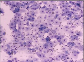
compared to the contralateral atrophic one. Fig. 3. Case 2. Epithelial cells with signs of squamous metaplasia collected from a prostatic cyst. May-Grünwald-Giemsa stain, ×250.
hyperechoic capsule. In the other quadrant, another isoechoic formation of smaller dimensions that resembled testicular parenchyma was also observed. The prostate had increased in volume (4.7 × 4.8 × 7.8 cm), and contained a non-homogeneous parenchyma that included small anechoic areas likely to be cysts. Abdominal radiographs confirmed these findings and, together with the thoracic findings, did not reveal metastases.
Hematological analysis indicated moderate anemia and leukocytosis (hematocrit: 25.7%, hemoglobin: 11.90 g/dL, RBC: 4.37 × 10
6/μL, WBC: 21.60 × 10
3/μL). Endocrine evaluation showed alterations in sexual steroid levels with increases in estrogen [progesterone (P
4) = 0.39 ng/mL, 17 beta-estradiol (E
2) = 17.90 pg/mL, testosterone (T) = 0.66 ng/mL]. Suspecting a Sertoli cell tumor in the retained testis, an exploratory midline laparotomy was carried out to locate and remove both testes.
The left testis, greatly increased in size, presented multiple spermatic cord torsions (Fig. 1B). The right testis, moderately increased in size, was also neoplastic. Grossly, the left testis (10.5 × 6.5 × 5.5 cm) had a significant profile alteration and multilobular appearance; cross-sectionally, it appeared to be completely replaced by spongy neoplastic tissue. The right testis (5 × 4 × 2.5 cm) had a pyramidal shape and was almost completely replaced by lobular compact neoplastic tissue that was yellowish in color. Histopathological sections confirmed the presence of a Sertoli cell tumor with tubular and pseudofollicular patterns in both gonads.
Post-operative treatment was administered including anti-inflammatory drugs for 3 days and antibiotics for 7 days. After 1 month, the clinical symptoms significantly improved. Specifically, the prostate size was greatly reduced, hematological values appeared to recover, and sexual hormones returned to baseline levels (E
2and T were not detected. P
4= 0.14 ng/mL). Total remission of skin
symptoms was observed 3 months after surgery.
Case history 2: An 8-year-old male Rottweiler dog weighing 35 kg was admitted for severe hematuria, weight loss, and progressive apathy that had developed during the preceding 5 months. Clinical signs included non-pruritic bilateral symmetrical alopecia in the flanks and thighs. The prepuce was edematous and pendulous, and moderate gynecomastia was also evident. The abdomen was extremely sensitive to palpation and a nodular mass was detected in the caudal quadrant. The left testis had decreased in size and consistency, and was noticeably close to the external inguinal ring. The prostate appeared enlarged and painful upon digital palpation.
Ultrasound examination of the caudal right abdominal quadrant revealed a ‘complex mass’ structure. The prostate measured 6.9 × 6.8 × 10.5 cm, and had a non-homogeneous echogenicity with cystic formations up to 2.2 cm in diameter. Hematological analysis revealed anemia and leukocytosis (hematocrit: 24.0%, hemoglobin: 8.10 g/dL, RBC: 3.36 × 10
6/μL, WBC: 33.30 × 10
3/μL). Endocrine evaluation showed alterations in sexual steroids with increased estrogen levels (P
4= 1.32 ng/mL, E
2= 23.05 pg/mL, T = 0.20 ng/mL). Blood transfusion was necessary to lower anesthetic risk before surgery.
After removing the left testicle, a midline laparotomy identified the right neoplastic testis, associated with double torsion of the spermatic cord, in the abdomen. Following this procedure, the prostatic cyst content was aspirated and the abdomen was closed. Grossly, the right testis (2.5 × 1.5
× 1 cm) was atrophic while the left (4.5 × 3.5 × 2.7 cm) was
round in shape and appeared to be completely replaced by
neoplastic tissue with a spongy appearance and yellowish-
gray in color (Fig. 2). Histological sections confirmed the
diagnosis of a Sertoli cell tumor with a pseudotubular
pattern characterized by highly vacuolized elements and
Testicular diseases in dogs 209