ISSN 0378-6471 (Print)⋅ISSN 2092-9374 (Online)
http://dx.doi.org/10.3341/jkos.2016.57.7.1102
Original Article
한국인의 건강검진에서 발견되는 대사증후군의 구성요소와 망막 혈관 변화와의 관계
Association between Metabolic Syndrome and Retinal Vascular Changes in Koreans based on Health Check-ups
정경도⋅김재석
Kyeong Do Jeong, MD, Jae Suk Kim, MD, PhD
인제대학교 상계백병원 안과학교실
Department of Ophthalmology, Inje University Sanggye Paik Hospital, Seoul, Korea
Purpose: To evaluate the associations between components of metabolic syndrome and retinal vascular changes in a Korean population based on data collected at health check-ups.
Methods: Fundus photographs of 381 patients participating in a health check-up were examined to identify central retinal artery equivalent (CRAE), central retinal vein equivalent (CRVE), and arteriovenous ratio (AVR) by IVAN software. Retinal hemor- rhage, arteriovenous nicking, and retinal exudate were also noted. The association between metabolic syndrome and each com- ponent was then analyzed.
Results: Significant associations were shown between metabolic syndrome and CRAE (p = 0.032), central obesity and CRAE (p = 0.037), triglyceride and CRAE (p = 0.011), and triglyceride and AVR (p = 0.005), in addition to central obesity and arterio- venous nicking (odds ratio [OR] = 2.68, p = 0.013), central obesity and retinal exudate (OR = 2.30, p = 0.038), serum glucose and retinal hemorrhage (OR = 8.06, p = 0.030), and blood pressure and arteriovenous nicking (OR = 2.78, p = 0.007).
Conclusions: Metabolic syndrome showed a significant relationship with retinal artery diameter. Central obesity showed the greatest relationship with retinal vascular changes among each of the components of metabolic syndrome.
J Korean Ophthalmol Soc 2016;57(7):1102-1108
Keywords: Metabolic syndrome, Retinal artery, Retinal hemorrhage, Retinal vein
■Received: 2016. 3. 10. ■ Revised: 2016. 4. 30.
■Accepted: 2016. 6. 23.
■Address reprint requests to Jae Suk Kim, MD, PhD
Department of Ophthalmology, Inje University Sanggye Paik Hospital, #1342 Dongil-ro, Nowon-gu, Seoul 01757, Korea Tel: 82-2-950-1096, Fax: 82-2-935-6904
E-mail: [email protected]
ⓒ2016 The Korean Ophthalmological Society
This is an Open Access article distributed under the terms of the Creative Commons Attribution Non-Commercial License (http://creativecommons.org/licenses/by-nc/3.0/) which permits unrestricted non-commercial use, distribution, and reproduction in any medium, provided the original work is properly cited.
대사증후군은 심혈관 질환과 제2형 당뇨병의 위험도를 증가시키는 복부 비만, 고혈당증, 고혈압, 이상지혈증 등의 대사 위험요인이 군집성으로 존재하는 상태이다.1-3 대사 증 후군의 유병률은 연구들마다 차이가 있지만 미국 성인에서 는 24%, 한국 성인에서는 약 11-19%로 보고되고 있다.4,5
대사증후군 및 각 요소는 심혈관 질환과 당뇨병 발병률의 위험도, 동맥경화를 증가시킬 수 있다고 알려져 있으며, 최 근에는 미세혈관에도 영향을 줄 수 있다고 보고되고 있다.6-8 망막은 인체에서 미세혈관을 직접 관찰할 수 있는 유일 한 조직이다. 안저 검사로 망막과 망막 혈관을 직접 관찰하 여 미세 혈관의 병적 변화를 알 수 있다. 특히 고해상도의 안저 사진은 망막 혈관의 주행과 방향, 굵기, 동정맥 교차, 출혈, 삼출물 등의 병적 변화를 쉽게 관찰할 수 있어 망막 혈관 질환의 진단에 도움이 된다. 이전 연구에서 고혈압, 심부전, 관상동맥질환, 뇌졸중, 당뇨, 고혈당 등이 망막 혈 관의 직경에 미치는 영향에 대해서 보고가 되었으며,9-21 미
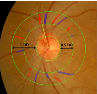
Figure 1. Fundus photograph centered at the optic disc of left
eye, grader analysis measures arteries and veins, the largest six arteries (red) and veins (blue) are used to calculate the cen- tral retinal artery equivalent and central retinal vein equivalent by the “Big 6” method. Yellow line is the vessel indicator of the IVAN. DD = disc diameter.국, 일본, 중국에서는 Atherosclerosis Risk in Communities (ARIC) study, Funagata study 등을 통해 대사증후군이 망 막 혈관 직경 및 망막 출혈, 동정맥 함요, 삼출물 등의 망막 미세 혈관 변화와 관련 있다고 보고되었다.18,22,23
최근에는 종합건강검진 시 대사증후군과 관계된 검사를 시행하여 조기에 발견하고 있으며, 대부분 안과 질환에 대 한 선별검사로 안저 사진을 촬영하고 있다. 본 연구에서는 한국인에서 대사증후군의 구성요소와 망막혈관의 변화에 대해 기존에 보고된 바가 없기에, 건강검진 자료를 활용하 여 대사증후군의 구성요소와 망막 혈관 변화와의 관계를 알아보고자 하였다.
대상과 방법
2014년 1월 1일부터 2014년 4월 30일까지 인제대학교 상계백병원 종합건강검진센터에서 건강 검진을 위하여 내 원한 환자 중 무산동안저촬영을 시행한 321명을 대상으로 후향적으로 의무기록을 분석하였고, 본원 임상시험 윤리위 원회로부터 심사면제 판정을 받았다. 과거력상 다른 전신 질환을 진단 받은 적이 없는 20-80세를 대상으로 하며, 기 존에 망막변성, 망막혈관폐쇄, 나이관련황반변성 등의 망막 질환, 포도막염, 유리체 출혈, 유리체 혼탁 등의 매체 혼탁 으로 안저 사진 판독이 어려운 경우는 제외하였다.
대상자들은 암실에서 안저 사진 촬영기(TRC-NW200, Topcon, Tokyo, Japan)로 양안에서 황반을 중심으로 45° 범위의 안 저를 촬영하였다. 촬영한 안저 사진은 이미지 관리 프로그 램(IMAGEnet2000, Topcon, Tokyo, Japan)을 이용하여 압 축하지 않은 1,500 × 1,500 픽셀 크기의 Tagged Image File (TIF)로 추출하였다. 모든 안저 사진의 분석은 환자의 정보 를 모르는 1명의 검사자에 의하여 시행되었다. 검사자는 안 저 사진에서 망막 출혈 유무, 동정맥 함요 유무, 망막 삼출 물 유무를 확인하였다.
망막 혈관 직경은 반자동화 망막혈관 분석 컴퓨터 프로 그램(Interactive Vessel Analysis, IVAN; University of Wisconsin-Madison, Madison, WI, USA)을 이용하여 디지 털 안저 사진을 분석하여 측정하였다. 프로그램은 안저 사 진에서 시신경 유두를 인식하여 시신경 유두 경계로부터 0.5에서 1 유두 직경 사이의 범위에 있는 망막 혈관을 자동 으로 찾아낸다. 시신경 유두의 위치가 잘못 되거나 망막 혈 관의 종류와 범위가 틀린 경우에는 수동으로 보정하였으며, 인식된 망막 혈관은 자동으로 직경이 측정되고 측정치에 대한 분석이 이루어진다. Leung et al24의 연구에서 양안의 망막 혈관 직경에 유의한 차이가 없었다는 보고를 참고하 여, 본 연구에서는 우안의 안저 사진을 분석하는 것을 원칙
으로 하고, 우안의 안저 사진 화질이 분석에 적합하지 않은 경우 좌안의 안저 사진을 분석하였다. 망막 혈관 직경의 계 산은 분석 범위 내의 망막 동맥과 정맥 중 각각의 가장 굵 은 6개의 직경을 계산하는 Big 6 법을 이용하여 이루어졌 다.25,26 망막 동맥의 직경은 central retinal artery equivalent (CRAE), 망막 정맥의 직경은 central retinal vein equivalent (CRVE), arteriovenous ratio (AVR)는 CRAE/CRVE로 망막 혈관의 동정맥 비율을 나타내는 값으로 IVAN 프로그램은 망막 혈관을 설정하면 CRAE, CRVE, AVR을 자동으로 측 정해준다(Fig. 1).
키와 몸무게는 신발을 벗은 상태로 가벼운 옷차림으로 측정하였으며 허리 둘레는 배꼽 위치에서 측정하였다. 모든 대상자는 10시간 공복을 시행 후 혈액을 채취하였으며 혈 당, 중성지방, 고밀도 콜레스테롤은 혈액검사장비(AU5800, Beckman Coulter, Brea, CA, USA)를 이용하여 측정하였다.
혈압은 앉은 자세로 5분간 휴식을 시행한 후 자동혈압계 (FT-500, Jawon Medical, Guri, Korea)로 측정하였다. 대사 증후군의 정의는 National Cholesterol Education Program Adult Treatment Panel III (NCEP III)를 참고하여 다음 중 3가지 이상을 만족하는 경우로 정하였다.2
- 복부비만 central obesity: 남성 90 cm 이상, 여성 85 cm 이상
- 중성지방 triglyceride: 150 mg/dL 이상
Table 1. Demographics of participants with or without metabolic syndrome, defined with National Cholesterol Education Program
Adult Treatment Panel III (NCEP III) criteriaMetabolic syndrome
Present (N = 52) Absent (N = 269) p-value*
Age (years) 58.5 ± 10.8 54.6 ± 10.0 0.012
Gender (% male) 37 (71.2) 165 (59.5) 0.122
Height (kg) 165.8 ± 9.7 163.8 ± 8.7 0.135
Weight (cm) 73.9 ± 11.8 64.6 ± 10.2 <0.001
Body mass index (kg/m2) 26.8 ± 2.7 24.0 ± 2.6 <0.001
Waist circumference (cm) 89.9 ± 7.15 79.7 ± 8.5 <0.001
Systolic blood pressure (mm Hg) 129.4 ± 6.1 119.5 ± 9.7 <0.001
Diastolic blood pressure (mm Hg) 78.1 ± 5.6 74.3 ± 5.9 <0.001
Triglyceride (mg/dL) 189.2 ± 84.0 114.2 ± 66.8 <0.001
HDL cholesterol (mg/dL) 40.9 ± 7.6 51.5 ± 10.7 <0.001
Fasting glucose (mg/dL) 118.3 ± 36.0 94.5 ± 14.6 <0.001
Values are presented as mean ± SD unless otherwise indicated.
HDL = high density lipoprotein.
*Independent t-test.
Table 2. Relationship of retinal vascular calibers with metabolic syndrome factors
CRAE (μm) CRVE (μm) AVR
Waist circumference
≥90 cm (M), ≥85 cm (F) 144.63 ± 18.56 209.00 ± 25.30 0.70 ± 0.11
<90 cm (M), <85 cm (F) 150.85 ± 17.26 211.85 ± 20.42 0.72 ± 0.09
p-value 0.037 0.602 0.250
Triglyceride
≥150 mg/dL 144.80 ± 16.10 212.06 ± 20.27 0.69 ± 0.08
<150 mg/dL 151.07 ± 17.98 210.95 ± 22.02 0.72 ± 0.10
p-value 0.011 0.566 0.005
HDL cholesterol
≤40 mg/dL (M), ≤50 mg/dL (F) 149.45 ± 18.08 210.75 ± 21.52 0.71 ± 0.09
>40 mg/dL (M), >50 mg/dL (F) 149.50 ± 17.60 211.41 ± 21.63 0.71 ± 0.09
p-value 0.994 0.864 0.958
Glucose
≥110 mg/dL 146.49 ± 16.51 213.07 ± 24.06 0.69 ± 0.08
<110 mg/dL 149.93 ± 17.86 210.96 ± 21.21 0.72 ± 0.10
p-value 0.543 0.279 0.169
Blood pressure
≥130/85 mm Hg 147.77 ± 16.90 209.21 ± 25.03 0.71 ± 0.10
<130/85 mm Hg 150.65 ± 18.18 212.58 ± 18.83 0.71 ± 0.09
p-value 0.360 0.316 0.755
Metabolic syndrome
Yes 144.08 ± 15.83 209.99 ± 22.30 0.69 ± 0.08
No 150.54 ± 17.89 211.47 ± 21.46 0.72 ± 0.10
p-value 0.032 0.750 0.084
Values are presented as mean ± SD unless otherwise indicated. All components are adjusted for age and gender.
CRAE = central retinal artery equivalent; CRVE = central retinal vein equivalent; AVR = arteriovenous ratio; M = male; F = female; HDL
= high density lipoprotein.
- 고밀도 콜레스테롤 high density lipoprotein (HDL) cholesterol: 남성 40 mg/dL 미만, 여성 50 mg/dL 미만 - 공복혈당 fasting plasma glucose: 110 mg/dL 이상 - 혈압 blood pressure: 130/85 mmHg 이상
각 군 간의 CRAE, CRVE, AVR의 비교를 위해 공분산 분석, 각 군 간의 망막출혈 여부, 동정맥 함요 여부, 망막
삼출물 여부의 비교를 위해 로지스틱 회귀분석을 통계프로 그램 Statistical Analysis Software (SAS) version 5.1 (SAS Institute Inc., Cary, NC, USA)로 분석하였으며, p-value가 0.05 미만인 경우를 통계적으로 의미 있는 것으로 해석하 였다. 모든 분석은 연령과 성별을 보정하여 시행하였다.
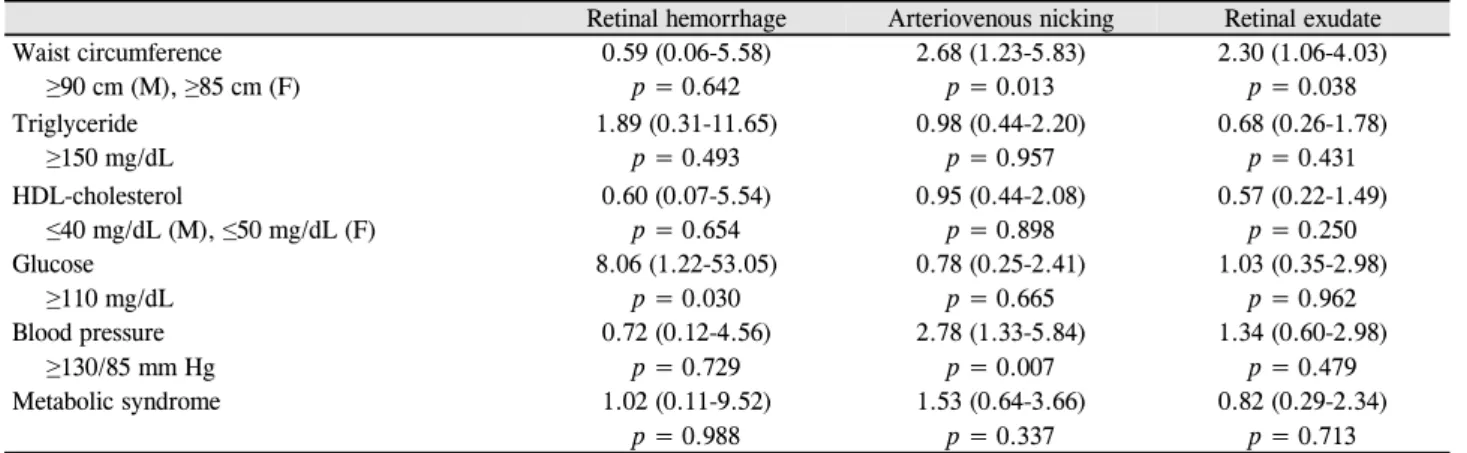
Table 3. Odds ratio (95% confidence interval) for components of metabolic syndrome
Retinal hemorrhage Arteriovenous nicking Retinal exudate
Waist circumference 0.59 (0.06-5.58) 2.68 (1.23-5.83) 2.30 (1.06-4.03)
≥90 cm (M), ≥85 cm (F) p = 0.642 p = 0.013 p = 0.038
Triglyceride 1.89 (0.31-11.65) 0.98 (0.44-2.20) 0.68 (0.26-1.78)
≥150 mg/dL p = 0.493 p = 0.957 p = 0.431
HDL-cholesterol 0.60 (0.07-5.54) 0.95 (0.44-2.08) 0.57 (0.22-1.49)
≤40 mg/dL (M), ≤50 mg/dL (F) p = 0.654 p = 0.898 p = 0.250
Glucose 8.06 (1.22-53.05) 0.78 (0.25-2.41) 1.03 (0.35-2.98)
≥110 mg/dL p = 0.030 p = 0.665 p = 0.962
Blood pressure 0.72 (0.12-4.56) 2.78 (1.33-5.84) 1.34 (0.60-2.98)
≥130/85 mm Hg p = 0.729 p = 0.007 p = 0.479
Metabolic syndrome 1.02 (0.11-9.52) 1.53 (0.64-3.66) 0.82 (0.29-2.34)
p = 0.988 p = 0.337 p = 0.713
All components are adjusted for age and gender.
M = male; F = female; HDL = high density lipoprotein.
결 과
검사 대상자 전체 321명의 평균 연령은 55.3세(최소 20 세, 최대 80세)이며, 남자가 197명, 여자가 124명이다. 대사 증후군 각 인자의 NCEP III 기준을 만족하는 대상자는 복 부비만 70명, 중성지방 81명, 고밀도 콜레스테롤 89명, 공 복혈당 41명, 혈압 129명으로 3가지 이상을 만족하는 경우 는 52명이다(Table 1).
전체 대상자의 망막 혈관 직경은 CRAE 149.49 ± 17.70 μm, CRVE 211.23 ± 21.57 μm, AVR 0.71 ± 0.09이다. 대사증 후군과 CRAE의 감소(p=0.032), 복부비만과 CRAE의 감소 (p=0.037), 중성지방과 CRAE의 감소(p=0.011), 중성지방과 AVR의 감소(p=0.005)는 유의한 관계가 있었으나 고밀도 콜레스테롤, 공복혈당, 혈압은 CRAE, CRVE, AVR과 의미 있는 관계를 보이지 않았다(Table 2).
전체 대상자들의 안저 사진에서 망막 출혈 5명, 동정맥 함요 36명, 망막 삼출물 29명이 관찰되었다. 대사증후군과 망막출혈, 동정맥 함요, 망막 삼출물의 유무는 의미 있는 관 계를 보이지 않았다. 복부비만과 동정맥 함요(교차비 2.68, p=0.013), 복부비만과 망막 삼출물(교차비 2.30, p=0.038), 공복혈당과 망막출혈(교차비 8.06, p=0.030), 혈압과 동정 맥 함요의 유무(교차비 2.78, p=0.007)는 유의한 관계를 보 였다(Table 3).
고 찰
The National Cholesterol Education Program’s ATP III 보고에서는 대사증후군을 여러 가지 대사 위험요인을 포함 하는 하나의 군집으로 보았으며, 이는 심혈관을 비롯한 전 신 혈관에 영향을 준다고 하였다.2 본 연구는 대사증후군이
망막 혈관에 미치는 영향을 알아보기 위해 건강검진을 시 행한 321명의 한국인을 대상으로 연구를 진행하였다. 망막 혈관과 아무런 관련성을 보이지 않은 고밀도 콜레스테롤을 제외한 대사증후군의 각 인자들을 살펴보면 다음과 같다.
복부비만은 CRAE의 감소, 동정맥 함요, 망막 삼출물의 유무와 연관성을 보였으며 중성지방은 CRAE와 AVR의 감 소와 유의한 관계를 보였다. 비만상태로 만든 Zucker rat을 이용한 동물 실험에서는 혈관내피 의존적인 혈관확장과 인 슐린에 의해 활성화되는 산화질소 합성이 억제되어 혈관 수축이 발생하며, 기능적인 변화 외에도 혈관벽의 경화로 인한 구조적인 변화가 동반되어 동맥의 협착이 발생한다고 보고하였다.27 또한 신체 용적의 증가와 동반되어 발생하는 전체 체혈량의 증가와 렙틴에 의한 미세혈관 자동조절의 손상 또한 망막 혈관에 영향을 줄 수 있다고 보고되었다.28
본 연구에서 공복혈당은 망막출혈의 유무는 관련성을 보 였으나 다른 인자들과는 의미 있는 관련성을 보이지 않았 다. Klein et al29은 당뇨환자에서 망막동맥이 더 가늘어진 다고 보고하였고 Tikellis et al30은 망막동맥이 더 굵어진다 고 보고하였다. Curtis et al31은 당뇨초기 단계에서 혈관평 활근 이온 통로의 혼란으로 망막동맥의 혈관 수축이 발생 하고 후기에는 혈관주위세포나 평활근의 사멸로 인한 자동 조절의 변화로 동맥혈관이 확장된다고 보고하였다. 당뇨환 자에서 망막정맥혈관 두께에 대한 이전 연구들은 대부분 증가하는 경향을 보였는데 고혈당, 저산소증, 염증반응, 그 리고 일산화질소 수치의 증가가 혈관 확장을 일으킨다고 알려져 있다.21 본 연구에서 공복혈당과 혈관 직경은 연관 성을 보이지 않았는데, 이는 본 연구의 검사 대상자들은 오 랜 기간 당뇨가 있던 것이 아니라 건강검진에서 공복혈당 의 상승이 처음 발견되었기 때문에 기존 연구들과 차이를 보인 것으로 생각된다.
혈압이 높은 군에서 동정맥 함요가 나타날 가능성이 높 으나 다른 인자들과 의미 있는 관계를 보이지 않았다. Liew et al19이 조사한 모든 연구들에서는 혈압이 높을수록 동맥 직경은 가늘어진다고 보고하였다. 고혈압이 정맥두께에 미 치는 영향은 이전 연구들에서 정맥두께를 넓힌다고 보고하 였지만 일관된 연관성을 나타내지 않았다.32 고혈압이 망막 혈관 두께 변화에 미치는 병리 생리학적 기전은 아직 정확 하게 밝혀져 있지 않다. 하지만 고혈압에서의 세동맥 두께 감소는 대동맥의 탄성감소와 연관된다고 알려져 있다.33 본 연구에서도 동맥 직경은 혈압이 높은 군에서 감소하는 경향 을 보였으나 의미 있는 차이를 보이지는 않았다(p=0.155).
이는 연구대상자들이 기존에 고혈압이 장기간이 있던 상태 가 아니라 혈압 상승이 처음 발견되었기 때문으로 생각된다.
대사증후군의 3가지 조건을 만족하는 경우에는 망막 동 맥 직경이 감소하는 것을 확인할 수 있는데 이는 일본, 중 국, 미국에서 시행한 연구와도 일치하는 부분이다. 망막 동 맥 직경 외에 일본의 Funagata study와 미국의 ARIC study 에서는 대사증후군이 있을 경우 망막 정맥 직경이 증가하 고 동정맥 함요와 망막병증이 발견될 가능성이 증가한다고 하였으나 본 연구와 중국의 연구에서는 망막 정맥 직경의 증가는 있으나 유의한 차이를 보이지는 않았다. 다른 연구 들과의 차이를 보이는 이유는 명확하지 않다. 다만 다른 연 구에서는 대사증후군의 진단을 위해 International Diabetes Federation (IDF) 또는 NECP III criteria를 각 국가의 사정 에 맞게 변형하여 사용하였는데 이는 본 연구에서 활용한 기준과 차이가 있다. 일본의 Funagata study는 공복혈당의 기준이 100 mg/dL 이상, 복부비만의 기준이 남성은 85 cm 이상, 여성은 90 cm 이상을 기준으로 두었으며, 중국의 연 구는 공복혈당의 기준이 100 mg/dL 이상, 복부비만의 기준 이 남성 90 cm 이상, 여성 80 cm 이상으로, 미국의 ARIC study는 복부비만의 기준이 남성 102 cm 이상, 여성 88 cm 이상을 대사증후군의 기준으로 삼아 본 연구와는 차이를 보인다. 이러한 대사증후군의 선정 기준의 차이 때문에 각 연구에서 대사증후군 세부 항목이 망막 미세 혈관 변화에 미치는 영향에 차이를 보이는 것으로 생각된다.18,22,23
본 연구의 결과를 종합하면 대사증후군과 각 인자들 중 에서 망막 미세 혈관 변화와 가장 많은 연관성을 보이는 것 은 복부비만이다. 이는 대상자 선정 시 기존에 당뇨, 고혈 압이 있던 사람을 제외하였기 때문으로 생각된다. 혈당과 혈압 상승은 검진 시 처음 발견된 것이므로 대상자가 장기 간에 영향을 받았을 가능성이 낮으며, 복부비만은 검진 전 부터 기존에 있었을 가능성이 높아 장기간의 망막 미세 혈 관의 변화를 반영할 수 있을 것으로 생각된다.
본 연구에는 몇 가지 제한점이 있다. 산동하지 않은 상
태에서 무산동안저촬영을 시행하여 매체 혼탁 등의 안구 자체적인 문제 때문이 아니라 사진 촬영 기술의 문제로 연 구에서 배제된 대상자들이 있다. 그리고 한쪽 눈을 대상으 로만 연구를 진행하였다. 비록 양안의 망막 혈관 직경은 전 반적으로 일치한다는 연구가 있으나,24 이는 망막출혈, 삼 출물 등과 같은 망막병증을 저평가했을 수 있다. 또한 본원 을 내원한 환자만을 대상으로 하여 다른 연구에 비해 대상 군의 수가 적어(ARIC study 11,265명18, Funagata study 1,961명22) 전체 인구의 특성을 반영하지 못한다. 그리고 본 연구에서 망막 혈관 분석에 사용한 IVAN 프로그램은 시신 경 유두 직경을 1,850 μm로 인식한 후 혈관의 직경을 구하 게 되어, 시신경 유두 직경이 1,850 μm보다 클 경우 실제 혈 관 직경보다 작게 측정되거나 시신경 유두 직경이 1,850 μm 보다 작을 경우 실제 혈관 직경보다 크게 측정이 될 수 있 다.26 또한 굴절 이상에 따른 안저 사진의 보정을 시행하지 않아 이러한 요인들이 혈관 직경의 측정에 영향을 주었을 수 있다.34
대사증후군은 망막 동맥 직경의 감소와 유의한 관계가 있다. 대사증후군의 각 인자들 중에서 복부비만은 망막 동 맥 직경의 감소 및 동정맥 함요, 망막 삼출물의 유무와 연 관이 있어 망막 미세 혈관 변화와 가장 많은 연관성을 보인 다. 따라서 특별한 병력이 없어 건강해 보이더라도 복부비 만이 있을 경우 망막 미세 혈관에 변화가 발생할 수 있음을 염두에 둬야 한다. 앞으로 많은 인구를 대상으로 다양한 인 자들을 분석하는 대규모 연구를 통하여 대사증후군의 구성 요소들이 망막 혈관 변화에 미치는 영향에 대하여 알아볼 필요가 있을 것으로 생각된다.
REFERENCES
1) Timar O, Sestier F, Levy E. Metabolic syndrome X: a review. Can J Cardiol 2000;16:779-89.
2) National Cholesterol Education Program (NCEP) Expert Panel on Detection, Evaluation, and Treatment of High Blood Cholesterol in Adults (Adult Treatment Panel III). Third report of the national cholesterol education program (NCEP) expert panel on detection, evaluation, and treatment of high blood cholesterol in adults (adult treatment panel III) final report. Circulation 2002;106:3143-421.
3) Grundy SM, Brewer HB Jr, Cleeman JI, et al. Definition of meta- bolic syndrome: report of the National Heart, Lung, and Blood Institute/American Heart Association conference on scientific is- sues related to definition. Circulation 2004;109:433-8.
4) Ford ES, Giles WH, Dietz WH. Prevalence of the metabolic syn- drome among US adults: findings from the third National Health and Nutrition Examination Survey. JAMA 2002;287:356-9.
5) Park JS, Park HD, Yun JW, et al. Prevalence of the metabolic syn- drome as defined by NCEP - ATPⅢ among the urban Korean population. Korean J Med 2002;63:290-8.
6) Klein BE, Klein R, Lee KE. Components of the metabolic syn- drome and risk of cardiovascular disease and diabetes in Beaver Dam. Diabetes Care 2002;25:1790-4.
7) Golden SH, Folsom AR, Coresh J, et al. Risk factor groupings re- lated to insulin resistance and their synergistic effects on sub- clinical atherosclerosis: the atherosclerosis risk in communities study. Diabetes 2002;51:3069-76.
8) de Jongh RT, Serné EH, IJzerman RG, et al. Impaired micro- vascular function in obesity: implications for obesity-associated microangiopathy, hypertension, and insulin resistance. Circulation 2004;109:2529-35.
9) Wong TY, Kamineni A, Klein R, et al. Quantitative retinal venular caliber and risk of cardiovascular disease in older persons: the car- diovascular health study. Arch Intern Med 2006;166:2388-94.
10) Ikram MK, de Jong FJ, Vingerling JR, et al. Are retinal arteriolar or venular diameters associated with markers for cardiovascular dis- orders? The Rotterdam Study. Invest Ophthalmol Vis Sci 2004;
45:2129-34.
11) Wong TY, Islam FM, Klein R, et al. Retinal vascular caliber, car- diovascular risk factors, and inflammation: the multi-ethnic study of atherosclerosis (MESA). Invest Ophthalmol Vis Sci 2006;47:
2341-50.
12) McGeechan K, Liew G, Macaskill P, et al. Meta-analysis: retinal vessel caliber and risk for coronary heart disease. Ann Intern Med 2009;151:404-13.
13) Ikram MK, de Jong FJ, Bos MJ, et al. Retinal vessel diameters and risk of stroke: the Rotterdam Study. Neurology 2006;66:1339-43.
14) McGeechan K, Liew G, Macaskill P, et al. Prediction of incident stroke events based on retinal vessel caliber: a systematic review and individual-participant meta-analysis. Am J Epidemiol 2009;
170:1323-32.
15) Jeganathan VS, Sabanayagam C, Tai ES, et al. Retinal vascular cal- iber and diabetes in a multiethnic Asian population. Microcirculation 2009;16:534-43.
16) Islam FM, Nguyen TT, Wang JJ, et al. Quantitative retinal vascular calibre changes in diabetes and retinopathy: the Singapore Malay eye study. Eye (Lond) 2009;23:1719-24.
17) Ikram MK, Janssen JA, Roos AM, et al. Retinal vessel diameters and risk of impaired fasting glucose or diabetes: the Rotterdam study. Diabetes 2006;55:506-10.
18) Wong TY, Duncan BB, Golden SH, et al. Associations between the metabolic syndrome and retinal microvascular signs: the Atherosclerosis Risk In Communities study. Invest Ophthalmol Vis Sci 2004;45:2949-54.
19) Liew G, Sharrett AR, Wang JJ, et al. Relative importance of sys- temic determinants of retinal arteriolar and venular caliber: the atherosclerosis risk in communities study. Arch Ophthalmol 2008;
126:1404-10.
20) Klein R, Klein BE, Knudtson MD, et al. Are inflammatory factors related to retinal vessel caliber? The Beaver Dam Eye Study. Arch Ophthalmol 2006;124:87-94.
21) Nguyen TT, Wang JJ, Sharrett AR, et al. Relationship of retinal vascular caliber with diabetes and retinopathy: the Multi-Ethnic Study of Atherosclerosis (MESA). Diabetes Care 2008;31:544-9.
22) Kawasaki R, Tielsch JM, Wang JJ, et al. The metabolic syndrome and retinal microvascular signs in a Japanese population: the Funagata study. Br J Ophthalmol 2008;92:161-6.
23) Yuan Y, Ikram MK, Vingerling JR, et al. Retinal vascular caliber and metabolic syndrome in a Chinese population. Intern Med J 2012;42:1014-22.
24) Leung H, Wang JJ, Rochtchina E, et al. Computer-assisted retinal vessel measurement in an older population: correlation between right and left eyes. Clin Experiment Ophthalmol 2003;31:326-30.
25) Hubbard LD, Brothers RJ, King WN, et al. Methods for evaluation of retinal microvascular abnormalities associated with hyper- tension/sclerosis in the Atherosclerosis Risk in Communities Study. Ophthalmology 1999;106:2269-80.
26) Knudtson MD, Lee KE, Hubbard LD, et al. Revised formulas for summarizing retinal vessel diameters. Curr Eye Res 2003;27:143-9.
27) Frisbee JC. Remodeling of the skeletal muscle microcirculation in- creases resistance to perfusion in obese Zucker rats. Am J Physiol Heart Circ Physiol 2003;285:H104-11.
28) Oren S, Grossman E, Frohlich ED. Arterial and venous compliance in obese and nonobese subjects. Am J Cardiol 1996;77:665-7.
29) Klein R, Klein BE, Moss SE, et al. Retinal vascular caliber in per- sons with type 2 diabetes: the Wisconsin Epidemiological Study of Diabetic Retinopathy: XX. Ophthalmology 2006;113:1488-98.
30) Tikellis G, Wang JJ, Tapp R, et al. The relationship of retinal vas- cular calibre to diabetes and retinopathy: the Australian Diabetes, Obesity and Lifestyle (AusDiab) study. Diabetologia 2007;50:
2263-71.
31) Curtis TM, Gardiner TA, Stitt AW. Microvascular lesions of dia- betic retinopathy: clues towards understanding pathogenesis? Eye (Lond) 2009;23:1496-508.
32) Kawasaki R, Cheung N, Wang JJ, et al. Retinal vessel diameters and risk of hypertension: the Multiethnic Study of Atherosclerosis.
J Hypertens 2009;27:2386-93.
33) Cheung N, Sharrett AR, Klein R, et al. Aortic distensibility and ret- inal arteriolar narrowing: the multi-ethnic study of atherosclerosis.
Hypertension 2007;50:617-22.
34) Hemminki V, Kähönen M, Tuomisto MT, et al. Determination of retinal blood vessel diameters and arteriovenous ratios in systemic hypertension: comparison of different calculation formulae. Graefes Arch Clin Exp Ophthalmol 2007;245:8-17.
= 국문초록 =
한국인의 건강검진에서 발견되는 대사증후군의 구성요소와 망막 혈관 변화와의 관계
목적: 한국인의 건강검진에서 발견되는 대사증후군의 구성요소와 망막 혈관 변화와의 관계를 알아보고자 하였다.
대상과 방법: 381명을 대상으로 망막 출혈, 동정맥 함요, 망막 삼출물의 유무 및 IVAN 프로그램으로 측정한 망막 동맥 직경, 망막 정맥 직경, 동정맥 비율과 대사증후군 및 각 인자들과의 관계를 분석하였다.
결과: 대사증후군, 복부비만, 중성지방은 망막 동맥 직경의 감소(p=0.032, 0.037, 0.011)와 유의한 관계가 있었으며, 중성지방은 동정 맥 비율의 감소(p=0.005)와 유의한 관계가 있었다. 복부비만과 동정맥 함요(교차비 2.68, p=0.013), 복부비만과 망막 삼출물(교차비 2.30, p=0.038), 혈당과 망막출혈(교차비 8.06, p=0.030), 혈압과 동정맥 함요(교차비 2.78, p=0.007)는 유의한 관계를 보였다.
결론: 대사증후군은 망막 동맥 직경의 감소와 유의한 관계가 있으며, 대사증후군의 구성요소 중 복부비만은 망막 미세 혈관 변화와 가장 많은 연관성을 보인다.
<대한안과학회지 2016;57(7):1102-1108>