ABSTRACT
Cereblon (CRBN), a negative modulator of AMP-activated protein kinase (AMPK), is highly expressed in the retina. We confirmed the expression of CRBN in ARPE-19 human retinal cells by Western blotting. We also demonstrated that CRBN knock-down (KD) could effectively downregulate IL-6 and MCP-1 protein and gene expression in LPS-stimulated ARPE-19 cells. Additionally, CRBN KD increased the phosphorylation of AMPK/acetyl- coenzyme A carboxylase (ACC) and the expression of heme oxygenase-1 (HO-1) in ARPE-19 cells. Furthermore, CRBN KD significantly reduced LPS-induced nuclear translocation of NF-κB p65 and activation of NF-κB promoter activity. However, these processes could be inactivated by compound C (inhibitor of AMPK) and zinc protoporphyrin-1 (ZnPP-1;
inhibitor of HO-1). In conclusion, compound C and ZnPP-1 can rescue LPS-induced levels of proinflammatory cytokines (IL-6 and MCP-1) in CRBN KD ARPE-19 cells. Our data demonstrate that CRBN deficiency negatively regulates proinflammatory cytokines via the activation of AMPK/HO-1 in the retina.
Keywords: AMP-activated protein kinase; Cereblon; Cytokines; Heme oxygenase-1;
Inflammation; Retina
INTRODUCTION
Inflammation is an adaptive response of body tissues to exogenous stimuli, including pathogens and anything causing cell damage. The inflammatory response involves complex biological interactions among molecular mediators, including immune cells, within a stimulus's microenvironment. Retinitis—inflammation of the retina—may be caused by a variety of infectious agents and can permanently damage the retina, leading to blindness (1).
LPS stimulation in the retina can lead to the production of several inflammatory cytokines,
Original Article
Received: Mar 26, 2020 Revised: Jun 19, 2020 Accepted: Jun 20, 2020
*Correspondence to Kyung Jin Lee
Department of Convergence Medicine, Asan Institute for Life Sciences, Asan Medical Center, University of Ulsan College of Medicine, 88 Olympic-ro 43-gil, Songpa-gu, Seoul 05505, Korea.
E-mail: [email protected] Myeong Gu Yeo
Department Integrative Medical Sciences, Nambu University, 23 Cheomdanjungang-ro, Gwangsan-gu, Gwangju, 62271, Korea.
E-mail: [email protected]
†These authors contributed equally to this work.
Copyright © 2020. The Korean Association of Immunologists
This is an Open Access article distributed under the terms of the Creative Commons Attribution Non-Commercial License (https://
creativecommons.org/licenses/by-nc/4.0/) which permits unrestricted non-commercial use, distribution, and reproduction in any medium, provided the original work is properly cited.
ORCID iDs Yun Kyu Kim
https://orcid.org/0000-0002-8403-945X Kyung Jin Lee
https://orcid.org/0000-0002-5282-5683 Conflicts of Interest
The authors declare no potential conflicts of interest.
Yun Kyu Kim 1,†, Soo Chul Chae2,3,†, Hun Ji Yang1, Da Eun An1, Sion Lee1, Myeong Gu Yeo2,*, Kyung Jin Lee 1,3,*
1 Department of Convergence Medicine, Asan Institute for Life Sciences, University of Ulsan College of Medicine, Asan Medical Center, Seoul 05505, Korea
2Department of Integrative Medical Sciences, Nambu University, Gwangju 62271, Korea
3Department of Life Science, Hanyang University, Seoul 04763, Korea
Cereblon Deletion Ameliorates Lipopolysaccharide-induced
Proinflammatory Cytokines through 5′-Adenosine Monophosphate-
Activated Protein Kinase/Heme
Oxygenase-1 Activation in ARPE-19 Cells
Abbreviations
ACC, acetyl-CoA carboxylase; AMPK, AMP- activated protein kinase; CC, compound C;
CRBN, cereblon; HO-1, heme oxygenase 1;
p, phosphor; KD, knock-down; ZnPP-1, zinc protoporphyrin-1.
Author Contributions
Conceptualization: Lee KJ; Data curation: Kim YK, Yang HJ, An DE, Lee S, Yeo MG, Lee KJ;
Formal analysis: Lee KJ; Methodology: Kim YK, Yang HJ, An DE, Lee S; Software: Kim YK;
Writing - original draft: Kim YK, Chae SC, Yeo MG; Writing - review & editing: Lee KJ.
including interleukin-6 (IL-6) and MCP-1 in human (2) and animal models (3). In contrast, blocking LPS activity downregulates ocular inflammatory responses (4). It is well known that the NF-κB and activator protein-1 pathways are the main signaling pathways activated during the inflammatory response.
Cereblon (CRBN) is a protein encoded by the CRBN gene, which has been associated with mild mental retardation in humans (5). CRBN is a substrate receptor for cullin-4 RING E3 ligase and has been shown to be involved in several different functions, including embryonic development (6) and the potassium channel regulation (7). The disruption of CRBN has been shown to enhance AMP-activated protein kinase (AMPK) activity in the heart (8), liver (9), and skeletal muscle (10). Recently, AMPK has been shown to protect against organ injury by suppressing inflammation in a septic animal model (11).
Heme oxygenase-1 (HO-1) is an important downstream effector of AMPK that suppresses the secretion of inflammatory cytokines from LPS-stimulated retinal tissue. The effects of CRBN on retinal AMPK activation and HO-1 expression, and CRBN's role in intraocular injury in a model of systemic inflammation, have not been reported.
We investigated how the absence of CRBN affects AMPK and HO-1 in the retina and how it augments protection against intraocular injury.
MATERIALS AND METHODS
Cells, Abs, and reagents
ARPE-19 cells were cultured in DMEM supplemented with 10% FBS, penicillin, and 100 µg/
ml streptomycin (Gibco BRL, Gaithersburg, MD). Abs against AMPK, phospho (p)-AMPK, acetyl-CoA carboxylase (ACC), and p-ACC were purchased from Cell Signaling Technology (Beverly, MA, USA), and Abs against HO-1, β-actin, scrambled siRNAs, and siCRBN were obtained from Santa Cruz Biotechnology (Santa Cruz, CA, USA). The CRBN Ab was from Sigma-Aldrich (St. Louis, MO, USA). The enhanced chemiluminescence Western blotting detection reagent was purchased from Amersham (Buckinghamshire, UK). All other chemicals, including LPS (from Escherichia coli 0111:B4), zinc protoporphyrin-1 (ZnPP-1), and compound C (CC) were purchased from Sigma-Aldrich.
Western blot analysis
RPE homogenates and ARPE-19 cell lysates were subjected to SDS-polyacrylamide gel electrophoresis and transferred to polyvinylidene difluoride membranes. The membranes were incubated with primary rabbit Abs, and signals were developed by enhanced chemiluminescence (Pierce Biotechnology, Waltham, MA, USA) and analyzed using an ImageQuant™ LAS 4000 biomolecular imager (GE Healthcare Life Sciences, Waukesha, WI, USA) and the bundled Multi Gauge 3.0 software. Western blot values in the figures represent the relative densities of the bands normalized to that of β-actin.
RNA interference
Human CRBN-specific siRNA (nucleotide: 558-579; #1, 701–721; #2) was purchased from Genolution Pharmaceuticals Inc. (Seoul, Korea). ARPE-19 cells were transfected with RNAIMAX reagent (Invitrogen, Carlsbad, CA, USA) using 1 μM CRBN-siRNAs or scrambled siRNA. CRBN expression was determined by Western blot at 24 h post-transfection.
ELISA
IL-6 and MCP-1 levels in culture medium were determined using ELISA kits (R&D Systems, Minneapolis, MN, USA) according to the manufacturer's instructions. Briefly, polyclonal rat anti-human cytokine Abs were used as primary Abs, and biotinylated polyclonal goat anti-rat Abs were used for detection. A standard curve was generated for each assay, and colorimetric changes were determined at 450 nm.
RNA preparation and RT-PCR analysis
Total RNA was isolated from cells using TRIzol (Invitrogen/Thermo Fisher Scientific, Waltham, MA, USA). The RNA was reverse-transcribed using the IScript cDNA synthesis kit (Bio-Rad Laboratories, Hercules, CA, USA). Real-time PCR was performed on an iCycler iQ (Bio-Rad Laboratories) with iQ SYBR Green Supermix (Bio-Rad Laboratories). The following primer sequences were used: human IL-6 sense, 5′-GGTACATCCTCGACGGCATCT-3′
and antisense, 5′-GTGCCTCTTTGCT-GCTTTCAC-3′; human MCP-1 sense,
5′-CATTGTGGCCAAGGAG- ATCTG-3′ and antisense, 5′-ATCTCTGCTGTTGTCCCAAAC-3′;
human ribosomal protein S18 sense, 5′- TTTGCGAGTACTCAACACCAACA-3′; and antisense, 5′-CCTCTTGGTGA GGTCAAT GTCTG-3′.
Transfection and luciferase activity assays
For transient transfections, cells were seeded at 5×10⁵ cells/well in a 48-well plate. The expression vectors that contained the NF-κB luciferase reporter construct (pNF-κB-LUC;
Stratagene, Grand Island, NY, USA) or pRL-SV40 plasmid (Renilla luciferase expression for normalization) (Promega, Madison, WI, USA) were transfected with 0.5 µL of serum- and antibiotic-free Lipofect AMINE 2000 reagent (Invitrogen). After 6 h, the medium was replaced with basal medium. Cells were treated with LPS for 24 h and then lysed. Relative luciferase activities were generated by normalizing promoter-driven firefly luciferase activity with the level of Renilla luciferase activity.
Statistical analysis
All experiments were repeated at least three times. The results are expressed as the mean±standard deviation, and the data were analyzed by one-way ANOVA followed by Student's t-test to detect significant differences. The Kaplan-Meier method was used to compare differences in mortality rates between groups; p-values <0.05, p-values <0.01 were considered statistically significant.
RESULTS
CRBN knock-down (KD) had diminished IL-6 and MCP-1 expression in LPS- stimulated ARPE cells
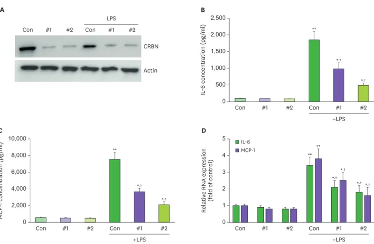
CRBN expression in the mouse retina has previously been reported (12). Therefore, we first confirmed the presence of CRBN protein in the retinal tissue and ARPE-19 cells (Fig. 1A).
Next, we compared the amount of CRBN between CRBN-siRNA- and CRBN-scrambled-treated ARPE-19 cells by Western blot assay. CRBN expression was diminished in CRBN-siRNA- treated cells (Fig. 1A). First, there was no difference in the level of IL-6 and MCP-1 between control group (scramble) to siRNA CRBN group (Fig. 1B-D).
It was well known that IL-6 and MCP-1 are representative cytokines that initiate retinal
inflammation (13). The IL-6 and MCP-1 levels were diminished when ARPE-19 cells were treated
with siCRBN RNA #2 (Fig. 1B and C). Moreover, IL-6 and MCP-1 mRNA expression levels also decreased relative to LPS-stimulated controls (Fig. 1D). These findings support the hypothesis that CRBN protein deficiency initiates the inflammatory process and immune cell recruitment.
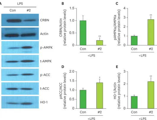
CRBN KD enhanced p-AMPK, p-ACC, and HO-1 expression in ARPE-19 cells
In a previous study, we reported that CRBN deficiency increased phosphorylated AMPK and ACC activity, as well as HO-1 expression, in mouse peritoneal macrophages (11). Thus, we checked whether CRBN KD increased AMPK and ACC activity in ARPE-19 cells. There were no significant differences in the amounts of total AMPK or ACC between control and CRBN KD (siCRBN RNA #2-treated) ARPE-19 cells (Fig. 2A and B). However, the amounts of phosphorylated AMPK and ACC in CRBN KD cells were higher than in control cells (Fig. 2A, C and D). The expression of HO-1 was significantly increased in KD cells (Fig. 2E). These results suggest that CRBN deficiency enhances AMPK pathway activation and, subsequently, HO-1 expression in LPS-stimulated ARPE-19 cells.A
#2
#1 Con
#2
#1 Con
CRBN Actin
LPS B
0 2,500
1,500 2,000
1,000 500
IL-6 concentration (pg/ml)
Con #1 #2 Con
+LPS
#2
#1
**
*,†
*,†
Con #1 #2 Con
+LPS
#2
#1 C
0 10,000
6,000 8,000
4,000 2,000
MCP-1 concentration (pg/ml) **
**
**
*,†
*,†
*,†
*,†
*,† *,†
Con #1 #2 Con
+LPS
#2
#1 D
0 5
3 4
2 Relative RNA expression (fold of control) 1
MCP-1 IL-6
Figure 1. Effects of CRBN on LPS-induced diminution of IL-6 and MCP-1 in ARPE cells.
(A) Expression of CRBN by Western blot in ARPE-19 cells. (B) ARPE-19 cells were pretreated with siCRBN RNA #1,2 at the indicated concentrations for 1 h and then stimulated with LPS (100 ng/ml) for 24 h. The culture medium was collected and subjected to ELISA to measure the concentration of IL-6. (C) ARPE-19 cells were pretreated with siCRBN RNA#1,2 at the indicated concentrations for 1 h and then stimulated with LPS (100 ng/ml) for 24 h. The culture medium was collected and subjected to ELISA to measure the concentration of MCP-1. (D) Analysis to determine expression of IL-6 and MCP-1. All experiments were performed in triplicate.
*p<0.05, **p<0.01 vs. treated sample. †p<0.05 vs control sample.
CRBN is negatively associated with NF- κB
The NF-κB pathway is considered a prototypical proinflammatory signaling pathway and is characterized by the activation of NF-κB and proinflammatory factors, including cytokines, chemokines, and adhesion molecules.
NF-κB expression diminished when ARPE-19 cells were transfected with an NF-κB luciferase reporter and CRBN and treated with LPS. To assess the role of CRBN in inflammatory signaling after NF-κB activation, we treated ARPE-19 cells with CRBN-siRNA, LPS, CC, and ZnPP-1 (Fig. 3). The LPS-stimulated, siRNA-treated ARPE-19 cells showed diminished NF-κB expression relative to controls (Fig. 3A). The cells treated with CC and ZnPP, however, showed increased NF-κB expression (Fig. 3B).
These results suggest that the CRBN gene regulates NF-κB expression as well as
downregulation of AMPK and HO-1 during NF-κB expression in CRBN-deficient retinitis.
CC and ZnPP upregulated IL-6 and MCP-1 in CRBN-deficient retinitis
To investigate IL-6 and MCP-1 expression in CRBN KO ARPE-19 cells, the cells were stimulated with LPS, then CC and ZnPP-1 function were evaluated (Fig. 4). First, there was no difference in the level of IL-6 and MCP-1 between control group to CC-treated or ZnPP-treated group (Fig. 4A and B). The LPS-stimulated CRBN KD ARPE-19 cells showed decreased IL-6
expression, but those treated with CC and ZnPP-1 showed increased IL-6 expression (Fig. 4A).
A
#2 Con
CRBN Actin p-AMPK t-AMPK p-ACC t-ACC HO-1
LPS B
0 1.0 1.5
CRBN/Actin (relative protein levels) 0.5
Con #2
+LPS
C
0 2 4 3
pAMPKa/AMPKa (relative protein levels) 1
Con #2
+LPS D
0 1.0 2.0 1.5
pACC/ACC (relative protein levels) 0.5
Con #2
+LPS
E
0 3
2
HO-1/Actin (relative protein levels) 1
Con #2
+LPS
**
* **
**
Figure 2. Expression of AMPK, ACC, and HO-1 in ARPE-19 cells with siCRBN RNA#2.
(A) ARPE-19 cells were pretreated with siCRBN RNA#2 and treated with LPS (100 ng/ml) for 24 h and then lysed.
Western blotting was carried out to evaluate the activation of AMPK and ACC and the expression of HO-1. (B) The relative ratio of CRBN to actin was calculated in ARPE-19 cells. (C) The relative ratio of pAMPKa to total AMPKa was calculated in ARPE-19 cells. (D) The relative ratio of pACC to total ACC was calculated in ARPE-19 cells. (E) The expression of HO-1 relative to actin in ARPE-19 cells.
t-, total; CON, control.
These findings suggest that AMPK inhibition with CC upregulates IL-6 expression, and HO-1 inhibition with ZnPP affects IL-6 expression in the absence of CRBN. MCP-1 was expression also affected by CC and ZnPP-1 (Fig. 4B). These findings suggest that the CRBN gene is crucial in AMPK/HO-1 signaling in the control of IL-6 expression during retinal inflammatory signaling.
A
0 5
3 4
2 NF-κB p65/Lamin B (relative protein levels) 1
NF-κB p65 Lamin B
#2 LPS
Com C ZnPP
−
−
−
−
+
−
−
−
+ +
−
−
+ + +
−
+ +
− +
**
** **
*,†
B
0 8
4 6
2
NF-κB activity (fold of control)
#2 LPS Com C ZnPP
−
−
−
−
+
−
−
− + +
−
− + + +
− + +
− +
−
− +
−
−
− +
−
−
−
− +
**
** **
*,†
*
Con #2
LPS
Com C ZnPP LPS+#2 Con
Figure 3. Effects of CRBN on NF-κB activation in LPS-stimulated ARPE cells.
(A) ARPE-19 cells were treated with LPS, LPS+siCRBN, and LPS+siCRBN+CC or PS+siCRBN+ZnPP for 24 h, and nuclear extracts were subjected to Western blotting to determine the NF-κB subunit p65 expression levels. (B) ARPE-19 cells transiently transfected with NF-κB-luciferase reporter plasmid was treated with siCRBN, Compound C, ZnPP-1, and LPS for 24 h, and subjected to a luciferase assay.
*p<0.05, **p<0.01 vs. treated sample −: no treatment; +: LPS-treated. †p<0.05 vs control sample.
B
0 10,000
6,000 8,000
4,000 2,000
MCP-1 concentration (pg/ml)
A
0 2,500
1,500 2,000
1,000 500
IL-6 concentration (pg/ml)
#2 LPS Com C ZnPP
−
−
−
−
+
−
−
− + +
−
− + + +
− + +
− +
−
− +
−
−
−
− +
#2 LPS Com C ZnPP
−
−
−
−
+
−
−
− + +
−
− + + +
− + +
− +
−
− +
−
−
−
− +
**
** **
*,†
**
** **
*,†
Figure 4. Effect of the HO-1 inhibitor ZnPP-1 on LPS-induced siCRBN upregulation of IL-6 and MCP-1 activation in ARPE-19.
(A) ARPE-19 cells were treated with Compound C, ZnPP, LPS, LPS+siCRBN, and LPS+siCRBN+CC or LPS+siCRBN+ZnPP for 24 h. The culture medium was collected and subjected to ELISA to measure the concentration of IL-6. (B) ARPE-19 cells were treated with Compound C, ZnPP, LPS, LPS+siCRBN, and LPS + siCRBN+CC or LPS+siCRBN+ZnPP for 24 h. The culture medium was collected and subjected to ELISA to measure the concentration of MCP-1. All experiments were performed in triplicate.
*p<0.05, **p<0.01 vs. treated sample. †p<0.05 vs control sample.
DISCUSSION
Previous research has shown that CRBN is highly expressed in the mouse retina (12), as confirmed by this study. We also confirmed that CRBN protein is expressed in the human retinal cell line, ARPE-19.
Uveitis is a common intraocular inflammatory disease. A well-studied model for acute infectious uveitis relies on inflammation induced by intravenous injection with LPS. This produces various inflammatory cytokines, including IL-6, TNF-α, and MCP-1 (14,15), which lead to prominent features as iris hyperemia and leukocyte infiltration (16).
In uveitis, IL-6 is a multifunctional proinflammatory cytokine; it is involved in selectively recruiting monocytes, neutrophils, and lymphocytes (17). MCP-1 is a key chemokine involved in regulating the migration and infiltration of monocytes and macrophages (18).
Our data demonstrate that CRBN deficiency can downregulate LPS-induced inflammatory cytokines (IL-6 and MCP-1) expression. Recently, CRBN was shown to acts as a
multifunctional protein in various diseases models, including cardiovascular disease, fatty liver, and sepsis. For all of these diseases, the ablation of CRBN increased AMPK levels and protected against organ damage. Additionally, AMPK was shown to modulate inflammation in diseases, including uveitis (19). Our data indicate that CRBN deficiency upregulates the expression of phosphorylated AMPK, ACC, and HO-1, which controls the expression of anti-inflammatory proteins by regulating AMPK activation in ARPE-19 cells. The regulatory pathway from CRBN to HO-1 might play a similar role in protecting against uveitis, where HO-1 is also a key mediator (20).
In association with intraocular injury and uveitis, NF-κB is an important transcription factor responding to endotoxin stimulation (21). NF-κB activity is essential for regulating the secretion of proinflammatory cytokines, such as IL-6, TNF-α and MCP-1. Notably, in our study, LPS-induced NF-κB transcriptional activity and proinflammatory cytokines (IL-6, MCP-1) expression were suppressed by CRBN depletion via AMPK/HO-1 signaling.
In the future, the effects of CRBN deficiency on other cell types or ophthalmic tissues should be evaluated to clarify the role of CRBN in the pathogenesis of uveitis.
In summary, this study suggests that CRBN suppresses AMPK signaling and HO-1 expression and that CRBN has a role in promoting the inflammatory response in the retina.
ACKNOWLEDGEMENTS
This research was supported by the Basic Science Research Program through the National Research Foundation of Korea (NRF) and funded by the Ministry of Education, Science and Technology (2018R1D1A1B07050192, 2017R1D1A1A02018653). It was also supported by a grant (1631040) from the National R&D Program for Cancer Control, Ministry of Health and Welfare, Republic of Korea, as well as a grant from Nambu University, 2016.
REFERENCES
1. Wang AL, Knight DK, Vu TT, Mehta MC. Retinitis pigmentosa: review of current treatment. Int Ophthalmol Clin 2019;59:263-280.
PUBMED | CROSSREF
2. Carreño E, Portero A, Herreras JM, García-Vázquez C, Whitcup SM, Stern ME, Calonge M, Enríquez- de-Salamanca A. Cytokine and chemokine tear levels in patients with uveitis. Acta Ophthalmol 2017;95:e405-e414.
PUBMED | CROSSREF
3. Pollreisz A, Rafferty B, Kozarov E, Lalla E. Klebsiella pneumoniae induces an inflammatory response in human retinal-pigmented epithelial cells. Biochem Biophys Res Commun 2012;418:33-37.
PUBMED | CROSSREF
4. Tode J, Richert E, Koinzer S, Klettner A, Pickhinke U, Garbers C, Rose-John S, Nölle B, Roider J.
Intravitreal injection of anti-interleukin (IL)-6 antibody attenuates experimental autoimmune uveitis in mice. Cytokine 2017;96:8-15.
PUBMED | CROSSREF
5. Higgins JJ, Pucilowska J, Lombardi RQ, Rooney JP. A mutation in a novel ATP-dependent Lon protease gene in a kindred with mild mental retardation. Neurology 2004;63:1927-1931.
PUBMED | CROSSREF
6. Ito T, Ando H, Handa H. Discovery of the target for immunomodulatory drugs (IMiDs). Rinsho Ketsueki 2016;57:556-562.
PUBMED | CROSSREF
7. Jo S, Lee KH, Song S, Jung YK, Park CS. Identification and functional characterization of cereblon as a binding protein for large-conductance calcium-activated potassium channel in rat brain. J Neurochem 2005;94:1212-1224.
PUBMED | CROSSREF
8. Kim J, Lee KM, Park CS, Park WJ. Ablation of cereblon attenuates myocardial ischemia-reperfusion injury.
Biochem Biophys Res Commun 2014;447:649-654.
PUBMED | CROSSREF
9. Lee KM, Yang SJ, Kim YD, Choi YD, Nam JH, Choi CS, Choi HS, Park CS. Disruption of the cereblon gene enhances hepatic AMPK activity and prevents high-fat diet-induced obesity and insulin resistance in mice. Diabetes 2013;62:1855-1864.
PUBMED | CROSSREF
10. Ko JR, Seo DY, Park SH, Kwak HB, Kim M, Ko KS, Rhee BD, Han J. Aerobic exercise training decreases cereblon and increases AMPK signaling in the skeletal muscle of STZ-induced diabetic rats. Biochem Biophys Res Commun 2018;501:448-453.
PUBMED | CROSSREF
11. Gil M, Kim YK, Kim HY, Pak HK, Park CS, Lee KJ. Cereblon deficiency confers resistance against polymicrobial sepsis by the activation of AMP activated protein kinase and heme-oxygenase-1. Biochem Biophys Res Commun 2018;495:976-981.
PUBMED | CROSSREF
12. Hohberger B, Enz R. Cereblon is expressed in the retina and binds to voltage-gated chloride channels.
FEBS Lett 2009;583:633-637.
PUBMED | CROSSREF
13. Leung KW, Barnstable CJ, Tombran-Tink J. Bacterial endotoxin activates retinal pigment epithelial cells and induces their degeneration through IL-6 and IL-8 autocrine signaling. Mol Immunol 2009;46:1374-1386.
PUBMED | CROSSREF
14. Paeng SH, Park WS, Jung WK, Lee DS, Kim GY, Choi YH, Seo SK, Jang WH, Choi JS, Lee YM, et al.
YCG063 inhibits Pseudomonas aeruginosa LPS-induced inflammation in human retinal pigment epithelial cells through the TLR2-mediated AKT/NF-κB pathway and ROS-independent pathways. Int J Mol Med 2015;36:808-816.
PUBMED | CROSSREF
15. Zanon CF, Sonehara NM, Girol AP, Gil CD, Oliani SM. Protective effects of the galectin-1 protein on in vivo and in vitro models of ocular inflammation. Mol Vis 2015;21:1036-1050.
PUBMED
16. Kuryltsiv NB, Halei KM. The role of interleukins and their inhibitors in the development of autoimmune uveitis. Wiad Lek 2019;72:716-722.
PUBMED
17. Lopalco G, Fabiani C, Sota J, Lucherini OM, Tosi GM, Frediani B, Iannone F, Galeazzi M, Franceschini R, Rigante D, et al. IL-6 blockade in the management of non-infectious uveitis. Clin Rheumatol 2017;36:1459-1469.
PUBMED | CROSSREF
18. Tuaillon N, Shen DF, Berger RB, Lu B, Rollins BJ, Chan CC. MCP-1 expression in endotoxin-induced uveitis. Invest Ophthalmol Vis Sci 2002;43:1493-1498.
PUBMED
19. Kalariya NM, Shoeb M, Ansari NH, Srivastava SK, Ramana KV. Antidiabetic drug metformin suppresses endotoxin-induced uveitis in rats. Invest Ophthalmol Vis Sci 2012;53:3431-3440.
PUBMED | CROSSREF
20. Ohta K, Kikuchi T, Arai S, Yoshida N, Sato A, Yoshimura N. Protective role of heme oxygenase-1 against endotoxin-induced uveitis in rats. Exp Eye Res 2003;77:665-673.
PUBMED | CROSSREF
21. Ando Y, Keino H, Kudo A, Hirakata A, Okada AA, Umezawa K. Anti-inflammatory effect of
dehydroxymethylepoxyquinomicin, a nuclear factor-κB inhibitor, on endotoxin-induced uveitis in rats in vivo and in vitro. Ocul Immunol Inflamm 2020;28:240-248.
PUBMED | CROSSREF