Ji-Woong Choi,†,|| Seoung Min Bong†, Seung Won Yang†, Hyonchol Jang‡, SangYoun Park§, Seung Jun Kim#, and Byung Il Lee†,*
†Biomolecular Function Research Branch, Division of Convergence Technology, National Cancer Center, Goyang, Gyeonggi 410-769, Korea. *E-mail: bilee@ncc.re.kr
‡Division of Cancer Biology, Research Institute, National Cancer Center, Goyang, Gyeonggi 410-769, Korea
§School of Systems Biomedical Science, Soongsil University, Seoul 156-743, Korea
#Medical Proteomics Research Center, Korea Research Institute of Bioscience and Biotechnology, Daejeon 305-333, Korea (Received June 10, 2015; Accepted July 20, 2015)
Key words: Laforin, EPM2A, Baculovirus, Insect cells, Phosphatase, Glycogen, Lafora disease
Lafora disease (LD; OMIM 254780) is an autosomal recessive neurodegenerative disorder, which leads to demen- tia, epilepsy, myoclonus, and consequentially to death.1 The feature of LD is the accumulation of Lafora bodies which are hyperphosphorylated glycogen-like tangles in the cytoplasm.2 Together with mutations in the NHLRC1 (EPM2B) gene, mutations in the Laforin gene are directly related to a severe autosomal recessive LD (EPM2). Many Laforin mutations have been found in Lafora disease patients.1b Laforin is a member of dual specificity phospha- tases (DSP) which directly remove the phosphate group from glycogen.3 The DSP domain of Laforin has phos- phatase activities against both phosphorylated protein and glucan.3 Laforin is encoded by the EPM2A (epilepsy of progressive myoclonus type 2A) gene where mutations are found in about half of LD patients.4
The Laforin protein is comprised of an N-terminal car- bohydrate-binding module (CBM) and a C-terminal DSP domain.5 CBM of Laforin is classified into CBM20 family by evolutional relationship, polypeptide fold, and affinity to carbohydrates and glyco-conjugates in the Carbohydrate- Active Enzyme (CAZy) database.6 CBM of Laforin has been reported to bind to glycogen, lafora body, and even plant amylopectin.5,7 Human Laforin is a unique phosphatase in human which contains a CBM whose usual substrates are carbohydrates such as glycogen.8
Laforin also has DSP domain at the C-terminus. This DSP domain belongs to a member of protein tyrosine phos- phatase family of cysteine-dependent phosphatases.9 The general substrates of Laforin are C2 and C3 phosphomon-
oesters of oligosaccharides,10 however, recombinant Laforin purified from E. coli elicits phosphatase activities against artificial substrates such as para-nitrophenylphosphate (pNPP) and 3-O-methylfluorescein phosphate (OMFP) as observed in other DSP domain phosphatases.11
Herein, the application of baculovirus/insect cell expres- sion system in the production of wild-type and phospha- tase active site mutant was investigated where Laforin proteins were expressed in insect cells using baculovirus and compared with that from the E. coli expression system. The phosphatase activities of purified recombinant Laforin against various substrates such as pNPP, OMFP, and glycogen were exam- ined.
In this study, the Laforin coding DNA sequence was cloned into bacterial expression vector and expressed in E. coli.
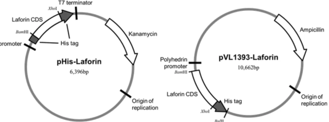
Using this construct, the polyhistidine-tagged Laforin was also cloned into baculoviral transfer vector pVL1393 (Fig. 1).
Both plasmids have the polyhistidine tags for purification using a Ni-NTA resin. However, purification of polyhis- tidine-tagged Laforin was not successful using a Ni-NTA resin as previously reported by Dukhande et al.12
Overexpressed recombinant Laforins in E. coli (B-Laforin) and in insect cells (I-Laforin) were successfully purified by affinity chromatography with an amylose resin (Fig. 2), which utilizes Laforin itself with a carbohydrate-binding module (CBM). While the purified recombinant Laforin using the Ni-NTA resin was prone to aggregation, purified recombi- nant Laforin especially the one expressed in the insect cells was relatively stable. Laforin was eluted off at 500 mM maltose containing buffer (Fig. 2A and 2B). Considering that the maltose binding protein (MBP)-tagged proteins usu- ally elutes at 10 mM maltose containing buffers, interac-
||Current address: Wide River Institute of Immunology, Seoul National University, Hongcheon, Gangwon 250-812, Republic of Korea.
tions between the CBM of Laforin and the amylose resin seem to be very strong. Moreover, the addition of maltose improved the protein stability of purified Laforin as pre- viously reported.13 Size exclusion chromatography results showed a single chromatogram peak (Fig. 2C), and the eluted Laforin associated into a dimer as concluded from a dynamic light scattering experiment (Fig. 2D). The native
molecular weight of I-Laforin as estimated by dynamic light- scattering analysis was ~64 kDa with 12.7% polydispersity.
That of B-Laforin was ~86 kDa with 12.6% polydispersity.
Therefore, the results indicated that human Laforin expressed in insect cells exists as dimers in solution (the calculated monomer weight including the fusion tag is 38,497 Da for I-Laforin and 41,087 Da for B-Laforin, respectively).
Figure 1. Construction of human Laforin cDNA cloned into the expression vector pHis and pVL1393. pHis vector is modified from pET32 (Novagen).
Figure 2. Purification and characterization of B-Laforin and I-Laforin. SDS-PAGE stained with Coomassie Blue of fractions from (A) E. coli strain Rosetta 2 (DE3) and (B) Sf9 cells (NC, negative control (uninduced E. coli or uninfected Sf9 cells); I, IPTG-induced E.
coli or Baculovirus-infected Sf9 cells; S, soluble fraction; M, Amylose resin elution; G, fraction from SuperdexTM 200 pg column) (C) The chromatogram of B-Laforin and I-Laforin in SuperdexTM 200 column. (D) DLS analysis of B-Laforin and I-Laforin in buffer con- taining 100 mM Tris-HCl pH 8.5.
Generally, recombinant proteins using the E. coli expres- sion system gives higher purification yield compared to using the insect cell expression system. However, bacu- lovirus/insect cell expression system for the recombinant human Laforin showed higher expression level and puri- fication yield than the E. coli expression system. The purifi- cation yields of baculovirus expressed wild type and C266S mutant Laforins respectively were 5.7 and 3.4 fold higher than that of from the E. coli expression system (Table 1).
We performed in vitro phosphatase assays using the purified Laforins. Because recombinant Laforin purified from E. coli is well known to dephosphorylate pNPP and OMFP, we analyzed and compared the phosphatase activ- ities of B-Laforin and I-Laforin. Unexpectedly, the phos- phatase activity of B-Laforin (WT) was ~450 times higher than that of I-Laforin (WT) when pNPP was used as substrate.
Likewise, OMFP phosphatase activity of B-Laforin (WT) was ~37 times higher than that of I-Laforin (WT). Because Laforin directly dephosphorylates glucans in glycogen metabolism, we next investigated the phosphatase activity of B-Laforin and I-Laforin against glycogen. A branched poly- mer of glucose, glycogen, contains a small amount of covalently linked phosphate for determining its branching structure.3 The released phosphates from glycogen were monitored using the malachite green reagent for the mea- surement of phosphatase activity. Contrary to the above assay results, the I-Laforin dephosphorylated glycogen more efficiently than the B-Laforin (Fig. 3C). Taken together,
biochemical properties. While the phosphatase activity against the general substrates such as pNPP and OMPF was higher for the B-Laforin, the I-Laforin had higher activity against glycogen which is one of Laforin’s real substrate. This effect is probably due to the higher substrate selectivity in I-Laforin. In general, insect cell expression has advan- tages in protein folding and post-translational modifica- tion.14 The difference in protein folding during protein expression may be one of the reasons behind different bio- chemical properties in I- and B-Laforin. Even though we failed to detect any phosphorylation in I-Laforin, post-trans- lational modification can be another cause of different biochemical properties in the two recombinant Laforins.
Of note, Ser25 of Laforin is known to be phosphorylated by AMP-activated protein kinase (AMPK) and the critical role for modulating Laforin phosphatase activity and inter- action with binding partners of Laforin.15
Since Laforin genes of Lafora disease patients have many mutations,1b structural and biochemical studies on the various Laforin mutations from the Lafora disease would be helpful for understanding the molecular basis of the disease. Very recently, crystal structure of nearly full-length Laforin was determined and it provided the functional mechanism of Laforin and the structural basis of disease.16 Although crystal structure of Laforin was determined using the E. coli expressed recombinant Laforin, I-Laforin can be more preferable over B-Laforin for future biochemical studies because I-Lafo- rin exhibited enhanced substrate specificity in our studies.
Figure 3. Analysis of phosphatase activities of B-Laforin and I-Laforin. Phosphatase activities of B-Laforin and I-Laforin were mea- sured against the substrates of pNPP (A), OMPF (B), and glycogen (C). Error-bars indicate means ± standard deviations from at least three independent experiments.
EXPERIMENTAL METHODS
Cloning and Mutagenesis
The gene covering the full regions of human Laforin were amplified by polymerase chain reaction (PCR) and cloned into pHis (modified from Novagen’s pET32) vector using the BamHI/XhoI restriction enzyme sites. The resulting recom- binant Laforin contains polyhistidine tags at its N- and C- terminus (MHHHHHHGSLVPRSENLYFQGS for N-ter- minus and LEHHHHHHHH for C-terminus). The phospha- tase active site mutant (C266S) was obtained by applying the QuikChangeTM method. For the generation of baculovi- rus, wild type and mutant genes covering the Laforin and the C-terminal fused polyhistidine tag were amplified by PCR and cloned into pVL1393 baculovirus transfer vector (AB Vector) using BamHI/BglII restriction enzyme sites (Fig. 1).
Expression of Human Laforin in E. coli
The recombinant proteins were overexpressed in E. coli strain Rosetta 2(DE3) (Novagen). Transformed cells with the pHis-Laforin plasmid were grown in Terrific Broth to an OD600 of 0.5 at 37oC and protein expression was induced by 0.1 mM isopropyl-D-thiogalactopyranoside at 18oC.
Cells were further cultured at 18oC for 48 h after protein induction, and were harvested by centrifugation at 10,000×g for 10 min.
Preparation of Recombinant Baculovirus and Expres- sion of Human Laforin in Sf9 Cells
Spodoptera frugiperda (Sf9) cells were grown at 27oC in Sf-900TM II SFM medium (Life Technologies) supple- mented with 100 units/ml penicillin, 100 µg/ml streptomy- cin, and 1% fetal bovine serum (Life Technologies). The baculovirus transfer vectors pVL1393-Laforin and the lin- earized baculoviral genomic DNA vector (ProGreenTM, AB vector) were co-transfected into Sf9 cells using ProFectinTM reagent (AB vector). Recombinant baculoviruses generated by homologous recombination were harvested at 3 days post transfection and were amplified to produce high-titer virus stocks. The expression of Laforin was assessed at 2~3 days post infection by Western blot, using an anti-His tag antibody (Applied Biological Materials).
Purification of Recombinant Human Laforin
All of the protein purification steps were performed under ice-cold conditions. Cell pellets were resuspended and soni- cated under hypotonic lysis buffer (20 mM 4-(2-hydroxyethyl)- 1-piperazineethanesulfonic acid (HEPES) pH 7.4, 10 mM
sodium chloride, and 1 mM ethylenediaminetetraacetic acid (EDTA), 10 mM 2-mercaptoethanol, 10% (v/v) glycerol, and 1 mM phenylmethylsulfonyl fluoride). The crude lysate was centrifuged at 40,000×g for 1 h at 4oC. The resulting supernatant was applied to an amylase column (New England BioLabs) and washed with wash buffer (20 mM HEPES pH 7.4, 150 mM sodium chloride, 1 mM EDTA, 10 mM 2-mercaptoethanol, 10% (v/v) glycerol, and 10 mM malt- ose). The recombinant Laforin was further eluted with elu- tion buffer (20 mM HEPES pH 7.4, 150 mM sodium chloride, 1 mM EDTA, 10 mM 2-mercaptoethanol, 10% (v/v) glyc- erol, and 500 mM maltose). The eluted protein was pooled and concentrated prior to loading on to SuperdexTM 200 HiLoadTM 16/60 prep-grade column (GE Healthcare) equili- brated with the buffer of 20 mM HEPES pH 7.4, 300 mM sodium chloride, 1 mM EDTA, 10 mM 2-mercaptoethanol, and 500 mM maltose. The homogeneity of the purified recombinant Laforin was examined by SDS-PAGE with Coomassie Blue staining. The final protein concentration was determined by Bradford assay (Sigma).
Measurement of Dynamic Light Scattering
Dynamic light-scattering experiments were performed using a DynaPro-titan instrument (Wyatt technology). The data were measured at 25oC at 0.125 mg/ml protein in the buffer of 100 mM Tris-HCl pH 8.5.
Phosphatase Activity Assay
pNPP, OMPF, and glycogen were used as substrates for measuring phosphatase activities of purified recombinant Laforins. The pNPP reaction mixture was 0.1 M sodium acetate, 0.05 M Bis-Tris pH 6.0, 2 mM dithiothreitol, and 50 mM pNPP. The OMFP mixture was 0.1 M Tris-HCl pH 8.0, 40 mM sodium chloride, 2 mM dithiothreitol, and 0.5 mM OMFP. 10 µg of recombinant Laforins were added to each reaction mixtures and stored at 30oC for 20 min.
The absorbance of the reaction products were measured at 405 nm for pNPP assay and at 477 nm for OMFP assay using a microplate reader (Molecular Devices). The phosphatase assay with glycogen was carried out under the reaction mixture of 100 mM sodium acetate, 50 mM Bis-Tris pH 6.0, 2 mM dithiothreitol, and 1 mg/ml glycogen at 37oC. The released phosphate was measured with malachite green reagent (Cayman Chemical) using the same microplate reader at 620 nm. Data were expressed as means ± standard deviation of at least three independent experiments. Statistical dif- ferences between groups were analyzed using one-way anal- ysis of variance (ANOVA).
REFERENCES
1. (a) Gentry, M. S.; Roma-Mateo, C.; Sanz, P., Laforin, a protein with many faces: glucan phosphatase, adapter pro- tein, et alii. The FEBS Journal 2013, 280(2), 525; (b) Gentry, M. S.; Dixon, J. E.; Worby, C. A., Lafora disease: insights into neurodegeneration from plant metabolism. Trends Biochem. Sci. 2009, 34(12), 628.
2. Ganesh, S.; Puri, R.; Singh, S.; Mittal, S.; Dubey, D., Recent advances in the molecular basis of Lafora’s progressive myoclonus epilepsy. J. Hum. Genet. 2006, 51(1), 1.
3. Tagliabracci, V. S.; Turnbull, J.; Wang, W.; Girard, J. M.;
Zhao, X.; Skurat, A. V.; Delgado-Escueta, A. V.; Minassian, B. A.; Depaoli-Roach, A. A.; Roach, P. J., Laforin is a gly- cogen phosphatase, deficiency of which leads to elevated phosphorylation of glycogen in vivo. Proceedings of the National Academy of Sciences of the United States of America 2007, 104(49), 19262.
4. Minassian, B. A.; Lee, J. R.; Herbrick, J. A.; Huizenga, J.;
Soder, S.; Mungall, A. J.; Dunham, I.; Gardner, R.; Fong, C. Y.; Carpenter, S.; Jardim, L.; Satishchandra, P.; Andermann, E.; Snead, O. C., 3rd; Lopes-Cendes, I.; Tsui, L. C.; Delgado- Escueta, A. V.; Rouleau, G. A.; Scherer, S. W., Mutations in a gene encoding a novel protein tyrosine phosphatase cause progressive myoclonus epilepsy. Nat. Genet. 1998, 20(2), 171.
5. Wang, J.; Stuckey, J. A.; Wishart, M. J.; Dixon, J. E., A unique carbohydrate binding domain targets the lafora disease phosphatase to glycogen. J. Biol. Chem. 2002, 277(4), 2377.
6. Cantarel, B. L.; Coutinho, P. M.; Rancurel, C.; Bernard, T.; Lombard, V.; Henrissat, B., The Carbohydrate-Active EnZymes database (CAZy): an expert resource for Gly- cogenomics. Nucleic Acids Res. 2009, 37 (Database issue), D233.
7. Ganesh, S.; Tsurutani, N.; Suzuki, T.; Hoshii, Y.; Ishihara,
2006, 7(11), 833.
9. Alonso, A.; Sasin, J.; Bottini, N.; Friedberg, I.; Friedberg, I.; Osterman, A.; Godzik, A.; Hunter, T.; Dixon, J.; Mustelin, T., Protein tyrosine phosphatases in the human genome.
Cell 2004, 117(6), 699.
10. Tagliabracci, V. S.; Heiss, C.; Karthik, C.; Contreras, C. J.;
Glushka, J.; Ishihara, M.; Azadi, P.; Hurley, T. D.; DePaoli- Roach, A. A.; Roach, P. J., Phosphate incorporation during glycogen synthesis and Lafora disease. Cell Metab. 2011, 13(3), 274.
11. Girard, J. M.; Le, K. H.; Lederer, F., Molecular characteriza- tion of laforin, a dual-specificity protein phosphatase impli- cated in Lafora disease. Biochimie 2006, 88(12), 1961.
12. Dukhande, V. V.; Rogers, D. M.; Roma-Mateo, C.; Donderis, J.; Marina, A.; Taylor, A. O.; Sanz, P.; Gentry, M. S., Laforin, a dual specificity phosphatase involved in Lafora disease, is present mainly as monomeric form with full phosphatase activity. PloS one 2011, 6(8), e24040.
13. Brewer, M. K.; Husodo, S.; Dukhande, V. V.; Johnson, M.
B.; Gentry, M. S., Expression, purification and characteriza- tion of soluble red rooster laforin as a fusion protein in Escherichia coli. BMC biochemistry 2014, 15(1), 8.
14. Jarvis, D. L., Baculovirus Expression Vectors. In The Baculo- viruses; Plenum Press: New York, 1997, Miller, L.K., Ed.;
pp 389-431.
15. Roma-Mateo, C.; Solaz-Fuster Mdel, C.; Gimeno-Alcaniz, J.
V.; Dukhande, V. V.; Donderis, J.; Worby, C. A.; Marina, A.;
Criado, O.; Koller, A.; Rodriguez De Cordoba, S.; Gentry, M. S.; Sanz, P., Biochem. J. 2011, 439(2), 265.
16. Raththagala, M.; Brewer, M. K.; Parker, M. W.; Sherwood, A. R.; Wong, B. K.; Hsu, S.; Bridges, T. M.; Paasch, B.
C.; Hellman, L. M.; Husodo, S.; Meekins, D. A.; Taylor, A.
O.; Turner, B. D.; Auger, K. D.; Dukhande, V. V.; Chakravar- thy, S.; Sanz, P.; Woods, V. L., Jr.; Li, S.; Vander Kooi, C. W.;
Gentry, M. S., Mol. Cell 2015, 57(2), 261.