• 검색 결과가 없습니다.

Corresponding author: Sung Ho Lee, Department of Thoracic and Cardiovascular Surgery, Korea University Medical Center, Korea University College of Medicine, 73 Inchon-ro, Seongbuk-gu, Seoul 136-705, Korea

N/A
N/A
Protected

Academic year: 2021

Share "Corresponding author: Sung Ho Lee, Department of Thoracic and Cardiovascular Surgery, Korea University Medical Center, Korea University College of Medicine, 73 Inchon-ro, Seongbuk-gu, Seoul 136-705, Korea"

Copied!
4
0
0

로드 중.... (전체 텍스트 보기)

전체 글

(1)

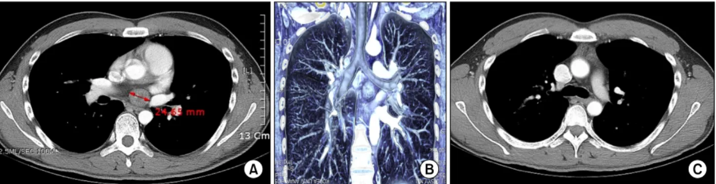
Korean J Thorac Cardiovasc Surg 2014;47:298-301 □ Case Report □ http://dx.doi.org/10.5090/kjtcs.2014.47.3.298 ISSN: 2233-601X (Print) ISSN: 2093-6516 (Online)

− 298 −

Department of Thoracic and Cardiovascular Surgery, Korea University Medical Center, Korea University College of Medicine Received: August 2, 2013, Revised: October 14, 2013, Accepted: October 15, 2013, Published online: June 5, 2014

Corresponding author: Sung Ho Lee, Department of Thoracic and Cardiovascular Surgery, Korea University Medical Center, Korea University College of Medicine, 73 Inchon-ro, Seongbuk-gu, Seoul 136-705, Korea

(Tel) 82-2-920-5837 (Fax) 82-2-2-928-8793 (E-mail) [email protected]

C

The Korean Society for Thoracic and Cardiovascular Surgery. 2014. All right reserved.

CC

This is an open access article distributed under the terms of the Creative Commons Attribution Non-Commercial License (http://creative- commons.org/licenses/by-nc/3.0) which permits unrestricted non-commercial use, distribution, and reproduction in any medium, provided the original work is properly cited.

Resection of Intrapericardial Schwannoma Co-Existing with Thymic Follicular Hyperplasia through Sternotomy

without Cardiopulmonary Bypass

Jae Ho Chung, M.D., Jae Seung Jung, M.D., Ph.D., Sung Ho Lee, M.D., Ph.D., Kwang Taik Kim, M.D., Ph.D., Kanghoon Lee, M.D., Seung Hun Lee, M.D.

A 35-year-old man was admitted to Korea University Anam Hospital for evaluation of intermittent chest pain.

Computed tomography of the chest showed enlargement of a previously identified anterior mediastinal mass and al- so a well-defined, circumscribed mass in the subcarinal area, surrounded by the roof of the left atrium, right pul- monary artery, and the carina. Complete resection of the intrapericardial tumor was performed through median ster- notomy without cardiopulmonary bypass. Pathologic examination identified the tumor as schwannoma, of an ancient type, diffusely positive for the S-100 antigen. Unlike other reported cases, grossly, the tumor did not seem to be involved with any nerve.

Key words: 1. Schwannoma 2. Mediastinal neoplsms 3. Pericardium

4. Sternotomy 5. Benign neoplasm

CASE REPORT

A 35-year-old male patient with intermittent chest pain was admitted to Korea University Anam Hospital for further eval- uation of a mediastinal mass that was found on computed to- mography (CT) at another hospital. He had a history of Graves disease treated with radioactive iodine and methima- zole and endoscopic sinus surgery for chronic sinusitis with a nasal polyp. The initial physical examination and chest X-ray were non-specific and electrocardiography showed ST seg- ment elevation in leads V1–3, but no irregular rhythms. CT of the chest showed diffuse enlargement of the anterior me-

diastinal mass and a well-defined, circumscribed, heteroge- neous soft-tissue attenuation mass in the subcarinal area (Fig.

1). It was 2.5×2.5×2 cm in size and was surrounded by the roof of the left atrium, right pulmonary artery, and carina.

Echocardiography revealed a hyperechogenic mass in the pos-

terior left atrium, but its relationship with the left atrium was

not clear. Considering the possibility of neural or cardiac in-

volvement, cardiac magnetic resonance imaging (MRI) was

performed. A well-defined, intermediate-signal-intensity, 2.4-cm

mass was noted in the middle mediastinum, at the roof of the

left atrium. A relatively preserved regional fat plane was

seen.

(2)

Intrapericardial Schwannoma Co-Existing with Thymic Follicular Hyperplasia

− 299 −

Fig. 1. (A) Constrast enhanced chest computed tomography (CT) and (B) three dimensional CT reconstruction showing the well-demarcated margin of the mass and its location between the posterior wall of the left atrium, right pulmonary artery, and the carina. (C) A diffusely enlarged anterior mediastinal mass was also seen in chest CT.

Fig. 2. Intraoperative findings. (A) About 2.5×2.5×2 cm in size round tumor was located just posterior to the roof of the left atrium, and between the aorta and the main pulmonary artery. (B) Macroscopic view of the tumor.

Under general anesthesia, the patient underwent median sternotomy and total thymectomy. Then, the pericardium was opened and the major vessels exposed; cardiopulmonary by- pass (CPB) was readied in case it was required. The mass and its surrounding structures were explored carefully. A 2.5-cm round tumor was located just posterior to the roof of the left atrium, between the aorta and the main pulmonary artery. No significant surrounding tissue invasion was ob- served; therefore, we decided to continue the operation with- out CPB. The plane between the ascending aorta and the main pulmonary artery was dissected carefully, and a soft, smooth, grey, ovoid tumor with scant vascularity was seen surrounded by the roof of the left atrium, right pulmonary ar- tery, and carina. Unlike the reported cases, the tumor did not

seem to involve any nerve grossly and the fat plane kept the tumor in place (Fig. 2). The tumor was removed completely, without injuring the surrounding tissues. After removing the intrapericardial tumor, the thymic mass and its surrounding mediastinal fat tissues were removed as an extended thymectomy.

Pathologically, the intrapericardial tumor was identified as

an ancient-type schwannoma, diffusely positive for the S-100

antigen (Fig. 3). The cut surface revealed a well-demarcated

white-and-gray fibrotic mass with a multifocal hemorrhagic

appearance. The anterior mediastinal mass was reported to be

thymic follicular hyperplasia. The postoperative course was

uneventful, and the patient continues to do well 2 months

postoperatively without any complications.

(3)

Jae Ho Chung, et al

− 300 −

Fig. 3. Histopathologic findings. (A) The spindle cells show palisading arrangement (H&E, ×100). (B) The tumor cells are diffusely and strongly positive for S100 immunohistochemical stain in nucleus and cytoplasm (H&E, ×200).

DISCUSSION

Schwannomas, also called neurilemmomas, are benign en- capsulated tumors derived from Schwann cells [1,2]. They are the most common tumor of peripheral nerves and grow from peripheral nerves or nerve roots in an eccentric fashion, with the nerve itself usually incorporated within the capsule [1].

Schwannomas are the most common benign posterior media- stinal neurogenic tumors. These are typically asymptomatic but can produce signs of nerve compression, paralysis, Pancoast’s syndrome, and Horner’s syndrome. Intrapericardial neurogenic tumors are extremely rare, and most arise from the cardiac branches of the vagus nerve or the cardiac plexus.

Mediastinal neurogenic tumors, which comprise 19% to 39% of all mediastinal tumors, develop mostly from media- stinal peripheral nerves, sympathetic and parasympathetic gan- glia, and embryonic remnants of the neural tube. They are most frequently located in the posterior mediastinum. Of the posterior mediastinal neurogenic tumors, schwannomas are the most common benign tumors. They originate from Schwann cells and affect patients of both sexes, predominantly in the third and fourth decades of life [3]. Mediastinal benign schwannomas are most often asymptomatic, although signs of nerve compression, paralysis, Pancoast’s syndrome, and Horner’s syndrome can occur.

Among mediastinal neurogenic tumors, intrapericardial neu-

rogenic tumors are extremely rare and arise mostly from the cardiac branches of the vagus nerve and the cardiac plexus [4]. Unlike other cases, despite our pre- and intraoperative studies, the tumor in our patient seemed to lack neural in- volvement, either grossly or microscopically. Neither pre- operative cardiac MRI nor echocardiography showed involve- ment of the tumor with any nerve. Intraoperatively, the tumor was surrounded only by fat tissues and encased between the left atrium, right pulmonary artery, and carina.

Among the many possible surgical approaches, including thoracotomy and video-assisted thoracic surgery, we chose median sternotomy to approach the tumor, considering the tu- mor’s intrapericardial location and because there was also an anterior mediastinal tumor. The thymic tissue and pericardial fat were removed easily by extended thymectomy, in combi- nation with the removal of the intrapericardial mass.

Although CPB has been used in some reported cases for bet- ter hemodynamic stability and visualization of the tumor [4,5], it was avoided since the tumor was small and did not show any direct involvement of the surrounding cardiac wall, vessels, or carina. Consequently, the tumor was removed easi- ly, but carefully, with minimal handling of the heart and without injuring the surrounding tissues.

Unlike malignant neurogenic tumors, which usually mani-

fest symptoms of cough, dyspnea, chest wall pain, and nerve

compression, most benign neurogenic tumors are asympto-

(4)

Intrapericardial Schwannoma Co-Existing with Thymic Follicular Hyperplasia

− 301 − matic and are usually discovered incidentally. Our patient had atypical chest wall pain in the left anterior chest, which was evaluated to rule out a cardiac origin. As there was no spe- cific evidence of cardiac pain, chest CT was done. Compared with the reported mediastinal or intrapericardial tumors, the tumor was small, round, and well-demarcated. Despite their small size and absence of signs of cardiac compression, sur- gical resection is the standard of care for neurogenic tumors, since they can continue to grow and subsequently compress the cardiac chambers, resulting in cardiac dysfunction, ar- rhythmias, or even thromboembolism [6,7]. Therefore, the tu- mor in our patient was resected completely, and we expect a good long-term prognosis in keeping with the literature.

We present an intrapericardial schwannoma that was not directly associated with the cardiac wall or the surrounding tissues, combined with a thymoma. Both were removed suc- cessfully via sternotomy without CPB.

CONFLICT OF INTEREST

No potential conflict of interest relevant to this article was reported.

REFERENCES

1. Pilavaki M, Chourmouzi D, Kiziridou A, Skordalaki A, Zarampoukas T, Drevelengas A. Imaging of peripheral nerve sheath tumors with pathologic correlation: pictorial review.

Eur J Radiol 2004;52:229-39.

2. Skovronsky DM, Oberholtzer JC. Pathologic classification of peripheral nerve tumors. Neurosurg Clin N Am 2004;15:

157-66.

3. Marchevsky AM. Mediastinal tumors of peripheral nervous system origin. Semin Diagn Pathol 1999;16:65-78.

4. Hashimoto T, Eguchi S, Nakayama T, Ohzeki H, Hayashi J.

Successful removal of massive cardiac neurilemoma with cardiopulmonary bypass. Ann Thorac Surg 1998;66:553-5.

5. Forbes AD, Schmidt RA, Wood DE, Cochran RP, Munkenbeck F, Verrier ED. Schwannoma of the left atrium:

diagnostic evaluation and surgical resection. Ann Thorac Surg 1994;57:743-6.

6. Cooley DA. Surgical treatment of cardiac neoplasms: 32-year experience. Thorac Cardiovasc Surg 1990;38 Suppl 2:176-82.

7. Sirlak M, Uymaz OK, Tasoz R, Erden E, Ozyurda U, Akalin H. Primary benign schwannoma of the heart.

Cardiovasc Pathol 2003;12:290-2.

수치

Fig. 1. (A) Constrast enhanced chest computed tomography (CT) and (B) three dimensional CT reconstruction showing the well-demarcated  margin of the mass and its location between the posterior wall of the left atrium, right pulmonary artery, and the carina
Fig. 3. Histopathologic findings. (A) The spindle cells show palisading arrangement (H&E, ×100)

참조

관련 문서