202
Immune Network
Inducible Protein-10 (IP-10), Monokine Induced by Interferon-γ (Mig) and Interleukin-8 (IL-8) mRNA in Kawasaki Disease
Young-Hwan Lee1, Won-Duk Kim2 and Hee-Sun Kim3
1Department of Pediatrics, College of Medicine, Yeungnam University, Daegu, 2Department of Pediatrics, College of Medicine, Dongguk University, Gyeongju, and 3Department of Microbiology, College of Medicine, Yeungnam University, Daegu, Korea
ABSTRACT
Background: Kawasaki disease is an acute febrile illness with systemic vasculitis which primarily affects children, We examined the production of leptin in plasma and gene expressions of CXC chemokines in peripheral blood mononuclear cells from patients with Kawasaki disease. Methods: Consecutive 39 samples from 13 patients according to the different clinical stages (acute, subacute, convalescent) of Kawasaki disease were collected. The plasma leptin levels according to clinical stages of Kawasaki disease were examined by ELISA and the expression of IP-10, Mig and IL-8 mRNAs in 39 samples (13 samples of each stage) from 13 cases were examined by RT-PCR. Results: There were not significant changes of plasma leptin levels according to the clinical stages of Kawasaki disease. The mean values of plasma leptin concentrations during each of the stages (n=13, p>0.05, pg/ml) were 335.8±549.0 in acute, 358±347.6 in subacute, and 443.6±645.9 in convalescent stage. The mRNAs of IP-10, Mig, and IL-8 were expressed in 13/13 (100%), 2/13 (15%), 9/13 (69%) during acute stage, 13/13 (100%), 6/13 (46%), 13/13 (100%) during subacute stage, and 13/13 (100%), 4/13 (31%), 10/13 (77%) during the convalescent stage, respectively. In three patients, the production of leptin and expression of IP-10 mRNA were dramatically decreased according to the process of the clinical stages. In five patients with prominent cervical lymphadenopathy, the expression of IL-8 mRNA during the subacute stage was more elevated than the acute and convalescent stages. Conclusion: This data suggests that the production of leptin and the gene expressions of IP-10, Mig and IL-8 seem to have no significant correlation to the clinical stages of Kawasaki disease. However, expression patterns of IP-10, Mig and IL-8 mRNA may be related to the specific clinical manifestations, and the expression of IP-10 may also be correlated to leptin levels with pericardial involvement. (Immune Network 2002;2(4):202-207)
Key Words: Leptin, IP-10, Mig, IL-8, Kawasaki disease
Correspondence to: Hee-Sun Kim, Department of Microbiology, College of Medicine, Yeungnam University, 317-1 Daemyung- dong, Namgu, Daegu 705-717, Korea. (Tel) 82-53-620-4363, (Fax) 82-53-653-6628, (E-mail) heesun@med.yu.ac.kr
This study was supported by a 2001 research grant from the Chunma Medical Research Foundation, Korea.
Introduction
Kawasaki disease, first described by Kawasaki in 1967, is an acute systemic vasculitis occurring in early childhood (1). Although the clinical and epidemi- ological features suggest an infectious origin, its eti- ology remains unknown. The interaction between leu- kocytes and vascular cell walls contributes to the
pathogenesis of vasculitis, and it is important to understand the factors that recruit and activate leu- kocytes to the region of vasculitis in Kawasaki dis- ease. Many studies (2-5) suggest that cytokines play an important role in the onset of this disease. Re- cently, several studies (6-10) have been concerned about chemokines, a large family of structurally re- lated small proteins of 8~10 kDa sharing the ability to induce chemotaxis and tissue extravasation and to modulate various functions of leukocytes. Chemo- kines (11) are important for the recruitment of leu- kocytes to sites of infection, which is essential in host defense and may lead to clearance of inciting factors, and have been detected in body fluids and tissues in
Table I. Baseline characteristics and the concentrations of plasma leptin in patients with Kawasaki disease
Patients Sex Age (Mo) W (kg) H (cm) BMI Specific findings
P1 M 28 13 92 15.3
P2 M 10 10.8 73.8 20* PE, CL
P3 F 23 11.5 86 15.5 PE
P4 M 18 11.1 85 15.4
P5 F 17 12 82 17.9 CL
P6 F 10 7.8 69 16.3 CAD
P7 F 12 9.2 76.3 15.9
P8 M 36 16 93 18.6* Meningitis, CL
P9 F 36 15 96.9 16 CL
P10 F 12 9.6 78 15.7 CAD
P11 F 24 15 98 15.6
P12 M 24 17.5 99.9 17.5 LV, CL
P13 M 24 17.5 89 22.2* CAD
W: weight, H: height, BMI: body mass index, PE: pericardial effusion, CL: cervical lymphadenopathy, CAD: coronary artery dilatation, LV: decreased function of left ventricle.
*: obesity, >95% in BMI percentile chart in children.
a variety of pathologic conditions. However, chemo- kine interferon-γ(IFN-γ)-inducible protein-10 (IP-10) and monokine induced by IFN-γ (Mig) have not been investigated in this disease. In addition, there has not been any data produed about the relationship of plasma leptin to the expression of these chemo- kines and clinical stages in Kawasaki disease.
Leptin is a 16-kDa nonglycosylated peptide hor- mone encoded by the ob gene and synthesized exclu- sively in adipocyte (12). It appears to be important in the regulation of glucose metabolism, insulin secre- tion and body weight (13). The most well-known func- tion of leptin is considered to regulate food intake.
However, leptin is a pleiotropic molecule which af- fects cytokine production, the activation of monocytes/
macrophages, wound healing, angiogenesis and hema- topoiesis (14). Leptin production is increased acutely during infection and inflammation, and leptin can also directly activate the inflammatory response (15).
In the present study, we measured plasma levels of leptin and evaluated the expression of IP-10, Mig and IL-8 genes in peripheral blood mononuclear cells according to the clinical stages of Kawasaki disease.
Patients and Methods
Patients and controls. The study was composed of 13 consecutive patients (Table I) who met the diagnostic guidelines for Kawasaki disease as described else- where (1). All patients received a high dose single in- travenous gamma globulin (IVIG) infusion (Liv- gammaⓇ 2 gm/kg in 10 to 12 hours) in combination with oral aspirin (RhonalⓇ 60~100 mg/kg divided
into 3 equal doses) treatment in acute phase, and were
prospectively evaluated at Yeungnam University Hos- pital, Daegu, Korea. All patients underwent serial blood sampling and echocardiographic evaluation.
The duration of illness, at the time when the blood sampling and echocardiographic study was done, was as follows: acute stage - before treatment with IVIG and aspirin, subacute stage - 7 to 10 days after intra- venous gamma globulin treatment, and convalescent stage - 30 to 40 days after onset of the Kawasaki disease. None of the patients died. Informed consent was obtained from the parents of the patients in- cluded in this study.
Plasma, PBMCs and total RNA isolation. plasma and peripheral blood mononuclear cells (PBMC) were isolated by density centrifugation on a Ficoll-Hypaque gradient at 2,000 rpm for 30 min, then the total RNA was isolated with the use of Trizol solution as in- structed by the manufacturer. Briefly, after the addi- tion of 1 ml of Trizol and 200μL of chloroform followed by centrifugation, the aqueous phase was combined with an equal volume of isopropanol. The precipitated pellet was washed with 70% ethanol and suspended again in diethylpyrocarbonate (DEPC)-treated water. Plasma samples were frozen at -70oC until radioimmunoassay.
RIA for plasma leptin. RIA for the concentration of leptin was performed according to the manufacturer's instructions from Linco Systems (St. Charles, MO, USA). Briefly, 100μL of assay buffer was added to 100μL of each sample in duplicate. Then, each 100 μL of 125I-Human Leptin and 100μL of Human Leptin antibody was added. The mixture was incu-
bated for 24 hours at 4oC. Then, 1 mL of cold pre-
0 200 400 600 800 1000 1200
plasma leptin Conc. (pg/ml) p10
p11 p12 p13
0 200 400 600 800 1000 1200 1400 1600 1800 2000
plasma leptin Conc. (pg/ml) p4
p5 p6 p7 p8 p9
C B
0 200 400 600 800 1000 1200 1400 1600 1800 2000
plasma leptin Conc. (pg/ml) p1
p2 p3
A
acute subacute convalescent
acute subacute convalescent acute subacute convalescent
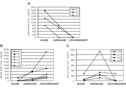
Figure 1. Three patterns of plasma leptin levels in patients with Kawasaki disease. Serial values before (acute), at 1~3 days (subacute) and at 2~3 weeks (convalescent) after the gamma globuline infusion are shown. A: acute, B: subacute, C: convalescent.
cipitating reagent was added and the mixture was further incubated for 20 minutes at 4oC. Bound and free ligands were separated by centrifugation. Leptin levels were counted in a gamma counter (Cobra II, Packard) for 1 minute.
Reverse transcriptase-polymerase chain reaction (RT-PCR).
One μg of total RNA per sample was reverse trans- cribed using Moloney murine leukemia virus reverse transcriptase (Perkin Elmer, Norwalk, CT, USA) and oligo dT priming according to the manufacturer's instruction, at 42oC for 15 minutes. Amplification with specific primers was performed in a Gene Amp PCR system 9600 (Perkin Elmer) for 31 cycles with a 45 s/94oC denaturation, 1 min/59oC annealing, 1 min/72oC extension profile in the case of IP-10; for 35 cycles with a 45 s/94oC denaturation, 45 s/61oC annealing, 45 s/72oC extension profile in the case of Mig; for 35 cycles with a 15 s/95oC denaturation, 30 s/60oC annealing, 1 min/72oC extension profile in the case of IL-8; for 40 cycles with a 1 min/94oC denaturation, 1 min/55oC annealing, 1.5 min/72oC extension profile in the case of TNF-α; for 30 cycles with a 30 s/95oC denaturation, 30 s/60oC annealing, 30 s/72oC extension profile in the case of GAPDH.
Amplification of mRNA for the housekeeping gene
GAPDH was used as an internal quality standard.
Amplified products were electrophoresed on 1.5~ 2% agarose gel stained with 0.5 g/mL ethidium bromide. The primer sequences were as follows:
GAPDH (178 bp) sense; 5'-acccactcctccacctttg-3', antisense; 5'-ctcttgtgctcttgctggg-3', IL-8 (300 bp) sense;
5'-atgacttccaagctggccgtg-3', antisense; 5'-ttatgaattctcag- ccctcttcaaaaacttctc-3', IP-10 (107 bp) sense; 5'-ggaacc tccagtctcagcacc-3', antisense; 5'-gcgtacggttctagagaga- ggtac-3', Mig (123 bp) sense; 5'-ttcctcttgg gcatcatc- ttgctg-3', antisense; 5'-ggtctttcaaggattgtaggtgga-3'.
Statistical analysis. Results were presented as mean±
SD. Statistical differences were analyzed by the paired t-test. Values were considered significant when p< 0.05.
Results
Plasma levels of leptin in Kawasaki disease. We first exam- ined the plasma leptin levels according to clinical stages of Kawasaki disease. The production of plasma leptin according to clinical stages showed 3 patterns, namely, descending (Fig. 1A), ascending (Fig. 1B), and reverse V shape (Fig. 1C). In descending pattern, two of three cases had pericardial effusion. Three of nine cases which showed ascending pattern had severe cervical lymphoadenopathy, and two of four
cases which showed reverse V shape pattern had
Table III. Summary of expression of chemokine genes in Kawa- saki disease
Acute Subacute Convalescent
IP-10 13/13 (100) 13/13 (100) 13/13 (100) Mig 2/13 (15) 6/13 (46) 4/13 (31) IL-8 9/13 (69) 13/13 (100) 10/13 (77)
Values given in parentheses are the percent of positive patient samples.
Table II. Mean values of plasma leptin
Acute Subacute Convalescent
335.8±549.0* 358.3±347.6† 443.6±645.9‡
n=13, mean±standard deviation (pg/mL)
*p=0.137, compared with subacute, †p=0.754, compared with convalescent, ‡p=0.212, compared with acute
P2
P9 P8
IL-8 P5
GAPDH a b c a b c
P12
Figure 3. Expression pattern of IL-8 during the 3 phases of Kawasaki patients with cervical lymphadenopathy. Total RNA was isolated from PBMC collected from Kawasaki patients, and RT-PCR was performed with primers indicated as described in materials and methods. GAPDH was used as internal standard.
a: acute, b: subacute, c: convalescent.
IP-10 P1 P2 P3
M a b c
GAPDH a b c
Figure 2. The expression profiles of IP-10 mRNA during the 3 phases in three patients with Kawasaki disease. P2 and P3 accompanied pericardial effusion. Total RNA was isolated from PBMC collected from Kawasaki patients, and RT-PCR was performed with primers indicated as described in materials and methods. GAPDH was used as an internal standard. M: DNA marker (100 bp DNA ladder) a: acute, b: subacute, c: con- valescent.
GAPDH IL-8 IP-10 Mig
a b c
Figure 4. Expression pattern of the genes for chemokines in a Kawasaki patient (P8) with meningitis. Total RNA was isolated from PBMC collected from Kawasaki patients, and RT- PCR was performed with primers indicated as described in materials and methods. GAPDH was used as an internal standard. a: acute, b:
subacute, c: convalescent.
coronary artery dilatation. However, as described in specific findings in Table I, there were not significant differences in patterns of leptin levels between spe- cific clinical findings. To gain a further insight into plasma leptin levels, the expression of leptin receptor, long form (OB-RL) from the PBMC in three cases which showed the descending pattern of plasma leptin (Fig. 1A) was examined. The expressions of OB-RL were gradually increased according to the clinical stages. The mean values of plasma leptin concentrations during each of the stages (n=13, p> 0.05, mean±SD, pg/ml) were 335.8±549.0 in acute, 358±347.6 in subacute, and 443.6±645.9 in conva- lescent stage (Table II). The gender, age, and body mass index (BMI) in all cases were not related to the patterns of leptin levels during the clinical stages.
Consequently, there was a negative correlation between the plasma leptin and clinical stages in Kawasaki disease.
Expression of chemokine IP-10, Mig, and IL-8 in Kawasaki disease. The expression of IP-10, Mig and IL-8 mRNAs in 39 samples (13 samples of each stage) from 13 cases were examined. Expressions of IP-10, Mig, IL-8 mRNA were detected in 13/13 (100%), 2/13 (15%), 9/13 (69%) during the acute stage, 13/
13 (100%), 6/13 (46%), 13/13 (100%) during the subacute stage, and 13/13 (100%), 4/13 (31%), 10/
13 (77%) during the convalescent stage, respectively (Table III). The lowest rate of expression was shown in Mig mRNA. We could not find any relationship between the clinical manifestations in patients and the
expression patterns of Mig mRNA during the clinical
stages. The expression of IP-10 mRNA was detec- table in all samples over the three clinical stages. In the group of descending pattern of plasma leptin le- vels, the expression of IP-10 mRNA was gradually decreased according to the clinical stages (Fig. 2).
Pericardial effusion was the distinguished clinical finding in two of three cases. In all 5 cases with massive cervical lymphadenopathy (lymph node size
>5 cm), the expression of IL-8 mRNA during the subacute stage was more elevated than acute and convalescent stages (Fig. 3). The production pattern
of plasma leptin in each of these cases was different,
as shown in Fig. 1. We could also find a specific pattern in the expressions of IP-10, Mig, IL-8 mRNA in one case with meninigitis. The expressions of all cytokine mRNAs during the subacute stage were more elevated than other stages (Fig. 4). In this case, the production of plasma leptin during the clinical stages showed an ascending pattern (Fig. 1B).
Discussion
In the present study, we analyzed the production of plasma leptin and the expression of CXC chemo- kine IP-10, Mig, and IL-8 mRNA according to the clinical stages in Kawasaki disease. Earlier studies (5-8,10) on the production or expression of che- mokines in Kawasaki disease have focused mainly on CC chemokines or IL-8. Moreover, a leptin study of Kawasaki disease has not been performed yet.
Although we found three patterns of plasma leptin kinetics according to clinical stages, there was no specific correlation between leptin levels and the clin- ical stages of Kawasaki disease. Increased serum leptin levels have been found in patients with alco- holic cirrhosis (16) or during pregnancy (17). In sepsis, either increases or no changes in leptin levels have been reported (18-20). However, no correlation between leptin levels and disease activity has been found in rheumatoid arthritis (21) and inflammatory bowel disease (22).
Kawasaki disease is associated with the activation of peripheral blood macrophages/monocytes, T cells, B cells, and clonal expansion of T cells, resulting in highly elevated levels of various cytokines (23-25).
Pathophysiologic roles of chemokines in Kawasaki disease are still poorly understood. CXC chemokines are mostly chemotactic for neutrophils, but CXC che- mokine IP-10 is a chemoattractant for monocytes and T cells, and Mig is chemotactic for tumor infiltrating lymphocytes and activated T cells. Wong et al (6) have reported that there was no obvious correlation between clinical stages of the Kawasaki disease and expression of chemokine RANTES, MCP-1 and MIP-1β. Our data with IP-10, Mig, and IL-8 also showed no obvious correlation between clinical stages. However, we observed that some expression patterns of IP-10 and IL-8 might be related to the specific clinical manifestations of Kawasaki disease.
In three patients especially, the expression pattern of IP-10 according to the clinical stages coincided with the pattern of plasma leptin production. Namely, both IP-10 expression and leptin production gradu- ally decreased according to the process of clinical stages. However, we could not find any specific find- ings in common among three patients except peri- cardial effusion in two of three cases.
IL-8 has been suggested as an indicator of high
risk for coronary lesions in Kawasaki disease (5). In our data, the expression of IL-8 mRNA in all 5 cases with severe cervical lymphadenopathy was over- expressed in subacute stages, compared with other stages. There have been various studies of IL-8 modulation with IVIG in Kawasaki diseases (8,10,26).
In a study by Asano et al (26), the expression of IL-8 was shown to increase after IVIG, and Terai et al (7) have reported that IVIG treatment correlated with a rapid decrease in the circulating levels of MCP-1 but not IL-8. In an In vitro study with human mono- cyte (27), IVIG has also been shown to enhance the expression of IL-8 mRNA and protein production.
All samples of the subacute stage in this study were obtained from the patients after IVIG therapy. How- ever, the IL-8 mRNA in other samples except 5 cases was not over-expressed during subacute stage, and in two cases with coronary artery dilatation, the IL-8 mRNA was expressed evenly during the three clinical stages. Therefore, further discrete study of the ki- netics of IL-8 in Kawasaki disease must be per- formed.
In a patient with meningitis, the expressions of IP-10, Mig, and IL-8 mRNA during subacute stage were more elevated than during other stages. Chemo- kine production has been demonstrated in the cere- brospinal fluid in infectious meningitis (28), and there is some suggestion that IL-8, MCP-1 and IP-10 are probably the most important chemokines in the pathophysiology of meningitis (29). But, we could not find any reports for a cytokine network between Kawasaki disease and meningitis. To confirm our data about Kawasaki disease with meningitis, study with more cases must be performed.
Although, the number of cases in this study was not sufficient, these results suggest that plasma leptin does not play a role as a factor participating in pathophysiologic functions in Kawasaki disease, however, the action mechanism of IVIG and addi- tional specific clinical manifestations may contribute to the expression patterns of chemokine IL-8 and IP-10, Mig in Kawasaki disease.
References
1. Kawasaki T: Acute febrile mucocutaneous syndrome with lymphoid involvement with specific desquamation of the fingers and toes: Clinical observations of 50 cases (in Japa- nese). Jpn J Allerg 16;178-222, 1967
2. Hirao J, Hibi S, Andoh T, Ichimura T: High levels of circulating interleukin-4 and interleukin-10 in Kawasaki Disease. Int Arch Allergy Immunol 112;152-156, 1997 3. Lin CY, Lin CC, Hwang B, Chiang BN: The changes of
interleukin-2, tumor necrotic factor and gamma-interferon production among patients with Kawasaki disease. Eur J Pediatr 150;179-182, 1991
4. Lin CY, Lin CC, Hwang B, Chiang B: Serial changes of
serum interleukin-6, interleukin-8, and tumor necrosis factor alpha among patients with Kawasaki disease. J Pediatr 121;924-926, 1992
5. Lin CY, Lin CC, Hwang B, Chiang BN: Cytokines predict coronary aneurysm formation in Kawasaki disease pa- tients. Eur J Pediatr 152;309-312, 1993
6. Wong M, Silverman ED, Fish EN: Evidence for RANTES, monocyte chemotactic protein-1β, and macrophage in- flammatory protein-1b expression in Kawasaki disease. J Rheumatol 24;1179-1185, 1997
7. Terai M, Jibiki T, Harada A, Terashima Y, Yasukawa K, Tateno S, Hamada H, Oana S, Niimi H, Matsushima K:
Dramatic decrease of circulating levels of monocyte che- moattractant protein-1 in Kawasaki disease after gamma globulin treatment. J Leuk Biol 65;566-572, 1999 8. Suzuki H, Noda E, Miyawaki M, Takeuchi T, Uemura S,
Koike M: Serum levels of neutrophil activation cytokines in Kawasaki disease. Pediatrics International 43;115-119, 2001
9. Asano T, Ogawa S: Expression of monocyte chemo- attractant protein-1 in Kawasaki disease: the anti-in- flammatory effect of gamma globulin therapy. Scand J Immunol 51(1);98-103, 2000
10. Gupta M, Noel GJ, Schaefer M, Friedman D, Bussel J, Johann-Liang R: Cytokine modulation with immune gamma-globulin in peripheral blood of normal children and its implications in Kawasaki disease treatment. J Clin Immunol 21(3);193-199, 2001
11. Wuyts A, Proost P, Damme JV: Interleukin-8 and other chemokines. In: Thomson A: The Cytokine Handbook, 3rd ed. P271-311, New York: Academic Press; 1998 12. Zhang Y, Proenca R, Maffei M, Barone M, Leopold L,
Friedman JM: Positional cloning of the mouse obese gene and its human homologue. Nature 372;425-432, 1994 13. Houseknecht KL, Portocarrero CP: Leptin and receptors:
Regulators of whole-body energy homeostssis. Dcmestic Animal Endocrinology 15(6);457-475, 1998
14. Fantuzzi G, Faggioni R: Leptin in the regulation of im- munity, inflammation, and hematopoiesis. J Leuk Biol 68;437-446, 2000
15. Zarkesh-Esfahani H, Pockley G, Metcalfe RA, Bidling- maier M, Wu Z, Ajami A, Weetman AP, Strasburger CJ, Ross RJM: High-dose leptin activaes human leukocytes via receptor expression on monocytes. J Immunol 167;4593- 4599, 2001
16. Henriksen JH, Holst JJ, Moller S, Brinch K, Bendtsen F:
Increased circulating leptin in alcoholic cirrhosis: relation to release and disposal. Hepatology 29;1818-1824, 1999 17. Butte NF, Hopkinson JM, Nicolson MA: Leptin in human
reproduction: Serum levels in pregnant and lactating women. J Clin Endocrinol Metab 82;585-589, 1997 18. Torpy DJ, Bornstein SR, Chrousos GP: Leptin and in-
terleukin-6 in sepsis. Horm Metab Res 30;726-729, 1998 19. Arnalich F, Lopez J, Codoceo R, Jimenez M, Madero R,
Montiel C: Relationship of plasma leptin to plasma cytokines and human survival in sepsis and septic shock.
J Infect Dise 180;908-911, 1999
20. Carlson GL, Saeed M, Little RA, Irving MH: Serum leptin concentrations and their relation to metabolic abnor- malities in human sepsis. Am J Physiol 276;E658-E662, 1999
21. Anders HJ, Rihl M, Heufelder A, Loch O, Schatten- kirchner M: Leptin serum levels are not correlated with disease activity in patients with rheumathoid arthritis.
Metabolism 48;745-748, 1999
22. Hoppin AG, Kaplan LM, Zurakowski D, Leichtner AM, Bousaros A: Serum leptin in children and young adults with inflammatory bowel disease. J Pediatr Gastroenterol Nutr 26;500-505, 1998
23. Furukawa S, Matsubara T, Yabuta K: Immunological pa- rameters in determining the severity of vascular damage during acute Kawasaki disease. Disease Mechanisms 149- 154, 1992
24. Koga M, Ishihara T, Takahashi M, Umezawa Y, Furukawa S: Activation of peripheral blood monocytes and macro- phages in Kawasaki desease: Ultrastructural and immuno- cytochemical investigation. Pathol Inter 48;512-517, 1998 25. Choi IH, Chwae YJ, Shim WS, Kim DS, Kwon DH, Kim
JD, Kim SJ: Clonal expression of CD8+ T cells in Kawa- saki disease. J Immunol 159;481-486, 1997
26. Asano T, Ogawa S: Expression of IL-8 in Kawasaki dis- ease. Clin Exp Immunol 122;514-519, 2000
27. de Souza VR, Carreno M-P, Kaveri SV, Ledur A, Sadeghi H, Cavaillon J-M, Kazatchkine MD, Haeffner-Cavaillon N: Selective induction of interleukin-1 receptor antagonist and interleukin-8 in human monocytes by normal poly- specific IgG (intravenous immunoglobulin). Eur J Immu- nol 25;1267-1273, 1995
28. Lahrtz F, Piali L, Spanaus KS, Seebach J, Fontana A:
Chemokines and chemotaxis of leukocytes in infectious meningitis. J Neuroimmunol 85(1);33-43, 1998
29. Grygorczuk S, Pancewicz S, Kondrusik M, Zajkowska J, Hermanowska-Szpakowicz T: Chemokines in meningitis of different etiologies. Pol Merkuriusz Lek 10(56);117-121, 2001