pISSN 2093-596X · eISSN 2093-5978
Article
Usefulness of Measuring Thyroid Stimulating Antibody at the Time of Antithyroid Drug Withdrawal for Predicting Relapse of Graves Disease
Hyemi Kwon1, Won Gu Kim1, Eun Kyung Jang1,2, Mijin Kim1, Suyeon Park1, Min Ji Jeon1, Tae Yong Kim1, Jin-Sook Ryu3, Young Kee Shong1, Won Bae Kim1
1Department of Internal Medicine, Asan Medical Center, University of Ulsan College of Medicine, Seoul; 2Department of Internal Medicine, Cancer Center, Dongnam Institute of Radiological & Medical Sciences, Busan; 3Department of Nuclear Medicine, Asan Medical Center, University of Ulsan College of Medicine, Seoul, Korea
Background: Hyperthyroidism relapse in Graves disease after antithyroid drug (ATD) withdrawal is common; however, measur- ing the thyrotropin receptor antibody (TRAb) at ATD withdrawal in order to predict outcomes is controversial. This study com- pared measurement of thyroid stimulatory antibody (TSAb) and thyrotropin-binding inhibitory immunoglobulin (TBII) at ATD withdrawal to predict relapse.
Methods: This retrospective study enrolled patients with Graves disease who were treated with ATDs and whose serum thyroid- stimulating hormone levels were normal after receiving low-dose ATDs. ATD therapy was stopped irrespective of TRAb positivi- ty after an additional 6 months of receiving the minimum dose of ATD therapy. Patients were followed using thyroid function tests and TSAb (TSAb group; n=35) or TBII (TBII group; n=39) every 3 to 6 months for 2 years after ATD withdrawal.
Results: Twenty-eight patients (38%) relapsed for a median follow-up of 21 months, and there were no differences in baseline clini- cal characteristics between groups. In the TSAb group, relapse was more common in patients with positive TSAb at ATD withdrawal (67%) than patients with negative TSAb (17%; P=0.007). Relapse-free survival was shorter in TSAb-positive patients. In the TBII group, there were no differences in the relapse rate and relapse-free survivals according to TBII positivity. For predicting Graves dis- ease relapse, the sensitivity and specificity of TSAb were 63% and 83%, respectively, whereas those of TBII were 28% and 65%.
Conclusion: TSAb at ATD withdrawal can predict the relapse of Graves hyperthyroidism, but TBII cannot. Measuring TSAb at ATD withdrawal can assist with clinical decisions making for patients with Graves disease.
Keywords: Graves disease; Hyperthyroidism; Immunoglobulins; Prognosis
INTRODUCTION
Autoantibodies to thyroid-stimulating hormone (TSH, also called thyrotropin) receptor (thyrotropin receptor antibody
[TRAb]) have two different main functions; stimulation or blocking of the TSH receptor (TSHR) [1-4]. Graves disease (GD) is an autoimmune disorder that is mediated by the TRAbs that activate TSHR, thereby stimulating thyroid hormone synthe-
Received: 27 January 2016, Revised: 15 February 2016, Accepted: 10 March 2016
Corresponding author: Won Gu Kim
Division of Endocrinology and Metabolism, Department of Internal Medicine, Asan Medical Center, University of Ulsan College of Medicine, 88 Olympic-ro 43-gil, Songpa-gu, Seoul 05505, Korea
Tel: +82-2-3010-5883, Fax: +82-2-3010-6962, E-mail: wongukim@amc.seoul.kr
Copyright © 2016 Korean Endocrine Society
This is an Open Access article distributed under the terms of the Creative Com- mons Attribution Non-Commercial License (http://creativecommons.org/
licenses/by-nc/4.0/) which permits unrestricted non-commercial use, distribu- tion, and reproduction in any medium, provided the original work is properly cited.
Endocrinol Metab 2016;31:300-310
http://dx.doi.org/10.3803/EnM.2016.31.2.300 pISSN 2093-596X · eISSN 2093-5978
sis, secretion, and thyroid cell growth [1,2]. The presence of cir- culating TRAbs is an important pathogenic indicator of GD [5- 7]. Administering an antithyroid drug (ATD) is an effective ther- apeutic modality for the treatment of GD patients [1,8]. ATDs have beneficial immunosuppressive effects and reduce the pro- duction of thyroid hormones, as well as maintain the euthyroid state while patients wait for spontaneous remission [1,8].
The main drawback of ATD treatment for GD hyperthyroid- ism is the relapse rate after the withdrawal of ATDs, which is as high as 50% to 60% [6,9]. A useful predictive parameter for relapse would help to prevent long and complicated drug regi- mens that carry potentially serious adverse effects. The ability to predict the course and relapse of GD would also facilitate the selection of patients who require early definitive therapy including radioactive iodine ablation (RAI) or surgery. Many clinical factors have been suggested for predicting relapse, such as age, sex, presence of a goiter, family history of GD, thyroid hormone levels, thyroid echogenicity on ultrasonogra- phy (US), the results of Doppler US, and technetium-99m (99mTc) pertechnetate uptake [3,10-13]. TRAbs measurement is known to be useful for predicting GD relapse because of the active pathogenic role of TRAbs [3,10,14-24].
Assays for TRAb detection include the competitive thyrotro- pin-binding inhibitory immunoglobulin (TBII) assay and the thyroid stimulatory antibody (TSAb) bioassay [3,4]. The TBII assay can detect immunoglobulins that inhibit the binding of radio-labeled TSH to TSHRs [4,25-27]. This assay measures not only thyroid-stimulating immunoglobulins (TSIs), but also thyroid-blocking immunoglobulins (TBIs) and neutral immu- noglobulins [4,25,26]. However, the TSAb bioassay measures the production of cyclic adenosine monophosphate (cAMP) in response to interactions between TSHR and TSI. It can distin- guish TSI from TBI and neutral TRAb [4,25,26]. TSI in a pa- tient’s serum results in the increased production of cAMP, whereas TBI inhibits cAMP production [3,4].
The aim of this study was to compare the value of TSAb and TBII measurements at the time of ATD withdrawal for predict- ing relapse in GD patients. This study evaluated GD hyperthy- roidism relapse during a median follow-up period of 21 months after ATD discontinuation.
METHODS
Patients
This retrospective study enrolled 74 GD patients who were treated between 2005 and 2012 at Asan Medical Center in
Seoul, Korea. Patients with newly diagnosed GD who were treated with ATDs for the first time were enrolled. The diagno- sis of GD was based on the following criteria: serum free thy- roxine (fT4) level >24.45 pmol/L, decreased serum TSH level
<0.3 mIU/L, increased serum TRAb levels (TSAb levels ≥ 140% or TBII titers ≥1.5 IU/L), increased uptake on 99mTc thy- roid scan, and the presence of the appropriate clinical features such as a diffuse goiter, thyroid-associated orbitopathy (TAO), or symptoms of hyperthyroidism. A goiter was defined as the abnormal enlargement of the thyroid gland and diagnosed at the time of initial physical examination. The TAO diagnosis was based on the symptoms (dry eyes, diplopia, visual loss, vi- sual field loss, and ocular pain) and signs (lid retraction, lid lag, glabellar furrows, proptosis). Patients who became pregnant during the follow-up period were excluded from this study.
Those who required RAI therapy due to intractable GD or ma- jor side effects associated with ATDs were also excluded. This study was approved by institutional review board of Asan Medical Center, Seoul, Korea.
Patients were divided into the TSAb or TBII groups. Patients in the TSAb group received follow-up using the TSAb bioas- say (n=35), and patients in the TBII group received follow-up using the TBII assay (n=39). ATD therapy and follow-up ex- aminations were performed using a uniform protocol using a dose titrating regimen, mostly with methimazole (MMI) throughout the study period. Some patients were changed to re- ceiving propylthiouracil (PTU) or carbimazole due to minor side effects associated with MMI. ATDs were discontinued when the serum fT4 and TSH levels of the patients were within the normal range for ≥6 months while receiving the minimum maintenance dose of ATDs (MMI ≤5 mg/day, carbimazole ≤ 10 mg/day, and PTU ≤100 mg/day), regardless of TRAb sta- tus. Median treatment duration with ATDs was 21 months (in- terquartile range [IQR], 10.0 to 27.5). All patients received fol- low-up using thyroid function tests (fT4, total triiodothyronine [T3], and TSH), and measurement of their TRAb levels (TSAb or TBII). These tests were performed at the time of ATD with- drawal and 3, 9, 15, and 21 months afterward.
Fifty-eight of 74 patients were treated with MMI, nine patients were treated with carbimazole, and seven patients were treated with PTU. Patients were considered to have relapsed if their fT4 levels were above the upper limit of the normal range (>24.45 pmol/L) and their serum TSH concentrations were low (<0.3 mIU/L).
Thyroid function tests
As previously described, the serum TSH concentrations (refer-
ence range, 0.3 to 4 mIU/L; lower detection limit, 0.01 mIU/L) were measured using an immunoradiometric assay (TSH- CTK-3; DiaSorin S.p.A., Saluggia, Italy) with a functional sen- sitivity of 0.07 mIU/L [28]. The serum fT4 level (normal range, 10.30 to 24.45 pmol/L) was measured using the fT4 radioim- munoassay (RIA) KIT (Immunotech, Prague, Czech Republic).
The serum total T3 level (reference range, 1.51 to 2.77 nmol/L) was measured by RIA using T3-CTK (DiaSorin S.p.A.).
TSAb assays
For the TSAb assays, the Thyretai TSI reporter Bio Assay (Di- agnostic Hybrids Inc., Athens, OH, USA) was used according to the manufacturer’s instructions. The Thyretai kit is a Mc4 assay. The specimen-to-reference ratio (SRR), which is defined as the mean specimen TSI/mean reference TSI, is equal to 100.
A specimen was considered positive if SRR was ≥140%.
TBII assays
For the TBII assays, the values were detected using the B.
R.A.H.M.S. TRAK human RIA (B.R.A.H.M.S. GmbH, Henni- gsdorf/Berlin, Germany) according to the manufacturer’s rec- ommendation. TBII titers ≥1.5 IU/L were considered positive.
The analytical sensitivity was 0.3 IU/L and the functional assay sensitivity was 1.0±0.2 IU/L.
Statistical analysis
The statistical analysis was conducted using using SPSS ver- sion 21.0 (IBM Co., Armonk, NY, USA). Graphs were pro- duced using Prism version 5.01 (GraphPad Software Inc., La Jolla, CA, USA). The continuous variables are presented as the mean±SD or medians with IQR. The continuous variables were compared between groups using the Student t test or Mann-Whitney test. The categorical variables are presented as numbers with percentages. The categorical variables were compared between groups using the chi-square test or Fisher exact test. The relapse-free survival (RFS) curves were calcu- lated using the Kaplan-Meier method and the log-rank test was used to compare RFSs according to TRAb titers. The hazard ratio (HR) and 95% confidence interval (CI) used to evaluate the risk of relapse in the TSAb group were derived using Cox proportional hazards modeling. The multivariate analysis in- cluded age, sex, presence of a goiter and orbitopathy, thyroid function at baseline, treatment duration of any ATDs, and TSAb. All P values were 2-sided, with P<0.05 considered sta- tistically significant.
RESULTS
Baseline characteristics
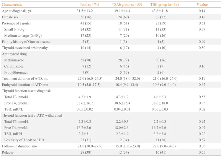
The baseline characteristics are listed in Table 1. The mean age of the 74 patients was 31.5±13.2 years, and 56 patients (76%) were female (Table 1). In the median follow-up period of 21.0 months (IQR, 10.0 to 27.5), 28 of 74 patients (38%) demon- strated GD relapse.
In the TSAb group (n=35), the mean age was 39.1±14.4 years, and 24 patients (69%) were female (Table 1). Eighteen pa- tients (51%) had a goiter, and six patients (17%) had TAO at the diagnosis of GD. Twenty-eight patients (72%) were treated with MMI, and the median treatment duration using ATDs was 24.0 months (IQR, 18.0 to 32.0). The median duration of euthyroid status while using the minimum dose of ATDs was 10.4 months (IQR, 9.9 to 13.4). Twelve patients (34%) relapsed during the median follow-up period of 15.0 months (IQR, 10.0 to 23.0).
In the TBII group (n=39), the mean patient age was 43.6±
11.8 years, and 32 patients (82%) were female (Table 1). Twen- ty-three patients (59%) had a goiter, and four patients (10%) had TAO at the diagnosis of GD. Thirty patients (86%) were treated with MMI, and the median treatment duration with ATDs was 21.0 months (IQR, 16.0 to 26.0). The median dura-
100 80 60 40 20 0
Relapse-free survival
Duration (mo) Total
10 20 30 40 50 60
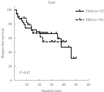
Fig. 1. Relapse-free survival of Graves disease patients after anti- thyroid drug withdrawal in the thyroid stimulatory antibody (TSAb) and thyrotropin-binding inhibitory immunoglobulin (TBII) groups. Among 74 patients, there was no significant differ- ence in the relapse-free survival between the TSAb and TBII groups.
TSAb (n=35) TBII (n=39)
P=0.87
tion of euthyroid with the minimal maintenance dose of ATDs was 10.6 months (IQR, 9.0 to 14.0). Sixteen patients (41%) re- lapsed during a median follow-up period of 22.0 months (IQR, 9.0 to 34.0).
There were no significant differences in age, sex, presence of a goiter, 99mTc uptake on the thyroid scans, family history of GD, presence of TAO, treatment duration with ATDs, duration of euthyroid status while using a minimal maintenance dose of ATDs, thyroid function at diagnosis (except initial serum TSH levels), TRAbs positivity at withdrawal of ATD, or RFS be- tween the TSAb and TBII groups (Table 1, Fig. 1).
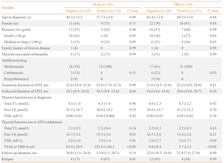
Clinical characteristics of the patients in the TSAb group according to TSAb at ATD withdrawal
In the TSAb group (n=35), 23 patients (66%) were negative
for TSAb and 12 patients were positive for TSAb at the time of ATD withdrawal. There were no significant differences in age, sex, presence of a goiter, 99mTc uptake on the thyroid scans, family history of GD, presence of TAO, treatment duration with ATDs, duration of the euthyroid status using the minimal maintenance dose of ATDs, and thyroid function at diagnosis between the TSAb-positive and -negative patients (Table 2).
The fT4 level at ATD withdrawal was higher in TSAb-positive patients (19.31±2.57 pmol/L) than TSAb-negative patients (16.73±2.57 pmol/L, P=0.01). There were significantly more cases of relapse in TSAb-positive patients (8 of 12 patients, 67%) than TSAb-negative patients (4 of 23 patients, 17%) dur- ing the median follow-up period of 15.0 months (IQR, 10.0 to 23.0; odds ratio, 9.5; 95% CI, 1.9 to 47.7; P=0.007) (Table 2).
Table 1. Baseline Characteristics of the Graves Disease Patients in the TSAb and TBII Groups
Characteristic Total (n=74) TSAb group (n=35) TBII group (n=39) P value
Age at diagnosis, yr 31.5±13.2 39.1±14.4 43.6±11.8 0.14
Female sex 56 (76) 24 (69) 32 (82) 0.18
Presence of a goiter 41 (55) 18 (51) 23 (59) 0.51
Small (<40 g) 24 (32) 11 (31) 13 (33) 0.77
Medium to large (≥40 g) 17 (23) 7 (20) 10 (26)
Family history of Graves disease 2 (3) 1 (3) 1 (3) 0.99
Thyroid associated orbitopathy 10 (14) 6 (17) 4 (10) 0.50
Antithyroid drug
Methimazole 58 (78) 28 (72) 30 (86)
Carbimazole 9 (12) 6 (15) 3 (9) 0.16
Propylthiouracil 7 (9) 5 (13) 2 (6)
Treatment duration of ATD, mo 22.0 (16.0–26.5) 24.0 (18.0–32.0) 21.0 (16.0–26.0) 0.19 Euthyroid duration of ATD, mo 10.5 (5.0–17.5) 10.4 (9.9–13.4) 10.6 (9.0–14.0) 0.67 Thyroid function test at diagnosis
Total T3, nmol/L 4.5±1.9 4.3±1.2 4.6±2.3 0.55
Free T4, pmol/L 38.6±16.7 38.6±15.4 38.6±18.0 0.95
TSH, mIU/L 0.05±0.02 0.04±0.01 0.06±0.03 0.02
Thyroid function test at ATD withdrawal
Total T3, nmol/L 2.2±0.3 2.2±0.3 2.2±0.3 0.92
Free T4, pmol/L 16.7±2.6 18.0±2.6 16.7±2.6 0.07
TSH, mIU/L 2.7±3.1 2.3±1.9 3.2±3.8 0.22
Positivity of TSAb or TBII 23 (31) 12 (34) 11 (28) 0.87
Follow-up duration, mo 21.0 (10.0–27.5) 15.0 (10.0–23.0) 22.0 (9.0–34.0) 0.05
Relapse 28 (38) 12 (34) 16 (41) 0.55
Values are expressed as mean±SD, number (%), or median (interquartile range).
TSAb, thyroid stimulatory antibody; TBII, thyrotropin-binding inhibitory immunoglobulin; ATD, antithyroid drug; T3, triiodothyronine; T4, thyrox- ine; TSH, thyroid-stimulating hormone.
Clinical characteristics of the patients in the TBII group according to TBII at ATD withdrawal
In the TBII group (n=39), 28 patients (72%) were negative for TBII and 11 patients (28%) were positive for TBII at the time of ATD withdrawal. There were no significant differences in age, sex, presence of a goiter, 99mTc uptake on thyroid scans, family history of GD, presence of TAO, treatment duration with ATDs, duration of euthyroid status using the minimal maintenance dose of ATDs, and thyroid function at diagnosis between the TBII-positive and -negative patients (Table 2).
There was no significant difference in the relapse rate between TBII-positive patients (4 of 11 patients, 36%) and -negative pa- tients (12 of 28 patients, 43%) during the median follow-up of
22.0 months (IQR, 9.0 to 34.0) (Table 2).
RFS according to TSAb or TBII positivity at ATD withdrawal
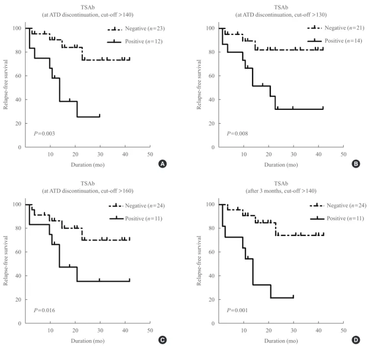
In the TSAb group, TSAb-positive patients demonstrated shorter RFS than TSAb-negative patients (P=0.003) (Fig. 2A).
When we applied various cut-off values for TSAb positivity at ATD withdrawal, TSAb-positive patients still demonstrated significantly shorter RFS in comparison with TSAb-negative patients (Fig. 2B, C). When we compared TSAb levels at 3 months after ATD withdrawal, the RFS of TSAb-positive pa- tients was also significantly shorter than TSAb-negative pa- tients (Fig. 2D).
Table 2. Clinical Factors in TRAb-Positive and -Negative Patients in the TSAb and TBII Groups
Variable TSAb (n=35) TBII (n=39)
Negative (n=23) Positive (n=12) P value Negative (n=28) Positive (n=11) P value
Age at diagnosis, yr 40.3±15.2 37.7±13.0 0.49 43.4±12.0 44.2±11.6 0.86
Female sex 15 (65) 9 (75) 0.71 22 (79) 10 (91) 0.65
Presence of a goiter 13 (57) 5 (42) 0.40 16 (57) 7 (64) 0.99
Small (<40 g) 10 (43) 1 (8) 0.05 10 (36) 3 (27) 0.65
Medium to large (≥40 g) 3 (13) 4 (33) 0.05 6 (21) 4 (36) 0.65
Family history of Graves disease 1 (4) 0 0.99 1 (4) 0 0.99
Thyroid-associated orbitopathy 4 (17) 2 (17) 0.99 3 (11) 1 (9) 0.99
Antithyroid drug
Methimazole 18 (78) 12 (100) 17 (61) 11 (100)
Carbimazole 3 (13) 0 0.11 6 (21) 0 0.03
Propylthiouracil 2 (9) 0 5 (18) 0
Treatment duration of ATD, mo 22.0 (18.0–32.0) 25.0 (17.0–31.3) 0.99 21.0 (14.5–25.8) 22.0 (16.0–28.0) 0.81 Euthyroid duration of ATD, mo 10.3 (9.9–24.0) 10.5 (9.8–13.8) 0.66 10.6 (9.6–14.6) 10.6 (10.0–20.7) 0.30 Thyroid function test at diagnosis
Total T3, nmol/L 4.3±1.0 4.3±1.6 0.96 4.6±2.3 4.7±2.2 0.92
Free T4, pmol/L 41.2±16.7 36.0±14.2 0.55 38.6±16.7 41.2±23.2 0.70
TSH, mIU/L 0.04±0.01 0.04±0.004 0.42 0.06±0.03 0.05±0.02 0.76
Thyroid function test at ATD withdrawal
Total T3, nmol/L 2.2±0.3 2.3±0.4 0.14 2.2±0.3 2.2±0.3 0.63
Free T4, pmol/L 16.7±2.6 19.3±2.6 0.01 16.7±2.6 15.4±2.6 0.21
TSH, mIU/L 2.4±2.0 1.9±1.6 0.41 3.0±2.7 3.6±7.1 0.69
TSAb or TBII levelsa 63.9±36.9 323.8±146.1 <0.05 0.8±0.4 3.7±3.5 <0.05
Follow-up duration, mo 20.0 (11.0–24.0) 13.0 (5.5–20.3) 0.14 22.0 (10.3–34.8) 23.0 (7.0–27.0) 0.49
Relapse 4 (17) 8 (67) 0.01 12 (43) 4 (36) 0.99
Values are expressed as mean±SD, number (%), or median (interquartile range).
TRAb, thyrotropin receptor antibody; TSAb, thyroid stimulatory antibody; TBII, thyrotropin-binding inhibitory immunoglobulin; ATD, antithyroid drug; T3, triiodothyronine; T4, thyroxine; TSH, thyroid-stimulating hormone.
aTSAb titer (%) and TBII (IU/L).
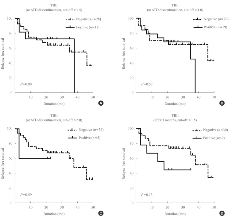
In the TBII group, there was no significant difference in RFS between TBII-positive and -negative patients at ATD with- drawal (using three different cut-off values) or at 3 months af- ter withdrawal (Fig. 3).
Diagnostic values of TSAb and TBII for predicting GD relapse
For predicting GD relapse, the sensitivity, specificity, positive
predictive value (PPV), negative predictive value (NPV), and accuracy of a positive TSAb value at ATD withdrawal were 63%, 83%, 78%, 69%, and 76%, respectively (Table 3). For a positive TBI, these values were 28%, 65%, 45%, 47%, and 50%, respectively (Table 3).
Factors associated with GD relapse in the TSAb group Patients who had higher TSAb titers at ATD discontinuation
100 80 60 40 20 0
100 80 60 40 20 0
100 80 60 40 20 0
100 80 60 40 20 0
Relapse-free survivalRelapse-free survival Relapse-free survivalRelapse-free survival
Duration (mo)
Duration (mo)
Duration (mo)
Duration (mo) Negative (n=23)
Negative (n=24)
Negative (n=21)
Negative (n=24) Positive (n=12)
Positive (n=11)
Positive (n=14)
Positive (n=11) (at ATD discontinuation, cut-off >140)TSAb
(at ATD discontinuation, cut-off >160)TSAb
(at ATD discontinuation, cut-off >130)TSAb
(after 3 months, cut-off >140)TSAb 10 20 30 40 50
10 20 30 40 50
10 20 30 40 50
10 20 30 40 50
Fig. 2. Relapse-free survival of the patients after antithyroid drug (ATD) withdrawal in the thyroid stimulatory antibody (TSAb) group according to TSAb levels. At ATD withdrawal using a cut-off value of (A) 140%, (B) 130%, (C) 160%, and (D) at 3 months after ATD withdrawal.
A
C
B
D P=0.003
P=0.016
P=0.008
P=0.001
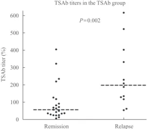
demonstrated more GD relapses (P=0.002). Patients with GD relapse demonstrated significantly higher TSAb titers at ATD discontinuation (P=0.002) (Fig. 4). The univariate and multi- variate analyses were performed to determine the factors asso- ciated with relapse in the TSAb group. The TSAb level at ATD withdrawal was the only significant factor associated with re- lapse on the univariate analysis (HR, 5.21; 95% CI, 1.53 to 17.71; P=0.008) (Table 4). On the multivariate analysis, the
TSAb level was a significant factor associated with relapse (HR, 6.68; 95% CI, 1.29 to 34.62; P=0.02) (Table 4).
DISCUSSION
In our current study, TSAb-positive patients demonstrated a higher risk for GD relapse after ATD withdrawal. The TSAb bioassay at ATD withdrawal was more useful for predicting the
100 80 60 40 20 0
100 80 60 40 20 0
100 80 60 40 20 0
100 80 60 40 20 0
Relapse-free survivalRelapse-free survival Relapse-free survivalRelapse-free survival
Duration (mo)
Duration (mo)
Duration (mo)
Duration (mo) Negative (n=28)
Negative (n=34)
Negative (n=20)
Negative (n=30) Positive (n=11)
Positive (n=5)
Positive (n=19)
Positive (n=9) (at ATD discontinuation, cut-off >1.5)TBII
(at ATD discontinuation, cut-off >2.0)TBII
(at ATD discontinuation, cut-off >1.0)TBII
(after 3 months, cut-off >1.5)TBII 10 20 30 40 50
10 20 30 40 50
10 20 30 40 50
10 20 30 40 50
Fig. 3. Relapse-free survival of the patients after antithyroid drug (ATD) withdrawal in the thyrotropin-binding inhibitory immunoglobu- lin (TBII) group according to TBII levels. At ATD withdrawal using a cut-off value of (A) 1.5, (B) 1.0, (C) 2.0, and (D) at 3 months after ATD withdrawal.
A
C
B
D P=0.99
P=0.59
P=0.57
P=0.13
relapse of GD hyperthyroidism in comparison with the TBII assay. This study is the first study demonstrating RFS of the GD patients in the TSAb (Mc4 assay) and TBII (second-gener- ation assay) groups. This study enrolled patients with newly di- agnosed GD who were initially treated by dose titrating regi- mens of ATDs, and they maintained an euthyroid state with a minimum dose of ATDs for ≥6 months. All patients were fol- lowed using the same protocol after ATD withdrawal. We found statistically significant differences between the TSAb and the TBII assay for predicting the GD hyperthyroidism re- lapse. The cut-off values for TSAb and TBII at ATD withdraw- al were the same as those used for diagnosing of GD.
Measuring TRAbs at the time of ATD withdrawal is useful
for predicting GD relapse [3,10,14-24]. GD is characterized by remission and relapse, like many autoimmune diseases. In a meta-analysis conducted 20 years ago, TRAb assays could not demonstrate a sufficient predictive value for GD relapse, main- ly due to the low sensitivity and specificity values of earlier as- says [6]. However, a recent study reported a significant correla- tion between serum TRAb levels at the end of MMI treatment and percentage of patients with recurrent hyperthyroidism (r=0.56, P<0.001) and time to recurrent hyperthyroidism (r=
–0.38, P=0.03) [29].
The TBII assay can detect immunoglobulins that inhibit the binding of radio-labeled TSH to TSHRs [25-27,30]. TBII as- says that initially used porcine TSHR were shown to have a Table 3. Diagnostic Values of TSAb and TBII at Antithyroid
Drug Withdrawal for Hyperthyroidism Relapse in Patients with Graves Disease
Vaiable
TSAb (n=35) TBII (n=39) Negative
(n=23) Positive
(n=12) Negative (n=28) Positive
(n=11)
Relapse 4 (17) 8 (67) 12 (43) 4 (36)
Diagnostic values, %
Sensitivity 63 28
Specificity 83 65
Negative predictive value 78 45
Positive predictive value 69 47
Accuracy 76 50
Values are expressed as number (%).
TSAb, thyroid stimulatory antibody; TBII, thyrotropin-binding inhibi- tory immunoglobulin.
Table 4. Cox Proportional Hazard Modeling for Predicting Graves Disease Relapse in the TSAb Group
Variable Univariate Multivariate
HR 95% CI P value HR 95% CI P value
Age at diagnosis, yr 0.99 0.95–1.03 0.65 - - NA
Male sex 1.04 0.31–3.47 0.06 - - NA
Goiter (medium to large) 2.07 0.62–6.94 0.24 - - NA
Thyroid associated orbitopathy 1.14 0.24–5.41 0.87 - - NA
Severe hyperthyroidism at diagnosisa 1.63 0.20–13.39 0.65 - - NA
Treatment duration of ATD 1.00 0.99–1.02 0.85 - - NA
Positive TSAb levels at ATD withdrawalb 5.21 1.53–17.71 0.008 6.68 1.29–34.62 0.02 TSAb, thyroid stimulatory antibody; HR, hazard ratio; CI, confidence interval; NA, not applicable; ATD, antithyroid drug.
aSevere hyperthyroidism at baseline was defined as serum free thyroxine level >64.4 pmol/L at baseline; bA specimen was considered positive if specimen-to-reference ratio was ≥140%.
600 500 400 300 200 100 0
TSAb titer (%)
TSAb titers in the TSAb group
Remission Relapse
Fig. 4. Distribution of thyroid stimulatory antibody (TSAb) titers in patients with remission and Graves disease relapse. The medi- ans of the TSAb titers were 61.1% and 203.5% in patients with remission and relapse, respectively (dotted lines).
P=0.002
sensitivity of 50% to 90% [27]. Second-generation assays, such as solid-phase enzyme-linked immunosorbent assays and radio receptor assay, demonstrated much higher sensitivities (90% to 99%) and specificities (95% to 100%) [27].
The TSAb biologic assays measure the production of cAMP when sera-containing TRAbs are exposed to TSHR on cell preparations [3]. The Mc4 assay, as a third-generation TSAb assay uses genetically engineered Chinese hamster ovary (CHO) Mc4 cells [25,26]. Because the substituted C-terminal area of TSHR includes epitopes for TBI, CHO Mc4 cells are capable of specifically detecting serum TSI without interfering with TBI [25,26]. A specimen was considered positive if SRR was ≥140%. Using a cut-off SRR of 140%, receiver operating characteristic analysis demonstrated a sensitivity of 93% and a specificity of 100% in 103 untreated GD patients and 80% and 93%, respectively, in 155 treated GD patients in two indepen- dent studies [31,32].
It remains controversial as to which assay is more useful for predicting GD relapse. Several studies have evaluated the value of the TBII assay for predicting GD relapse [16-18,20,21,29].
One prospective study showed that the median TRAb levels according to three different TBII assays performed at the time of ATD withdrawal were significantly higher in the relapse group than in the remission group (P<0.05 for each assay) [20].
However, another study showed no significant difference in RFS between patients with positive- and negative-TBII levels at 4 weeks after the withdrawal of ATDs [18].
In our present study, the TSAb bioassay was found to be more useful for predicting GD relapse than the TBII assay. This study demonstrated better RFS of the GD patients in the TSAb (Mc4 assay) than TBII (second-generation assay) groups. Re- cently, several studies reported the usefulness of the TSAb bio- assay for predicting relapse in GD patients. A prospective study with over 5 years of follow-up examinations reported that TSAb measurement using the Mc4 assay (Thyretain) can pre- dict hyperthyroidism relapse after ATD withdrawal [22]. The Mc4 assay demonstrated a trend toward improved NPV (82.6%
vs. 76.9%) and sensitivity (80.0% vs. 70.0%) in comparison with the M22 assay [22]. One retrospective study reported no significant differences in terms of sensitivity, specificity, PPV, and NPV between the M22 assay and the Mc4-TSAb assay for predicting GD relapse [14]. Using a high Mc4-TSAb cut-off value (230%), a better specificity (85%), and PPV (69%) were shown in comparison with the best cut-off value for the M22 assay [14]. These findings suggest that the TSAb bioassay is useful for predicting the relapse of GD hyperthyroidism. In our
current analyses, patients with positive TSAb demonstrated shorter RFS values than patients with negative TSAb (P=
0.003) (Fig. 2). In contrast, there was no significant difference in RFS between patients with positive and negative TBII titers (P=0.99) (Fig. 3). When we applied various cut-off values for TSAb levels, RFS was still significantly shorter in TSAb-posi- tive patients than TSAb-negative patients (Fig. 2).
In addition to the TRAb measurements, many factors such as age, sex, presence of a goiter, family history of GD, thyroid hormone levels, thyroid echogenicity, the results of Doppler US, and 99mTc uptake on thyroid scans are known to predict re- lapse [3,10-13]. However, according to our multivariate analy- sis, TSAb positivity was the only significant factor associated with relapse (Table 4).
This study has several limitations associated with its retro- spective design. First, we enrolled a relatively small number of patients, although these subjects achieved an euthyroid state for ≥6 months with the minimum maintenance dose of ATDs, and all patients were followed using a uniform protocol. There- fore, we were able to compare the relapse rate and RFS during the follow-up period. Second, we did not evaluate the TSAb and TBII levels in the same patients for direct comparison.
However, there was no significant difference in the baseline characteristics or RFS between the TSAb and TBII groups.
Third, this study was performed in an iodine-sufficient geo- graphical area, and the results cannot be generalized to other populations [33]. Finally, a second-generation TBII assay was used instead of a third-generation M22-based assay. Compar- ing the M22-based TRAb assay with the second-generation as- say, however, they had similar sensitivities and specificities, but the M22-based assay demonstrated significantly lower pre- cision in some ways including a higher intra-assay coefficient of variation [27,34,35].
In conclusion, TSAb assessment at the time of ATD with- drawal could be useful for predicting relapse in ATD-treated GD patients. However, TBII at ATD withdrawal cannot predict GD hyperthyroidism relapse. Measuring TSAb before ATD withdrawal could assist with clinical decision-making for GD patients.
CONFLICTS OF INTEREST
No potential conflict of interest relevant to this article was re- ported.
ACKNOWLEDGMENTS
This study was supported by the 2015 research fund of the Ko- rean Endocrine Society.
ORCID
Won Gu Kim http://orcid.org/0000-0002-8404-7759
REFERENCES
1. Bahn Chair RS, Burch HB, Cooper DS, Garber JR, Green- lee MC, Klein I, et al. Hyperthyroidism and other causes of thyrotoxicosis: management guidelines of the American Thyroid Association and American Association of Clinical Endocrinologists. Thyroid 2011;21:593-646.
2. Moon JH, Yi KH. The diagnosis and management of hyper- thyroidism in Korea: consensus report of the Korean Thy- roid Association. Endocrinol Metab (Seoul) 2013;28:275-9.
3. Kamath C, Adlan MA, Premawardhana LD. The role of thyrotrophin receptor antibody assays in Graves’ disease. J Thyroid Res 2012;2012:525936.
4. Kim WB. Clinical applications of thyrotropin binding inhib- itor immunoglobulin (TBII) assays. J Korean Endocr Soc 2008;23:174-8.
5. Weetman AP, McGregor AM. Autoimmune thyroid disease:
further developments in our understanding. Endocr Rev 1994;15:788-830.
6. Feldt-Rasmussen U, Schleusener H, Carayon P. Meta-anal- ysis evaluation of the impact of thyrotropin receptor anti- bodies on long term remission after medical therapy of Graves’ disease. J Clin Endocrinol Metab 1994;78:98-102.
7. Massart C, Hody B, Mouchel L, Edan G, Nicol M. Assays for thyrotropin-receptor binding and thyroid-stimulating an- tibodies in sera from patients with Graves’ disease. Clin Chem 1986;32:1332-5.
8. Cooper DS. Antithyroid drugs. N Engl J Med 2005;352:
905-17.
9. Mazza E, Carlini M, Flecchia D, Blatto A, Zuccarini O, Gamba S, et al. Long-term follow-up of patients with hy- perthyroidism due to Graves’ disease treated with methima- zole. Comparison of usual treatment schedule with drug discontinuation vs continuous treatment with low methima- zole doses: a retrospective study. J Endocrinol Invest 2008;
31:866-72.
10. Orgiazzi J, Madec AM. Reduction of the risk of relapse af-
ter withdrawal of medical therapy for Graves’ disease. Thy- roid 2002;12:849-53.
11. Zingrillo M, D’Aloiso L, Ghiggi MR, Di Cerbo A, Chiodini I, Torlontano M, et al. Thyroid hypoechogenicity after methim- azole withdrawal in Graves’ disease: a useful index for pre- dicting recurrence? Clin Endocrinol (Oxf) 1996;45:201-6.
12. Varsamidis K, Varsamidou E, Mavropoulos G. Doppler ul- trasonography in predicting relapse of hyperthyroidism in Graves’ disease. Acta Radiol 2000;41:45-8.
13. Prakash R. Prediction of remission in Graves’ disease treat- ed with long-term carbimazole therapy: evaluation of tech- netium-99m thyroid uptake and TSH concentrations as prognostic indicators. Eur J Nucl Med 1996;23:118-22.
14. Hwang S, Shin DY, Song MK, Lee EJ. High cut-off value of a chimeric TSH receptor (Mc4)-based bioassay may im- prove prediction of relapse in Graves’ disease for 12 months.
Endocrine 2015;48:89-95.
15. Maugendre D, Massart C. Clinical value of a new TSH binding inihibitory activity assay using human TSH recep- tors in the follow-up of antithyroid drug treated Graves’
disease. Comparison with thyroid stimulating antibody bio- assay. Clin Endocrinol (Oxf) 2001;54:89-96.
16. Schott M, Morgenthaler NG, Fritzen R, Feldkamp J, Wil- lenberg HS, Scherbaum WA, et al. Levels of autoantibodies against human TSH receptor predict relapse of hyperthy- roidism in Graves’ disease. Horm Metab Res 2004;36:92-6.
17. Quadbeck B, Hoermann R, Hahn S, Roggenbuck U, Mann K, Janssen OE. Binding, stimulating and blocking TSH re- ceptor antibodies to the thyrotropin receptor as predictors of relapse of Graves’ disease after withdrawal of antithyroid treatment. Horm Metab Res 2005;37:745-50.
18. Quadbeck B, Hoermann R, Roggenbuck U, Hahn S, Mann K, Janssen OE, et al. Sensitive thyrotropin and thyrotropin- receptor antibody determinations one month after discon- tinuation of antithyroid drug treatment as predictors of re- lapse in Graves’ disease. Thyroid 2005;15:1047-54.
19. Schott M, Eckstein A, Willenberg HS, Nguyen TB, Mor- genthaler NG, Scherbaum WA. Improved prediction of re- lapse of Graves’ thyrotoxicosis by combined determination of TSH receptor and thyroperoxidase antibodies. Horm Metab Res 2007;39:56-61.
20. Okamoto Y, Tanigawa S, Ishikawa K, Hamada N. TSH re- ceptor antibody measurements and prediction of remission in Graves’ disease patients treated with minimum mainte- nance doses of antithyroid drugs. Endocr J 2006;53:467-72.
21. Cappelli C, Gandossi E, Castellano M, Pizzocaro C, Agosti
B, Delbarba A, et al. Prognostic value of thyrotropin recep- tor antibodies (TRAb) in Graves’ disease: a 120 months prospective study. Endocr J 2007;54:713-20.
22. Giuliani C, Cerrone D, Harii N, Thornton M, Kohn LD, Dagia NM, et al. A TSHR-LH/CGR chimera that measures functional thyroid-stimulating autoantibodies (TSAb) can predict remission or recurrence in Graves’ patients under- going antithyroid drug (ATD) treatment. J Clin Endocrinol Metab 2012;97:E1080-7.
23. Takasu N, Yamashiro K, Komiya I, Ochi Y, Sato Y, Nagata A. Remission of Graves’ hyperthyroidism predicted by smooth decreases of thyroid-stimulating antibody and thy- rotropin-binding inhibitor immunoglobulin during antithy- roid drug treatment. Thyroid 2000;10:891-6.
24. Schott M, Minich WB, Willenberg HS, Papewalis C, Seissler J, Feldkamp J, et al. Relevance of TSH receptor stimulating and blocking autoantibody measurement for the prediction of relapse in Graves’ disease. Horm Metab Res 2005;37:741-4.
25. Barbesino G, Tomer Y. Clinical review: clinical utility of TSH receptor antibodies. J Clin Endocrinol Metab 2013;98:
2247-55.
26. Zophel K, Roggenbuck D, Schott M. Clinical review about TRAb assay’s history. Autoimmun Rev 2010;9:695-700.
27. Tozzoli R, Bagnasco M, Giavarina D, Bizzaro N. TSH re- ceptor autoantibody immunoassay in patients with Graves’
disease: improvement of diagnostic accuracy over different generations of methods. Systematic review and meta-anal- ysis. Autoimmun Rev 2012;12:107-13.
28. Jeon MJ, Kim WG, Jang EK, Choi YM, Lee YM, Sung TY, et al. Thyroglobulin level in fine-needle aspirates for preop- erative diagnosis of cervical lymph node metastasis in pa- tients with papillary thyroid carcinoma: two different cutoff values according to serum thyroglobulin level. Thyroid
2015;25:410-6.
29. Carella C, Mazziotti G, Sorvillo F, Piscopo M, Cioffi M, Pilla P, et al. Serum thyrotropin receptor antibodies concen- trations in patients with Graves’ disease before, at the end of methimazole treatment, and after drug withdrawal: evi- dence that the activity of thyrotropin receptor antibody and/
or thyroid response modify during the observation period.
Thyroid 2006;16:295-302.
30. Huh JE, Suk JH, Kim MK, Choi IJ, Son SM, Kim IJ, et al.
Clinical usefulness of the second generation TSH-binding inhibitory immunoglobulin assay using recombinant human TSH receptor in patients with Graves’ disease. J Korean Endocr Soc 2008;23:179-85.
31. Lytton SD, Li Y, Olivo PD, Kohn LD, Kahaly GJ. Novel chimeric thyroid-stimulating hormone-receptor bioassay for thyroid-stimulating immunoglobulins. Clin Exp Immu- nol 2010;162:438-46.
32. Lytton SD, Ponto KA, Kanitz M, Matheis N, Kohn LD, Kah- aly GJ. A novel thyroid stimulating immunoglobulin bioassay is a functional indicator of activity and severity of Graves’ or- bitopathy. J Clin Endocrinol Metab 2010;95:2123-31.
33. Sun X, Shan Z, Teng W. Effects of increased iodine intake on thyroid disorders. Endocrinol Metab (Seoul) 2014;29:
240-7.
34. Liu C, Hermsen D, Domberg J, Graeber C, Hautzel H, Duan Y, et al. Comparison of M22-based ELISA and hu- man-TSH-receptor-based luminescence assay for the mea- surement of thyrotropin receptor antibodies in patients with thyroid diseases. Horm Metab Res 2008;40:479-83.
35. Pedersen IB, Handberg A, Knudsen N, Heickendorff L, Laurberg P. Assays for thyroid-stimulating hormone recep- tor antibodies employing different ligands and ligand part- ners may have similar sensitivity and specificity but are not interchangeable. Thyroid 2010;20:127-33.