D I A B E T E S & M E T A B O L I S M J O U R N A L
This is an Open Access article distributed under the terms of the Creative Commons Attribution Non-Commercial License (http://creativecommons.org/licenses/by-nc/4.0/) which permits unrestricted non-commercial use, distribution, and reproduction in any medium, provided the original work is properly cited.
Copyright © 2018 Korean Diabetes Association http://e-dmj.org
Morphologic Comparison of Peripheral Nerves in Adipocyte Tissue from db/db Diabetic versus Normal Mice
Kyung Ae Lee, Na Young Lee, Tae Sun Park, Heung Yong Jin
Division of Endocrinology and Metabolism, Department of Internal Medicine, Chonbuk National University Medical School, Research Institute of Clinical Medicine of Chonbuk National University-Biomedical Research Institute of Chonbuk National University Hospital, Jeonju, Korea
Present study investigated the morphologic changes of autonomic nerves in the adipose tissue in diabetic animal model. Male obese type 2 diabetic db/db mice and age matched non-diabetic db/m control mice were used. Epididymal adipose tissue from di- abetic db/db mice with that from control heterozygous db/m mice was compared using confocal microscopy-based method to vi- sualize intact whole adipose tissue. Immunohistochemistry with tyrosine hydroxylase for sympathetic (SP), choline acetyltrans- ferase for parasympathetic (PSP), and protein gene product 9.5 (PGP 9.5) for whole autonomic nerves was performed. The quan- tity of immunostained portion of SP, PSP, and PGP 9.5 stained nerve fibers showed decreased trend in diabetic group; however, the ratio of SP/PSP of adipose tissue was higher in diabetic group compared with control group as follows (0.70±0.30 vs. 0.95±
0.25, P<0.05; normal vs. diabetic, respectively). Both SP and PSP nerve fibers were observed in white adipose tissue and PSP nerve fibers were suggested as more decreased in diabetes based on our observation.
Keywords: Adipose tissue; Diabetes mellitus; Neuropathy; Peripheral nerves
Corresponding author: Heung Yong Jin https://orcid.org/0000-0002-1841-2092 Division of Endocrinology and Metabolism, Department of Internal Medicine, Chonbuk National University Medical School, 20 Geonji-ro, Deokjin-gu, Jeonju 54907, Korea E-mail: mdjinhy@jbnu.ac.kr
Received: Nov. 8, 2017; Accepted: Jan. 15, 2018
INTRODUCTION
Adipose tissue is composed of adipocytes, periadipocytes, fi- brocytes, vascular endothelial cells, and immune cells [1].
Neurons have been postulated to play an important role in the regulation of adipose tissue. With respect to lipid mobilization, white adipose tissue (WAT) is innervated by sympathetic (SP) peripheral nerves; lipid metabolism is also related to peripheral nerve regeneration. On the other hand, autonomic neuropathy involving SP and parasympathetic (PSP) nerves is a common chronic complication in diabetes. Autonomic nerve fibers are prone to damage depending on their location and length. This damage is a subtype of diabetic neuropathy, which can also oc- cur in adipose tissue in relation with autonomic neuropathy or peripheral neuropathy [2,3], similar to enteric organs in the
human body. In obese patients, visceral WAT is associated with increased risk of type 2 diabetes mellitus; however, some pa- tients exhibit decreased fat mass as diabetes progresses al- though cardiac autonomic neuropathy can be associated with higher waist circumference [4]. In the later stages of diabetes progression, neuronal regulation of adipose tissue may be dif- ferent from that in the initial period of diabetes diagnosis.
However, the relationship between neuronal changes and dia- betes progression has not yet been studied. In particular, no studies have focused on the relationship between innervation state and neuronal regulation in adipose tissue. Therefore, it is important to clarify the complex relationship between adipose tissue and autonomic nerves. Therefore, we investigated the neuronal morphology changes in adipose tissue in db/db dia- betic mice and compared them with those in normal glucose-
Brief Report
Complication
https://doi.org/10.4093/dmj.2018.42.2.169 pISSN 2233-6079 · eISSN 2233-6087 Diabetes Metab J 2018;42:169-172
Lee KA, et al.
170 Diabetes Metab J 2018;42:169-172 http://e-dmj.org
metabolizing mice.
METHODS
All animal work was approved by the Institutional Animal Care and Use of Chonbuk National University Hospital (Ap- proval number: cuh-IACUC 2017-8). For this study, db/db mice with similar weights and blood glucose concentrations were assigned to the diabetic group and db/- mice showing normal glucose values were assigned to the control group.
Blood glucose levels were measured from the tail vein every week after 8 hours of fasting using a test strip and a Precision Xtra Plus® meter (Abbot Laboratories, MediSence Products, Bedford, MA, USA). Mice with blood glucose levels greater than 350 mg/dL were considered to be diabetic. A commer- cially available kit (NycoCard, Oslo, Norway) was used to mea- sure glycosylated hemoglobin (HbA1c) levels. All mice were sacrificed and peripheral nerve morphologic changes in adi- pose tissue were compared between the normal and diabetic groups. Adipose tissue was obtained from mouse epididymal fat at 8 weeks and stained for innervated nerve fibers using protein gene product 9.5 (PGP 9.5). Tyrosine hydroxylase (TH) and choline acetyltransferase (ChAT) were used to detect SP and PSP nerve fibers, respectively. Next, neurons were mea- sured quantitatively by determining the total innervated length of each SP and PSP nerve fiber. The values were compared be- tween the normal and diabetic groups. After measuring the to- tal length of each SP and PSP nerve fiber, the ratios of SP/PSP nerve fibers were compared between the normal and diabetic groups. All data are expressed as mean±standard deviation.
Data were analyzed for statistically significant differences be- tween the two groups using the t-test. Statistical significance was defined as P<0.05 with a 95% confidence interval. Statisti- cal analysis was performed using SPSS version 18.0 software (SPSS Inc., Chicago, IL, USA).
RESULTS
The mean weights of the normal and diabetic groups were significantly different (55.8±2.1 g vs. 50.8±1.8 g, respectively;
P< 0.05). Moreover, the mean blood glucose value was signifi- cantly higher in the diabetic group compared to the normal group (432±42 mg/dL vs. 98±10 mg/dL, respectively; P<0.05).
HbA1c levels showed a similar trend as blood glucose values (4.8%±0.4% vs. 12.2%±2.8%, normal vs. diabetic, respective-
ly; P<0.05). Adipocytes were smaller in diabetic mice, al- though the numbers of adipocytes were not assessed. Less in- nervated adipose tissue, as assessed by immunostaining with PGP 9.5, was observed in diabetic mice compared with normal glucose-metabolizing mice. This assessment was performed by measuring the occupied neuronal areas (100,806.4±29,323 µm2 vs. 58,457.5±38,323 µm2, normal vs. diabetes, respective- ly; P<0.05). The area of TH-stained SP nerve fibers was re- duced less than that of ChAT-stained PSP nerve fibers in the diabetic group; thus, the SP/PSP ratio was higher in the diabet- ic group than in the normal group (0.7±0.3 vs. 0.95±0.25, normal vs. diabetic, respectively; P<0.05) (Fig. 1).
DISCUSSION
Although adipose tissue has been traditionally thought of as an innervated endocrine gland, research has also focused on its role in metabolic disorders. Metabolic disorders have also been thought to be due to problematic peripheral nerve distribution in adipose tissue, although the roles of SP and PSP activity and the importance of their balance in adipose tissue have also been considered [5,6]. While peripheral nerves in adipose tis- sue are branches of autonomic nerve fibers, these fibers can also be classified as metabolically implicated nerve fibers or as anatomically distributed nerves without metabolic function.
However, metabolically important sensory neurons containing transient receptor potential channels are also distributed in ad- ipose tissue and play a role in energy balance [7].
Adipocytes contain both afferent and efferent peripheral nerves, which can communicate with the brain via diverse sig- nals, including leptin and adipokines. However, the precise pathways of communication have not yet been identified [8].
Adipose tissue can be classified according to its location and function, e.g., visceral fat versus subcutaneous fat and WAT versus brown adipose tissue (BAT). Among these types, excess visceral adipose tissue has been shown to be important in obe- sity-related diseases and in diabetes [9-11]. Currently, little is known regarding the roles of peripheral nerves in adipose tis- sue and their changes in diabetes with respect to metabolic outcomes or peripheral neuropathies. SP innervation and sen- sory nerves are included in BAT and WAT adipose tissue with vasculature innervation. In WAT, sensory innervation has been identified by immunohistochemical labeling of neuronal peptides such as substance P or calcitonin gene-related peptide [8]. SP innervation of the autonomic nerve system was dem-
Adipose tissue innervation in diabetes
Diabetes Metab J 2018;42:169-172 171 http://e-dmj.org
onstrated before its neuroanatomical and physiological roles in adipose tissue were shown [12,13]. Another study investigated PSP input and its functional implications in adipose tissue in the context of insulin sensitivity and glucose metabolism [14].
However, there is no clear cause-and-effect relationship be- tween adipose tissue and innervating peripheral nerves in dia- betes, especially because no studies have focused on the rela- tionships between diabetic neuropathy and fat mass or be- tween diabetic neuropathy and changes of adipose tissue func- tion. Similar to the diverse range of sensory neuropathies, sen- sory neuronal function in adipose tissue can also deteriorate.
This deterioration may influence adipocyte structure or func- tion via the adipocyte-brain conduit, if present. In addition, complications such as diabetic neuropathy or neuronal degen- eration can also harm adipose tissue function in diabetes.
We observed fewer peripheral nerves in adipose tissue in di- abetes; moreover, quantitative analysis revealed that PSP nerves were more prone to damage. Future studies should aim to identify the order in which adipose tissue and peripheral
nerve fibers are affected by the risk factors of diabetic periph- eral neuropathy (DPN) in diabetes. Moreover, in this study, small nerve fibers located in adipose tissue displayed more damage under diabetic conditions. However, the clinical sig- nificance of this neural damage was not determined. Therefore, it is unknown whether this damage affects adipokine regula- tion or whether it reflects deteriorated neuronal status in other tissue.
In summary, it has not been clear whether adipose tissue changes as diabetes complications develop and DPN progress- es. It has also been unclear whether DPN aggravation leads to adipose tissue degeneration. Moreover, besides central regula- tion, it is not clear whether neural regulation via the autonomic nerve system is related to adipose tissue, autonomic nerves in adipose tissue, and DPN. Therefore, further studies aiming to determine the exact functional and morphological associa- tions between autonomic nerves, somatic sensory nerves, and adipose tissue innervation in diabetes are warranted.
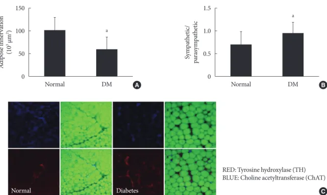
Fig. 1. Quantitative comparison of peripheral nerves in adipose tissue from db/db diabetic mice versus those from db/- control mice. (A) Quantity of PGP 9.5 (protein gene product 9.5)-positive immunostained nerve fibers, expressed as the total nerve fiber area. (B) Ratios of the occupied areas of sympathetic and parasympathetic nerve fibers. (C) Tyrosine hydroxylase (TH; red) and choline acetyltransferase (ChAT; blue) immunohistochemistry staining results of sympathetic and parasympathetic nerve fibers, respectively. Data are presented as mean±standard deviation (n=10 per group). DM, diabetes mellitus. aP<0.05 vs. the normal group.
150 100 50 0
1.5 1.0 0.5 0
Adipose innervation (103 μm2) Sympathetic/ parasympathetic
Normal DM Normal DM
a
a
A B
Normal Diabetes
RED: Tyrosine hydroxylase (TH) BLUE: Choline acetyltransferase (ChAT)
C
Lee KA, et al.
172 Diabetes Metab J 2018;42:169-172 http://e-dmj.org
CONFLICTS OF INTEREST
No potential conflict of interest relevant to this article was re- ported.
ACKNOWLEDGMENTS
This paper was partly supported by the fund of Clinical Medi- cine Research Institute of Chonbuk National University-Bio- medical Research Institute of Chonbuk National University Hospital.
REFERENCES
1. Kershaw EE, Flier JS. Adipose tissue as an endocrine organ. J Clin Endocrinol Metab 2004;89:2548-56.
2. Jang EH, Kim NY, Park YM, Kim MK, Baek KH, Song KH, Lee KW, Kwon HS. Influence of visceral adiposity on cardiovascu- lar autonomic neuropathy in patients with type 2 diabetes mel- litus. Diabetes Metab J 2012;36:285-92.
3. Bittel DC, Bittel AJ, Tuttle LJ, Hastings MK, Commean PK, Mueller MJ, Cade WT, Sinacore DR. Adipose tissue content, muscle performance and physical function in obese adults with type 2 diabetes mellitus and peripheral neuropathy. J Diabetes Complications 2015;29:250-7.
4. Voulgari C, Psallas M, Kokkinos A, Argiana V, Katsilambros N, Tentolouris N. The association between cardiac autonomic neuropathy with metabolic and other factors in subjects with type 1 and type 2 diabetes. J Diabetes Complications 2011;25:
159-67.
5. Fliers E, Romijn JA, Sauerwein HP, Kalsbeek A, Kreier F, Buijs RM. Adipose tissue: an innervated endocrine gland. Ned Tijd-
schr Geneeskd 2002;146:1976-9.
6. Romijn JA, Fliers E. Sympathetic and parasympathetic inner- vation of adipose tissue: metabolic implications. Curr Opin Clin Nutr Metab Care 2005;8:440-4.
7. Blaszkiewicz M, Townsend KL. Adipose tissue and energy ex- penditure: central and peripheral neural activation pathways.
Curr Obes Rep 2016;5:241-50.
8. Bartness TJ, Liu Y, Shrestha YB, Ryu V. Neural innervation of white adipose tissue and the control of lipolysis. Front Neuro- endocrinol 2014;35:473-93.
9. Kern PA, Ranganathan S, Li C, Wood L, Ranganathan G. Adi- pose tissue tumor necrosis factor and interleukin-6 expression in human obesity and insulin resistance. Am J Physiol Endo- crinol Metab 2001;280:E745-51.
10. Marette A. Molecular mechanisms of inflammation in obesity- linked insulin resistance. Int J Obes Relat Metab Disord 2003;
27 Suppl 3:S46-8.
11. Mokdad AH, Ford ES, Bowman BA, Dietz WH, Vinicor F, Bales VS, Marks JS. Prevalence of obesity, diabetes, and obesi- ty-related health risk factors, 2001. JAMA 2003;289:76-9.
12. Bamshad M, Aoki VT, Adkison MG, Warren WS, Bartness TJ.
Central nervous system origins of the sympathetic nervous system outflow to white adipose tissue. Am J Physiol 1998;275 (1 Pt 2):R291-9.
13. Penicaud L, Cousin B, Leloup C, Lorsignol A, Casteilla L. The autonomic nervous system, adipose tissue plasticity, and ener- gy balance. Nutrition 2000;16:903-8.
14. Kreier F, Fliers E, Voshol PJ, Van Eden CG, Havekes LM, Kals- beek A, Van Heijningen CL, Sluiter AA, Mettenleiter TC, Romijn JA, Sauerwein HP, Buijs RM. Selective parasympathet- ic innervation of subcutaneous and intra-abdominal fat--func- tional implications. J Clin Invest 2002;110:1243-50.