Reliability and Validity of Measurement Using SmartphoneBased Goniometer of Tibial External Rotation Angle in Standing Knee Flexion
In-cheol Jeon1, BHSc, PT, Oh-yun Kwon2, PhD, PT, Jong-hyuck Weon3, PhD, PT, Sung-min Ha4, PhD, PT, Si-hyun Kim1, MSc, PT
1Dept. of Physical Therapy, The Graduate School, Yonsei University,
2Dept. of Physical Therapy, College of Health Science, Yonsei University,
Dept. of Ergonomic Therapy, The Graduate School of Health and Environment, Yonsei University,
3Dept. of Physical Therapy, College of Tourism & Health, Joongbu University,
4Dept. of physical Therapy, Division of Health Science, Baekseok University
Abstract
1)The purpose of this study was to assess the intra-rater test-retest reliability of tibial external rotation angle measurement using a smartphone-based photographic goniometer, DrGoniometer (DrG) compared to a three-dimensional motion analysis system (Vicon). The current study showed an interchangeable method using DrG to measure the tibial external rotation angle in standing knee flexion at 90˚. Twelve healthy subjects participated in this study. A rest session was conducted 30 minutes later for within-day reliability and five days later for between-day intra-rater test-retest reliability. To assess the validity of the measurement using DrG, we used a three dimensional motion analysis system as a gold standard to measure the angle of tibial external rotation. Intra-class correlation coefficient (ICC) and the standard error of measurement (SEM) values were used to determine the within- and between- day intra-rater test-retest reliability of using DrG and a three dimensional motion analysis system. To assess validity, Pearson correlation coefficients were used for two measurement techniques. The measurement for tibial external rotation had high intra-rater test-retest reliability of within-day (ICC=.88) and between-day (ICC=.83) reliability using DrG and of within-day (ICC=.93) and between-day (ICC=.77) reliability using a three-dimentional motion analysis system. Tibial external rotation angle measurement using DrG was highly correlated with those of the three-dimensional motion analysis system (r=.86). These results represented that the tibial external rotation angle measurement using DrG showed acceptable reliability and validity compared with the use of three-dimensional motion analysis system.
Key Words: DrGoniometer; Reliability; Three-dimensional motion analysis system; Tibial external rotation angle; Validity.
Introduction
Tibial external rotation is shown while the tibia is twisted outward compared with the distal end of the femur in knee flexion (Sahrmann, 2011). The meas- urement of tibial external rotation is quite important for clinical evaluation of the knee because a greater than normal angle of tibial external rotation is af- fected by patellofemoral instability (Turner, 1994),
causing increased knee-valgus stress in a standing position and during gait (Turner and Smilliem, 1981).
In addition, tibial external rotation may increase with maturity and influencing lateral patellofemoral im- pingement (Fouilleron, 2010). Previous research found that the angle of tibial external rotation with respect to the femur was significantly increased in patients with patellofemoral pain, compared with healthy sub- jects (Bruce and Stevens, 2004; Eckhoff et al, 1997).
Corresponding author: Oh-yun Kwon [email protected]
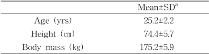
Mean±SDa
Age (yrs) 25.2±2.2
Height (㎝) 74.4±5.7
Body mass (㎏) 175.2±5.9
amean±standard deviation.
Table 1. General characteristics of the subjects (N=20) Since Nicol and Menelaus (1983) stated that the
alignment of the lower limb structure is affected by the angle of abnormal tibial rotation; tibial external rotation is a factor to evaluate with regard to knee instability associated with knee joint pain (Bruce and Stevens, 2004).
Several studies have described different methods for measuring the angle of tibial external rotation (Mikashima et al, 2006; Taussig, 1982; Turner and Smillie, 1981; Turner, 1994; von Porat et al, 2008;
Webster et al, 2010). Previous studies have shown the angle of tibial external rotation in patients with patellofemoral pain is more than 24.5°, while the an- gle in healthy subject is approximately 19°, as meas- ured using a tropometer (Taussig, 1982; Turner and Smillie, 1981; Turner, 1994). Mikashima et al (2006) have documented the value of normal tibial external rotation measured using computed tomography (CT) as 10° in knee extension. Sahrmann (2011) stated that the angle of tibial rotation in knee flexion at 90°
is between 25 and 57°. In addition, three-dimensional motion analysis techniques have become one of reli- able and traditional ways to assess dynamic knee motion for a variety of populations (von Porat et al, 2008; Webster et al, 2010). Three-dimensional motion analysis system (Vicon) has been used to calculate the kinematics of functional knee activity such as stepping, hopping, and knee bending motions on one leg standing (von Porat et al, 2006; von Porat et al, 2008). However, these methods were too expensive, time-consuming, and complex and these features made them less accessible for clinical settings (Lee, 2003). Currently, smartphone-based photographic go- niometer, DrGoniometer (DrG) (CDM S.r.l., Milano, Italy) applied in clinical practice has been increasing in popularity for a number of practitioners (Ferriero et al, 2011; Ferriero et al, 2013). Various smartphone applications with the ability to measure angles have been used to assess the range of motion (ROM) of peripheral joints in clinical settings and research (Vrtovec, 2009; Ferriero et al, 2011; Ferriero et al, 2013). These advantages of smartphones such as be-
ing time-saving, expedient, and portable could be more accessible for practitioners (Jacquot, 2012).
Also, their reliability has been proven through sev- eral recent studies (Al-Hadithy, 2012; Jacquot, 2012;
Qiao, 2012).
To our knowledge, no studies have investigated the reliability and validity of the tibial external rota- tion angle in standing knee flexion at 90° using DrG, compared to using a three-dimensional motion analy- sis system. The purposes of the present study were to (1) determine the intra-rater test-retest reliability of DrG and three-dimensional motion analysis sys- tem measurements for tibial external rotation in standing knee flexion at 90° between test days and (2) assess the validity of tibial external rotation an- gle measurement using DrG by comparing the find- ings with a three-dimensional motion analysis system. We hypothesized that (1) tibial external ro- tation measures using both DrG and a three dimen- sional motion analysis system would be reliable be- tween sessions and (2) the validity determined by correlating DrG measurement and the tibial external rotation angle, as measured by the three-dimensional motion analysis system.
Methods
Subjects
Twenty healthy male subjects were recruited by gathering a convenience sample of university stu- dents (Table 1). Inclusion criteria included: (1) nor- mal range of motion in ankle, knee, and hip joints, (2) no musculoskeletal and neurological abnormalities,
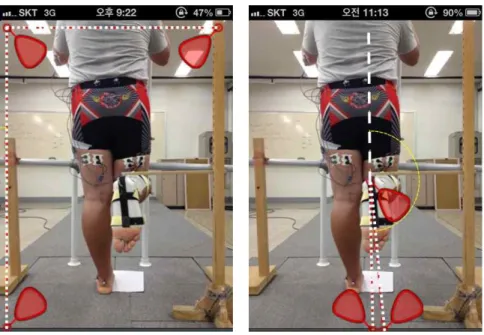
Figure 1. Three different markers with gravity sensor can be placed to calculate tibial external rotation angle performed in the frontal plane by DrGoniometer measurement.
(3) no history of knee and ankle surgery. Exclusion criteria included: (1) specific ankle and knee pain such as osteoarthritis, (2) a previous history of ankle or knee surgery, (3) patella instability, and (4) symptoms of meniscal or knee ligament and ankle ligament involvement. The investigator explained all of the procedures to the subjects in detail before the measurements were taken. Subjects also signed the informed consent form.
Instrument
DrG can measure the joint angle and observe all of the related data which has been developed to per- form a easier and faster measurement of joint angle (Ferriero et al, 2011). A circle highlights the center of the axis while a yellow pendulus indicated the gravity shaft. As a smartphone was placed perpen- dicular to the ground, the yellow pendurus showed a green color when it was close to perpendicular in the same reference position. The data on each angle cal- culated by placing three different markers with a gravity sensor can be stored for each subject. Each marker can be placed on the exact point of the heel (Figure 1). the picture can be taken with the gravity
sensor in respect to the ground and the same height between the smartphone lens and the joint axis to make precise reference indication. In this study, the line connecting from the second toe to the heel with a molded thermoplastic tibial splint was photographed for all the subjects 1 m from behind when perform- ing standing knee flexion at 90°.
Kinematic Data
Sixteen reflective markers of anatomical landmarks were placed over the anterior superior iliac spines, posterior superior iliac spines, lateral aspects of the thigh and leg, knee joint axis, lateral malleolus, heel, and forefoot (Webster et al, 2010). Kinematic data were sampled at the rate of 100 ㎐ with an eight camera three-dimensional motion analysis system (VICON MX system, Oxford Metrics Ltd., Oxford, UK). The diameter of reflective markers was 14 ㎜, and double-sided tape was used to be attached to the skin. Before data were collected, the manu- facturer’s instructions were used to calibrate this system. Vicon plug-in-gait biomechanical software was used to measure the kinematic profiles. The knee flexion period was divided into three phases.
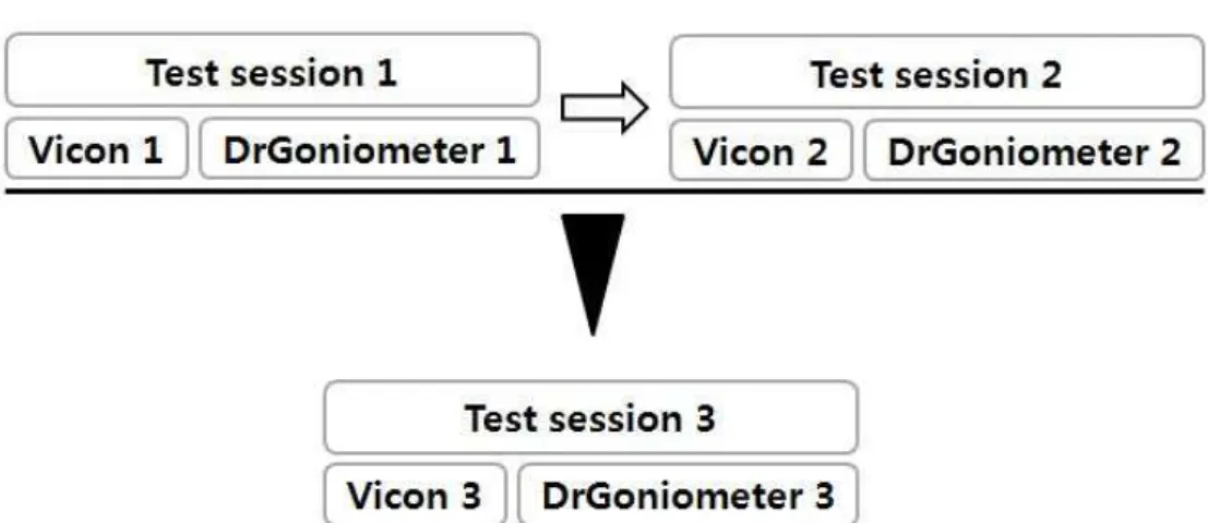
Figure 2. Flow chart of the progression of measurement through the study.
Each of the three periods of knee flexion in a stand- ing position involved placing a foot on the ground for 5 sec, being in motion for 5 sec, and being at the end range of knee flexion at 90° for 5 sec. Tibial external rotation angle was measured at the end range of knee flexion at 90° in the frontal plane. In addition, DrG pictures were taken at the end of the motion at 90° in the frontal plane for all of the sessions.
Procedures
Subject participated in session 1 and session 2 on the same day, and session 3 was conducted five days later (Figure 1). Each subject participated in two trials only for limbs on the right-sided limbs in session 1 since there is no significant difference be- tween left and right limbs in healthy subjects (Webster et al, 2010). Subjects were accustomed to performing the motion for sessions after practicing for 15 minutes. The examiner was familiarized with the experimental protocol to ensure standardization of the measurements using DrG and a three-dimen- sional motion analysis system. Since Sahrmann (2011) stated that the functional assessment of knee external rotation should be done while the partic- ipants position one of their knees at 90° of flexion while standing, Each subject performed knee flexion at 90° until the target bar touched the back of their calf in a standing position which allowed tibial ex-
ternal rotation to be performed in the frontal plane.
Each session was started with both of their feets at- tached and eyes looking forward. Subjects were sup- ported by holding two separate perpendicular shafts to minimize the weight shift. Furthermore, the ankle motion was restricted using the molded thermoplastic tibial splint (Alarm et al, 2011). Additionally, hip flexion during the tasks was limited by the fixation bar being placed in front of the thigh. Each subject was requested to sit for 30 min in the same place.
Reflective markers were remained in the same region (i.e., session 2 was conducted after 30 min of resting time once session 1 was completed). After the rest period, the subjects performed the same procedure for the limbs on the right side in session 2. These two sessions were performed to determine with- in-day reliability. All of the subjects returned five days later and were required to perform two trials of the same procedures for right limbs in session 3 (Figure 2). The markers placement was reapplied in session 3. The data of the last session was com- pared with session 1 to determine between-day reliability. One examiner placed the markers on all the subjects during all of their trials. DrG and a three-dimensional motion analysis system were used together at the same time for each trial.
Statistical Analysis
Through this process, mean measurement angles
Figure 3. Scatterplot of tibial external rotation angle measurement techniques (dashed line:
DrGoniometer measurement, solid line: Vicon measurement).
Within day (session 1 and 2) Between day (session 1 and 3)
ICC (95% CIb) ICC (95% CI)
DrGa .88 (.71-.96) .83 (.59-.94)
Vicon .93 (.83-.97) .77 (.43-.91)
aDrGoniometer, bconfidence intervals.
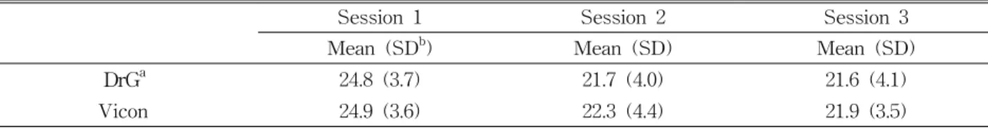
Table 2. Mean and standard deviation of the tibial external rotation angle (N=20)
Session 1 Session 2 Session 3
Mean (SDb) Mean (SD) Mean (SD)
DrGa 24.8 (3.7) 21.7 (4.0) 21.6 (4.1)
Vicon 24.9 (3.6) 22.3 (4.4) 21.9 (3.5)
aDrGoniometer, bstandard deviation.
Table 3. Mean and standard deviation of the tibial external rotation angle (Unit: ˚) with standard deviations (SD) of two trials were
calculated for each session. Within-day reliability was determined by the outcome of sessions 1 and 2.
Between-day reliability was determined by sessions 1 and 3. Intra-class correlation coefficients (ICC [3, 1]) and 95% confidence intervals (CI) were used to determine the within- and between-day intra-rater test-retest reliability of using DrG and a three di- mensional motion analysis system. In the inter- pretation, the ICC value of >.75 was used to indicate
“excellent,” .40∼.75 was “fair to good,” and .00∼.40 was “poor” (Crossley et al, 2004). The method used to provide information on absolute reliability was the
standard error of measurement (SEM), which is cal- culated using the following equation: [SEM=SD
. Where SD=the sample standard deviation. The smaller SEM is expressed, the more reliable the measurement (Atkinson and Mevill, 1998).
The mininal detectable difference (MDD) was calcu- lated using the formula [MDD95=1.96 * SEM * ] to determine the magnitude of change that would exceed the minimal error of the measurement at a 95% confidence level (Portney and Watkins, 2009).
Validity was calculated using Pearson’s correlation coefficient. The correlation coefficient (r) was defined as follows: >.75 was “excellent,” .5∼.75 was “good,”
.25∼.50 was “moderate,” and <.25 was “poor”
(Alanay et al, 2005). All statistical analyses were conducted using SPSS (ver. 18.0, SPSS Inc., IL, Chicago, USA).
Results
The intra-rater test-retest reliability of DrG and three-dimentional motion analysis system measure- ments was significantly good, as expected. The reli- ability values for the within-day ICC and be- tween-day ICC of measurements using DrG and the three-dimensional motion analysis system are dis- played in Table 2. The within-day (ICC=.93) value was higher than the between-day (ICC=.77) value in
Within day (session 1 and 2)
Between day (session 1 and 3)
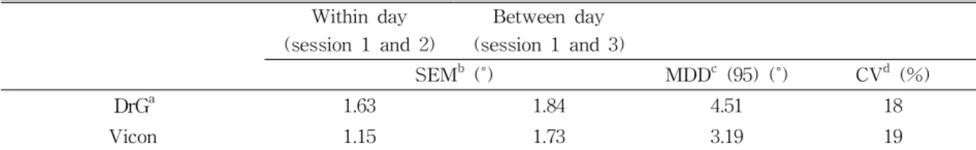
SEMb (˚) MDDc (95) (˚) CVd (%)
DrGa 1.63 1.84 4.51 18
Vicon 1.15 1.73 3.19 19
aDrGoniometer, bstandard error of measurement, cminimal detectable difference, dcoefficient of varience.
Table 4. Measurement error scores for tibial external rotation
the measurement of three-dimensional motion analysis. The between-day ICC value in DrG was lower than the within-day ICC (within ICC=.88; be- tween ICC=.83). SEM between DrG and the three-dimensional motion analysis is displayed in Table 4. This represented the average within-subject differences for both within- and between-day sessions. Also, the between-day ICC values for DrG and the three-dimensional motion analysis were dif- ferent (i.e., between-day ICC for DrG=.83; be- tween-day ICC for the Vicon=.77), which could in- dicate why between-day reliability values were low, especially for the kinematic measurement with the reflective skin markers (Brochard, 2011). In addition, measurements obtained from the ICC value with 95%CI of tibial external rotation, including the mean and SD values are presented in Table 2 and Table 3, respectively. A scatterplot was drawn to represent the relationship between DrG and a three-dimen- sional motion analysis measurements in Figure 3.
Discussion
The purpose of the current study was to de- termine the intra-rater test-retest reliability and val- idity of DrG and three-dimensional motion analysis measures of tibial external rotation in standing knee flexion at 90°. We believe that the present research is the first reported study to investigate the in- tra-rater test-retest reliability and validity of DrG and three-dimensional motion analysis system tech- niques to measure tibial external rotation. A three dimensional motion analysis system was the techni- que in which the advanced laboratory well used to
measure functional motion (Tashman et al, 2008).
The measurement using DrG saves time and is con- venient; thus, it is being used more frequently for clinical evaluations (Al-Hadithy, 2012; Jacquot, 2012) and research purpose with high ICC values of in- tra-rater and inter-rater reliability for upper limb measurements (Ferriero et al, 2011).
For intra-rater test-retest reliability, our study in- dicated that the value for within-day ICC was quite high compared with between-day ICC for both DrG and the three-dimensional motion analysis system. A previous study reported that within-day ICC had higher reliability than between-day ICC, which sup- ports our first hypothesis (Webster et al, 2010). The potential reason for the higher within-day ICC val- ues than between-day ICC values was possible adaptation in the specific tasks (Webster et al, 2010).
Altered motor performance might become the reason for being more repeatable performance of subjects on the task in within-day ICC compared to be- tween-day ICC because of the task performed on the same day. Webster et al (2010) noted that the be- tween-day errors resulted from subtle differences in marker placement between session 1 and session 3 in a three-dimensional motion analysis system. The result reported in this investigation was quite similar to the previous studies for lower limb measurements such as level gait (Kadaba, 1989), running (Ferber, 2002; Queen, 2006), landing (Ford, 2007), and pivoting (Webster et al, 2010) even though the tasks used in these previous studies were composed of active movements compared with current study.
However, our study aimed not only to compare within- and between-day reliability using DrG and a three-dimensional motion analysis system, but also
to provide the information that DrG is a highly reli- able method to measure tibial external rotation. Even though all of the subjects were instructed to follow the same action restricted by using a parallel bar in front of their thigh to control hip flexion and by us- ing vertical support to minimize weight shifting while standing for the same condition, a slight dif- ference between within-day and between-day reli- ability was shown. So, the findings in this study might be affected as a result of the task being re- peated on the same day which indicated the differ- ence between within day and between day reliability.
In addition, the result from a three dimensional mo- tion analysis system might be attributed to skin marker displacement in the same way (Webster et al, 2010).
In this study, we found that DrG provided valid datas that correlated with the findings from the three-dimensional motion analysis system for the measurement of tibial external rotation with the molded thermoplastic tibial splint. Similarly, several prior studies have indicated that DrG appears to be a reliable and accurate method in clinical practice and even for the research purpose of measuring the Cobb angle in kyphosis (Jacquot, 2012), the Cobb angle in scoliosis (Qiao, 2012), and hallux valgus (Ege, 2012). Several possible explanations for our results. First, the excellent validity of DrG and three-dimentional motion analysis measurements could be explained by the knee motion being well controlled as a result of being held in the molded thermoplastic tibial splint (Alam et al, 2011). A pre- vious study showed that tibial rotation is not accu- rately represented by foot rotation, and the degree of tibial external rotation is actually obscured by foot rotation (Alam et al, 2011; Costigan, 1992). The measurement using the tibial splint and the measure- ment performed directly on the tibia showed sig- nificant correlations at both knee external rotation (6˚
overestimated in the tibial splint compared with tibia directly measurmed at 90˚) (Alam et al, 2011). DrG measurement was performed by photographing the
line connecting subjects’ second toe to the middle of their heel with the molded thermoplastic tibial splint from behind 1 m in standing knee flexion at 90°. For this reason, it was important to control the measure- ment by limiting the motion of the foot (Alam et al, 2011) and to be performed with offering a perpendic- ular viewpoint and centering the lens on the target angle which influenced the measurement repeatability and accuracy in photographic-based measurements (Dunlevy, 2005; Lindqvist, 1998). However, since the measurement of the tibial external rotation angle with the molded thermoplastic tibial splint using DrG is not clinically accessible, the taping method may be an alternative method to address interrelated me- chanical fixation of ankle joints in clinical settings (Wilkerson, 2002). Second, another reason for the ex- cellent validity of knee flexion at 90° in standing was that the hip joint was controlled using a bar and a belt in front of the thigh to avoid unwanted hip flexion and rotation (Osternig, 1980). Osternig (1980) stated that measurement discrepancies in tibial external rotation have been attributed to the failure to control accompanying thigh motion. This implies that controlling thigh and foot motion during meas- urement would provide valid results for the measure- ment of functional motion such as tibial external rotation. This study had several limitations. First, one examiner was employed, as most of the studies progressed in the same manner. However, no clinical field can be reflected in these procedures, as the measurement can be performed by a number of therapists. Further study is required to employ more examiners to represent general clinical settings and to establish inter-rater test-retest reliability. Second, our results cannot be generalized to the patient pop- ulation because young, healthy male subjects partici- pated in this study. Therefore, further research is needed to investigate the reliability of tibial external rotation using DrG and a three-dimensional motion analysis system in individuals with knee-ankle pain and from various age groups. Third, the measure- ment technique using DrG for tibial external rotation
angle cannot be generalized to the measurement for other joints angle. Further study is needed to inves- tigate the reliability of other joints angle measure- ment using DrG.
Conclusion
This study demonstrated excellent intra-rater test-retest reliability and validity for the use of DrG and a three-dimensional motion analysis to measure the tibial external rotation angle in standing knee flexion at 90°. In summary, the findings suggested that DrG is an alternative method to three-dimen- sional motion analysis for measuring the tibial ex- ternal rotation angle. Thus, measurement of the tibial external rotation angle by DrG may provide useful information for the evaluation of lower extremity kinematics and treatment outcome. Based on these findings, we conclude that 1) tibial external rotation measurements using both DrG and a three-dimen- sional motion analysis system obtained on the same and different days can be reliable, and 2) DrG can be a interchangeable method with a three-dimen- sional motion analysis system to measure the tibial external rotation angle for clinical use.
References
Alam M, Bull AM, Thomas Rd, et al. Measurement of rotational laxity of the knee: In vitro compar- ison of accuracy between the tibia, overlying skin, and foot. Am J Sports Med. 2011;39(12):
2575-2581.
Alanay A, Cil A, Berk H, et al. Reliability and val- idity of adapted turkish version of scoliosis re- search society-22 (SRS-22) questionnaire. Spine (Phila Pa 1976). 2005;30(21):2464-2468.
Al-Hadithy N, Gikas PD, Al-Nammari SS.
Smartphones in orthopaedics. Int Orthop.
2012;36(8):1543-1547.
Atkinson G, Nevill AM. Statistical methods for as- sessing measurement error (reliability) in varia- bles relevant to sports medicine. Sports Med.
1998;26(4):217-238.
Brochard S, Lempereur M, Rémy-Néris O. Accuracy and reliability of three methods of recording scapular motion using reflective skin markers.
Proc Inst Mech Eng H. 2011;225(1):100-105.
Bruce WD, Stevens PM. Surgical correction of mis- erable malalignment syndrome. J Pediatr Orthop.
2004;24(4):392-396.
Costigan PA, Wyss UP, Deluzio KJ, et al.
Semiautomatic three-dimensional knee motion assessment system. Med Biol Eng Comput.
1992;30(3):343-350.
Crossley KM, Bennell KL, Cowan SM, et al.
Analysis of outcome measures for persons with patellofemoral pain: Which are reliable and val- id? Arch Phys Med Rehabil. 2004;85(5):815-822.
Dunlevy C, Cooney M, Gormely J. Procedural con- siderations for photographic-based joint angle measurements. Physiother Res Int. 2005;10(4):190 -200.
Eckhoff DG, Brown AW, Kilcoyne RF, et al. Knee version associated with anterior knee pain. Clin Orthop Relat Res. 1997;(339):152-155.
Ege T, Kose O, Koca K, et al. Use of the iPhone for radiographic evaluation of hallux valgus. Skeletal Radiol. 2013;42(2):269-273.
Ferber R, McClay Davis I, Williams DS 3rd, et al. A comparison of within- and between-day reli- ability of discrete 3D lower extremity variables in runners. J Orthop Res. 2002;20:1139-1145.
Ferriero G, Sartorio F, Foti C, et al. Reliability of a new application for smartphones (DrGoniometer) for elbow angle measurement. PM R. 2011;3(12):
1153-1154.
Ferriero G, Vercelli S, Sartorio F, et al. Reliability of a smartphone-based goniometer for knee joint goniometry. Int J Rehabil Res. 2013;36(2):146-151.
Ford KR, Myer GD, Hewett TE. Reliability of land- ing 3D motion analysis: Implications for longi-
This article was received March 8, 2013, was re- viewed March 8, 2013, and was accepted April 28, 2013.
tudinal analyses. Med Sci Sports Exerc.
2007;39(11):2021-2028.
Fouilleron N, Marchetti E, Autissier G, et al.
Proximal tibial derotation osteotomy for torsional tibial deformities generating patello-femoral disorders. Orthop Traumatol Surg Res.
2010;96(7):785-792.
Jacquot F, Charpentier A, Khelifi S, et al. Measuring the cobb angle with the iPhone in kyphoses: A reliability study. Int Orthop. 2012;36(8):1655-1660.
Kadaba MP, Ramakrishnan HK, Wootten ME, et al.
Repeatability of kinematic, kinetic, and electro- myographic data in normal adult gait. J Orthop Res. 1989;7(6):849-860.
Lee RY, Laprade J, Fung EH. A real-time gyro- scopic system for three-dimensional measure- ment of lumbar spine motion. Med Eng Phys.
2003;25(10):817-824.
Lindqvist B, Welander U, Mähler R. A three-dimen- sional photographic method for documentation and measurement of dental conditions. J Orofac Orthop. 1998;59(2):90-99.
Mikashima Y, Kimura M, Kobayashi Y, et al.
Clinical results of isolated reconstruction of the medial patellofemoral ligament for recurrent dis- location and subluxation of the patella. Acta Orthop Belg. 2006;72(1):65-71.
Osternig LR, Bates BT, James SL. Patterns of tibial rotary torque in knees of healthy subjects. Med Sci Sports Exerc. 1980;12(3):195-199.
Qiao J, Liu Z, Xu L, et al. Reliability analysis of a smartphone-aided measurement method for the cobb angle of scoliosis. J Spinal Disord Tech.
2012;25(4):E88-E92.
Queen RM, Gross MT, Liu HY. Repeatability of lower extremity kinetics and kinematics for standardized and self-selected running speeds.
Gait Posture. 2006;23(3):282-287.
Sahrmann SA. Movement System Impairment Syndromes of the Extremities, Cervical and
Thoracic Spines. 1st ed. Missouri, Mosby, 2011:424-425.
Tashman S, Collon D, Anderson K, et al. Abnormal rotational knee motion during running after an- terior cruciate ligament reconstruction. Am J Sports Med. 2004;32(4):975-983.
Taussig G. Tibial torsion. Rev Chir Orthop Reparatrice Appar Mot. 1982;68(1):20-23.
Turner MS. The association between tibial torsion and knee joint pathology. Clin Orthop Relat Res.
1994;(302):47-51.
Turner MS, Smillie IS. The effect of tibial torsion of the pathology of the knee. J Bone Joint Surg Br. 1981;63-B(3):396-398.
von Porat A, Henriksson M, Holmström E, et al.
Knee kinematics and kinetics during gait, step and hop in males with a 16 year old ACL in- jury compared with matched controls. Knee Surg Sports Traumatol Arthrosc. 2006;14(6):546- 554.
von Porat A, Holmström E, Roos E. Reliability and validity of videotaped functional performance tests in ACL-injured subjects. Physiother Res Int. 2008;13(2):119-130.
Vrtovec T, Pernus F, Likar B. A review of methods for quantitative evaluation of spinal curvature.
Eur Spine J. 2009;18(5):593-607.
Webster KE, McClelland JA, Wittwer JE, et al.
Three dimensional motion analysis of within and between day repeatability of tibial rotation dur- ing pivoting. Knee. 2010;17(5):329-333.
Wilkerson GB. Biomechanical and neuromuscular ef- fects of ankle taping and bracing. J Athl Train.
2002;37(4):436-445.