© 2011 Korean Breast Cancer Society http://ejbc.kr | pISSN 1738-6756
INTRODUCTION
Anthracycline and taxane have been very essential drugs in the treatment of breast cancer patients in neoadjuvant and adju- vant settings. In particular, anthracycline has been very widely used in spite of its major limitation, cardiotoxicity [1-3]. Some studies have reported that an anthracycline-based regimen had more favorable results in HER2 overexpression tumors.
Recently, it has been demonstrated that this result is mediated by topoisomerase IIα (TOP2A). Anthracycline binds to TOP2A and stabilizes DNA double strand breaks, resulting in cell cycle
arrest and apoptosis. The Top2A gene is located close to the HER2 gene on chromosome 17, and TOP2A gene co-amplica- tion has been found in about 40% of HER2-positive type breast cancers [4,5]. Nevertheless, anthracycline is still a fascinating chemotherapeutic regimen for HER2-positive type breast can- cer patients. Anthracycline-based chemotherapy has been used as a good neoadjuvant and adjuvant treatment choice for both HER2-positive type and triple negative breast cancers (TNBC).
It has been shown to produce higher pathological complete response (pCR) rates for TNBC in a neoadjuvant setting and better survival rates in an adjuvant setting [6-10].
Several studies have been conducted on the usefulness of the adenosine triphosphate-based chemotherapy response assay (ATP-CRA) in breast cancer [11-13]. This assay measures intra- cellular ATP, the basic energy source for living cells, which rap- idly disappears when cells lose viability. The ATP-CRA has an advantage of a high success rate in primary culture and require only a small number of cells [12-16]. Thus, it is widely used as a chemosensitivity assay modality for the treatment of various
Chemotherapy Response Assay Test and Prognosis for Breast Cancer Patients Who Have Undergone Anthracycline- and Taxane-Based Chemotherapy
Anbok Lee, Woosung Lim1, Byung-In Moon1, Nam-Sun Paik1, Suck-Hwan Koh2, Jeong-Yoon Song2
Department of Medicine, Kyung Hee University Graduate School, Seoul; 1Department of Surgery, Ewha Womans University School of Medicine, Seoul;
2Department of Surgery, Kyung Hee University School of Medicine, Seoul, Korea ORIGINAL ARTICLE
Purpose: A chemotherapy response assay test is performed to evaluate the degree of tumor growth inhibition by a chemothera- peutic agent. Several studies have been done on its usefulness;
however, to the best of our knowledge, only a few studies concern- ing the relationship between chemotherapy response assay test results and breast cancer patients’ prognoses have been con- ducted. Thus, we performed this study to analyze this relationship.
Methods: Among breast cancer patients who underwent curative surgery and neoadjuvant or adjuvant chemotherapy between August 2004 and December 2009, 102 were enrolled in this study.
Chemotherapeutic regimens for patients were doxorubicin plus taxane or doxorubicin plus cyclophosphamide followed by tax- ane. We divided these patients into two groups (sensitive group [n=19] and resistant group [n=83]) and analyzed the relationship between chemosensitivity results and patient prognosis. Results:
The sensitive group was associated with poor disease-free sur-
vival (DFS) (p=0.003) and overall survival (OS) (p<0.001). No sig- nificant differences were observed in tumor histology (p=0.548), tumor size (p=0.479), number of metastatic lymph nodes (p=
0.326), histologic grade (p=0.077), or nuclear grade (p=0.216) between the two groups. However, in respect to molecular sub- type, the HER2-positive type and triple negative breast cancer were more frequently observed in the sensitive group (p=0.001).
In a univariate and multivariate analysis for DFS, doxorubicin sen- sitivity was significantly associated with a poor prognosis (p<
0.05). Conclusion: Better chemosensitivity results are associated with a poor prognosis in breast cancer patients who have under- gone anthracycline- and taxane-based chemotherapy, however, examination of additional cases and the use of a longer study period are needed.
Key Words: Breast neoplasms, Doxorubicin, Prognosis, Sensitivity
Correspondence: Jeong-Yoon Song
Department of Surgery, Kyung Hee University Hospital at Gangdong, Sangil-dong, Gangdong-gu, Seoul 134-727, Korea
Tel: +82-2-440-6137, Fax: +82-2-440-7053 E-mail: [email protected]
The contents of this article was presented at the 26th Korean Breast Cancer Society Symposium.
Received: June 2, 2011 Accepted: October 14, 2011
Cancer
cancers in clinical settings. However, only a few studies have been performed concerning the relationship between chemo- therapy response assay test results and breast cancer patient prognosis [17]. We performed this study to evaluate the ATP- CRA results and prognoses for breast cancer patients who had undergone anthracycline and taxane based chemotherapy.
METHODS
From August 2004 to December 2009, we performed ATP- CRA for 257 patients with breast cancer at the Department of Surgery, Ewha Womans University Mok-dong Hospital, Seoul, Republic of Korea. Among these patients, we enrolled patients who had undergone curative surgery and had received doxo- rubicin plus taxane or doxorubicin plus cyclophosphamide followed by taxane chemotherapy. A total of 102 patients were included in this study. The mean follow-up duration was 29.8±
15.6 months.
For ATP-CRA, two specimens were taken per patient by true-cut biopsy or core needle biopsy. We stored these tissues in Hank’s Balanced Salt Solution (HBSS; Gibco BRL, Rockville, USA) composed of 100 IU/mL of penicillin (Sigma, St. Louis, USA), 100 mg/mL of streptomycin (Sigma), 100 mg/mL of gen- tamicin (Gibco BRL), 2.5 mg/mL of amphotericin B (Gibco BRL), and 5% fetal bovine serum (Gibco BRL). The stored tumor tissues were sent to a commercial laboratory center for deter- mination of their sensitivities to each chemotherapeutic regi- men. The most commonly used chemotherapeutic regimens and their concentrations were as follows: 5-FU (10 µg/mL), paclitaxel (8.5 µg/mL), docetaxel (3.7 µg/mL), doxorubicin (1.5 µg/mL), methotrexate (0.37 µg/mL), and cyclophosphamide (4-OH-cyclophosphamide, 1.2 µg/mL). If sufficient cancer cells were available to determine their sensitivity, these cells were tested with three treated drug concentrations (TDC; 0.2×, 1×, 5×). If not, their ATP–CRA results were determined using only one TDC (1×). Doxorubicin and taxane sensitivities were determined using a cell death ratio cut-off of 40% and 24%, respectively, on the basis of TDC (1×). Receiver operating characteristic (ROC) curves were used to determine these cut- off values.
The doxorubicin sensitive group was compared to the resis- tant group with respect to their clinical characteristics (age, op- eration methods, chemotherapy type, and chemotherapeutic regimen), pathologic results, disease-free survival (DFS), and overall survival (OS). In respect to estrogen receptor (ER) status, we defined ER-positive when more than 10% of cancer cells were stained with more than 1+ of intensity. In the case of HER2- positive, it was defined as c-erb-B2+++ or when the ratio of HER2 loci/chromosome 17 centromeres was more than 2.2.
Univariate and multivariate anaylsis were performed to identify whether ATP-CRA results could be an independent prognos- tic factor for DFS.
All data were analyzed using SPSS version 17.0 (SPSS Inc., Chicago, USA). Statistical significance was analyzed using the Student’s t-test and Pearson’s chi-square test. DFS and OS were calculated using the Kaplan-Meier method and log-rank test.
Cox’s proportional hazards regression model was used for uni- variate and multivariate analyses, and p-values <0.05 were considered statistically significant.
RESULTS
Patient characteristics
During the study period, a total number of 257 ATP-CRAs were performed in our hospital and among these, 102 were included in this study. We divided these patients into two groups according to doxorubicin sensitivity. There were 19 patients in the sensitive group with a mean age of 49.0±9.3 years (range, 29 to 69 years) and 83 patients in the resistant group with a mean age of 46.8±8.8 years (range, 30 to 69 years). No signifi- cant differences were noted with regard to patient age, opera- tion type, or chemotherapeutic regimen between the two groups.
However, there were more patients who received neoadjuvant chemotherapy (p=0.021), and more patients who were sensi- tive to taxane in the doxorubicin sensitive group (p=0.001, Table1).
Pathologic results according to doxorubicin sensitivity The pathologic results of all patients according to doxorubicin sensitivity have been presented in Table 2. No significant differ- Table 1. Patients’ demographics according to doxorubicin sensitivity
Variables Sensitive
group (n=19)
Resistant
group (n=83) p-value
Age (yr)* 49.0±9.3 46.8±8.8 0.365
Operation 0.430
BCS 5 (26.3) 32 (38.6)
MRM 14 (73.7) 51 (61.4)
Chemotherapy type 0.021
Neoadjuvant 9 (47.4) 17 (20.5)
Adjuvant 10 (52.6) 66 (79.5)
Chemotherapy regimen 0.097
AC followed by T 13 (68.4) 71 (85.5)
AT 6 (31.6) 12 (14.5)
Taxane sensitivity 0.001
Sensitive 13 (68.4) 21 (25.3)
Resistant 6 (31.6) 62 (74.7)
Values are presented as mean±SD or number (%).
BCS=breast conserving surgery; MRM=modified radical mastectomy; AC=
doxorubicin+cyclophophmide; T=taxane; AT=doxorubicin+taxane.
*Mean±SD.
ences were observed in histologic type, HER2-positive status,
histologic grade, nuclear grade, tumor size, and nodal status between the two groups. However, HER2-positive type cancer, TNBC, and ER-negative cancers were more frequently observed in the sensitive group (p<0.05). All patients had metastases of axillary lymph nodes (LNs) or lymphovascular invasion.
Table 2. Pathologic results according to doxorubicin sensitivity
Variables Sensitive
group (n=19)
Resistant
group (n=83) p-value
Histology 0.548
IDC 19 (100.0) 78 (94.0)
ILC 0 4 (4.8)
Others 0 1 (1.2)
Molecular subtype 0.001
Luminal A 2 (10.5) 51 (61.5)
Luminal B 5 (26.3) 10 (12.0)
HER2 type 3 (15.8) 8 (9.6)
TNBC 9 (47.4) 14 (16.9)
Estrogen receptor 0.006
Positive 7 (36.8) 61 (73.5)
Negative 12 (63.2) 22 (26.5)
HER2 status 0.082
Positive 8 (42.1) 18 (21.7)
Negative 11 (57.9) 65 (78.3)
Histologic grade 0.077
1 0 17 (20.5)
2 9 (47.4) 35 (42.2)
3 9 (47.4) 26 (31.3)
Unknown 1 (5.2) 5 (6.0)
Nuclear grade 0.216
1 0 10 (12.0)
2 8 (42.1) 37 (44.6)
3 10 (52.7) 32 (38.6)
Unknown 1 (5.2) 4 (4.8)
Tumor size (cm) 0.479
≤2 7 (36.8) 31 (37.3)
>2, ≤5 9 (47.4) 46 (55.4)
>5 3 (15.8) 6 (7.2)
No. of metastatic LNs 0.326
0 2 (10.5) 8 (9.6)
≤3 11 (57.9) 40 (48.2)
4-9 2 (10.5) 25 (30.2)
≥10 4 (21.1) 10 (12.0)
Values are presented as number (%).
IDC=invasive ductal carcinoma; ILC=invasive lobular carcinoma; TNBC=triple negative cancer; LNs=lymph nodes.
Disease free survival
Months from surgery
p=0.003 Resistant group Sensitive group
0 20 40 60 80
1.0
0.8
0.6
0.4
0.2
0.0
Figure 1. Disease-free survival according to doxorubicin sensitivity.
Overall survival
Months from surgery
p<0.001 Resistant group Sensitive group
0 20 40 60 80
1.0
0.8
0.6
0.4
0.2
0.0
Figure 2. Overall survival according to doxorubicin sensitivity.
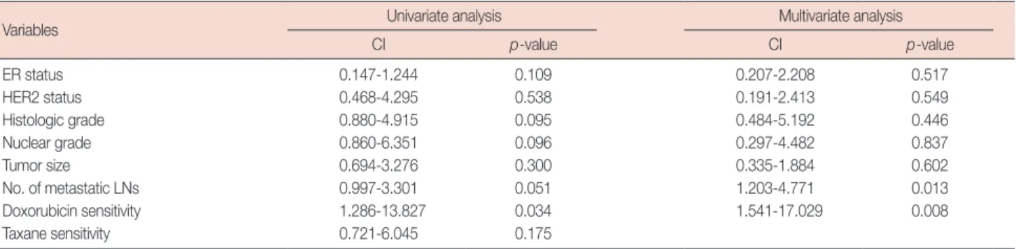
Table 3. Univariate and multivariate analysis for DFS in all patients
Variables Univariate analysis Multivariate analysis
CI p-value CI p-value
ER status 0.147-1.244 0.109 0.207-2.208 0.517
HER2 status 0.468-4.295 0.538 0.191-2.413 0.549
Histologic grade 0.880-4.915 0.095 0.484-5.192 0.446
Nuclear grade 0.860-6.351 0.096 0.297-4.482 0.837
Tumor size 0.694-3.276 0.300 0.335-1.884 0.602
No. of metastatic LNs 0.997-3.301 0.051 1.203-4.771 0.013
Doxorubicin sensitivity 1.286-13.827 0.034 1.541-17.029 0.008
Taxane sensitivity 0.721-6.045 0.175
DFS=disease free survival; CI=confidence interval; ER=estrogen receptor; LNs=lymph nodes.
Patient prognosis according to doxorubicin sensitivity In our study, a total of 14 cancer recurrences were observed, 7 of which were in the sensitive group. Among these, 3 patients died of breast cancer. We compared DFS and OS according to doxorubicin sensitivity. In the sensitive group, DFS and OS were significantly worse than those in the resistant group (Figures 1, 2).
Univariate analysis for DFS indicated that only doxorubicin sensitivity was significantly associated with high risk of recur- rence (p=0.006; confidence interval [CI], 1.550-12.994). With respect to taxane sensitivity, no significant effect was observed on DFS (p=0.175; CI, 0.721-6.045). We performed a multivar- iate analysis for significant factors in univariate analysis and established breast cancer prognostic factors, and according to the results, a higher nodal stage (p=0.013; CI, 1.203-4.771) and doxorubicin sensitivity (p=0.008; CI, 1.541-17.029) were asso- ciated with poor DFS (Table 3).
DISCUSSION
Anthracycline and taxane have been the most commonly used chemotherapeutic agents for breast cancer patients. In neoadjuvant treatment, it had been reported that anthracycline- based chemotherapy produced more superior treatment results in TNBC/basal like breast cancer (BLBC) compared to non- TNBC/BLBC. Furthermore, these results were improved by adding taxane to the anthracycline [8-10]. With respect to adjuvant settings, anthracycline-based chemotherapy has been effective for patients with HER2-positive type and positive TOP2A and also for TNBC patients when compared with CMF chemotherapy [18,19]. In HER2 over-expression tumors, the effect of anthracycline has been shown to be mediated by TOP2A. Anthracycline binds to TOP2A and stabilizes DNA double strand breaks, which results in cell cycle arrest and apop- tosis, and it has been known that the TOP2A gene is located close to the HER2 gene on chromosome 17. TOP2A gene co-amplification has been found in about 40% of HER2-posi- tive type breast cancers [4,5].
Among the various chemosensitive assay tests, ATP-CRA showed a relatively high diagnostic accuracy for predicting response to drugs, and this result was similar with or superior to those of other tests. The ATP-CRA measured intracellular ATP, which is a basic energy source for living cells, and which rapidly disappears when cells lose viability. This has an advan- tage of a high success rate in primary culture, in addition to requiring only a small number of cells. Therefore ATP-CRA has been widely used as a chemosensitivity assay modality for the treatment of various cancers [11-16]. Among studies on the correlation between ATP-CRA results and patient prognosis, Konecny et al. [20] reported that patients with chemosensitivity
had a favorable prognosis (progression-free survival [PFS], OS) compared to patients with chemoresistance in primary FIGO III ovarian cancer. In breast cancer, Ahn et al. [17] reported that the doxorubicin sensitive group had a lower early recurrence rate.
It has been previosly reported that in neoadjuvant chemo- therapy, TNBC/BLBC, and HER2-positive type are more sen- sitive to anthracycline and taxane chemotherapy. Despite this result, these cancer subtypes have worse prognoses among residual lesions after chemotherapy [7-10]. Furthermore, it has been suggested that ER-negative cancers showed a better response to primary chemotherapy than ER-positive cancers [21,22]. Actually, in our study, there were relatively more HER2- positive type, TNBC, ER-negative, and taxane-sensitive breast cancer patients in the sensitive group compared to those in the resistant group (Table 2). Thus, we think that doxorubicin sen- sitivity can reflect tumor biology, and more tumor types with a worse prognosis may be found in the doxorubicin sensitive group. For this reason, prognoses of patients in the sensitive group might be poorer than those of the resistance group. We determined doxorubicin and taxane sensitivity using cell death ratio cut-offs of 40% and 24%, respectively.
Ahn et al. [17] suggested that tumor recurrence within first two years after operation was associated with chemotherapy resistance and therefore, we determined cut-off values for doxo- rubicin and taxane using ROC curves based on their study.
However, these cut-off values have been a little bit different in every study, and a consensus value has yet to be established [12].
Thus, we believe that additional studies addressing this issue should be performed, as well as studies utilizing a larger num- ber of patients.
We performed univariate and multivariate analyses to deter- mine whether the doxorubicin sensitivity could be an indepen- dent prognostic factor. Univariate analysis for DFS indicated that only doxorubicin sensitivity was associated with poor DFS (p=0.006; CI, 1.550-12.994) whereas in the multivariate anal- ysis, doxorubicin sensitivity (p=0.008; CI, 1.541-17.029) and higher nodal stage (p=0.013; CI, 1.203-4.771) were associated with poor DFS.
In the present study, we could not find any significant rela- tionship between well known prognostic factors of breast cancer and prognosis except LN status. This may have been due to a small number of patients (n=102) and relatively short follow- up period (29.8±15.6 months). With respect to OS, we could not get a meaningful result because of the limited number of cancer-related deaths in the univariate and multivariate analysis.
There are some limitations in our study. This study was per- formed according to doxorubicin sensitivity, although all pa- tients were treated with combined or sequential chemotherapy (doxorubicin and taxane or doxorubicin and cyclophospha-
mide followed by taxane). As such, the way to reflect the ATP- CRA results of cyclophosphamide and taxane posed a chal- lenge. As the ATP-CRA is an in vitro test, we could not exactly predict the in vivo response of combined or sequential chemo- therapy. Thus, we included taxane sensitivity in the univariate analysis to confirm its effect on DFS to determine that there were no significant effects of taxane on DFS (p=0.175; CI, 0.721- 6.045). However, a study on this problem might be needed to determine a more precise relationship between ATP-CRA and patient prognosis. Another limitation of current study was the relatively small number of patients and short length of the study period, as previously mentioned. We feel that studies involving a large number of cases and employing a longer study period are needed to determine the relationship between the ATP- CRA and patient prognosis.
The main purpose of a chemosensitive test is to choose the most effective chemotherapeutic agent or exclude non-effec- tive agents for cancer patients. Additionally, if prognostic infor- mation for those patients is made available in this study, it may present a further benefit of this test. According to our study, the doxorubicin sensitive group was related with poor DFS and OS. Additionally, most of the patients in the sensitive group were HER2-positive types and TNBC. Considering these facts, we could predict the prognosis of patients who received anthra- cycline and taxane based chemotherapy with doxorubicin sensi- tivity on the basis of ATP-CRA results. However, as mentioned, higher numbers of patients and longer study periods are needed, as well as further studies on the precise cut-off values for drug sensitivity, and the most effective way to show the results of combined or sequential chemotherapy.
CONFLICT OF INTEREST
The authors declare that they have no competing interests.
REFERENCES
1. Yi SY, Ahn JS, Uhm JE, Lim do H, Ji SH, Jun HJ, et al. Favorable response to doxorubicin combination chemotherapy does not yield good clinical outcome in patients with metastatic breast cancer with triple-negative phenotype. BMC Cancer 2010;10:527.
2. Keam B, Im SA, Kim HJ, Oh DY, Kim JH, Lee SH, et al. Prognostic im- pact of clinicopathologic parameters in stage II/III breast cancer treated with neoadjuvant docetaxel and doxorubicin chemotherapy: paradoxi- cal features of the triple negative breast cancer. BMC Cancer 2007;7:203.
3. Saloustros E, Mavroudis D, Georgoulias V. Paclitaxel and docetaxel in the treatment of breast cancer. Expert Opin Pharmacother 2008;9:2603-16.
4. Biesaga B, Niemiec J, Ziobro M, Wysocka J, Kruczak A. Prognostic po- tential of topoisomerase IIα and HER2 in a retrospective analysis of early advanced breast cancer patients treated with adjuvant anthracycline
chemotherapy. Breast 2011;20:338-50.
5. Moretti E, Oakman C, Di Leo A. Predicting anthracycline benefit: have we made any progress? Curr Opin Oncol 2009;21:507-15.
6. Cheang MC, Voduc D, Bajdik C, Leung S, McKinney S, Chia SK, et al.
Basal-like breast cancer defined by five biomarkers has superior prognostic value than triple-negative phenotype. Clin Cancer Res 2008;14: 1368-76.
7. Bidard FC, Matthieu MC, Chollet P, Raoefils I, Abrial C, Dômont J, et al. p53 status and efficacy of primary anthracyclines/alkylating agent- based regimen according to breast cancer molecular classes. Ann Oncol 2008;19:1261-5.
8. Carey LA, Dees EC, Sawyer L, Gatti L, Moore DT, Collichio F, et al. The triple negative paradox: primary tumor chemosensitivity of breast cancer subtypes. Clin Cancer Res 2007;13:2329-34.
9. Liedtke C, Mazouni C, Hess KR, André F, Tordai A, Mejia JA, et al. Re- sponse to neoadjuvant therapy and long-term survival in patients with triple-negative breast cancer. J Clin Oncol 2008;26:1275-81.
10. Rouzier R, Perou CM, Symmans WF, Ibrahim N, Cristofanilli M, An- derson K, et al. Breast cancer molecular subtypes respond differently to preoperative chemotherapy. Clin Cancer Res 2005;11:5678-85.
11. Koechli OR, Avner BP, Sevin BU, Avner B, Perras JP, Robinson DS, et al.
Application of the adenosine triphosphate-cell viability assay in human breast cancer chemosensitivity testing: a report on the first results. J Surg Oncol 1993;54:119-25.
12. Kim HA, Yom CK, Moon BI, Choe KJ, Sung SH, Han WS, et al. The use of an in vitro adenosine triphosphate-based chemotherapy response as- say to predict chemotherapeutic response in breast cancer. Breast 2008;
17:19-26.
13. Choi SK, Jeong J, Lee SA, Hwang SH, Ahn SG, Jung WH, et al. Hetero- geneous chemosensitivity of breast cancer determined by adeonsine triphosphate based chemotherapy response assay. J Breast Cancer 2010;
13:180-6.
14. Sevin BU, Peng ZL, Perras JP, Ganjei P, Penalver M, Averette HE. Appli- cation of an ATP-bioluminescence assay in human tumor chemosensi- tivity testing. Gynecol Oncol 1988;31:191-204.
15. Andreotti PE, Cree IA, Kurbacher CM, Hartmann DM, Linder D, Harel G, et al. Chemosensitivity testing of human tumors using a microplate adenosine triphosphate luminescence assay: clinical correlation for cis- platin resistance of ovarian carcinoma. Cancer Res 1995;55:5276-82.
16. Kang SM, Park MS, Chang J, Kim SK, Kim H, Shin DH, et al. A feasibil- ity study of adenosine triphosphate-based chemotherapy response assay (ATP-CRA) as a chemosensitivity test for lung cancer. Cancer Res Treat 2005;37:223-7.
17. Ahn SG, Jeong J, Choi SK, Hwang SH, Lee SA, Jung WH, et al. Correla- tion of early systemic recurrence with in vitro adenosine triphophate- based chemotherapy response assay in stage II and III breast cancer pa- tients treated with doxorubicin-based chemotherapy. J Breast Cancer 2011;14(Suppl):S50-6.
18. Yamamoto Y, Iwase H. Clinicopathological features and treatment strat- egy for triple-negative breast cancer. Int J Clin Oncol 2010;15:341-51.
19. Freedman GM, Anderson PR, Li T, Nicolaou N. Locoregional recurrence of triple-negative breast cancer after breast-conserving surgery and radi- ation. Cancer 2009;115:946-51.
20. Konecny G, Crohns C, Pegram M, Felber M, Lude S, Kurbacher C, et al.
Correlation of drug response with the ATP tumorchemosensitivity assay in primary FIGO stage III ovarian cancer. Gynecol Oncol 2000;77:258-63.
21. Kuerer HM, Sahin AA, Hunt KK, Newman LA, Breslin TM, Ames FC, et al. Incidence and impact of documented eradication of breast cancer axillary lymph node metastases before surgery in patients treated with neoadjuvant chemotherapy. Ann Surg 1999;230:72-8.
22. Rouzier R, Pusztai L, Delaloge S, Gonzalez-Angulo AM, Andre F, Hess KR, et al. Nomograms to predict pathologic complete response and me- tastasis-free survival after preoperative chemotherapy for breast cancer.
J Clin Oncol 2005;23:8331-9.