Ultrasound Guided Local Endovascular Coiling of an Iatrogenic Superficial Temporal Artery Pseudoaneurysm
Christina Huang Wright1, James Wright1, Anish Badjatiya2, Sunil Manjila1, Steven Reed3, Robert Geertman4
1Department of Neurological Surgery, Case Western University, University Hospitals, Cleveland, OH, USA 2Case Western School of Medicine, Cleveland, OH, USA 3Department of Interventional Radiology, Metro Health Hospitals, Cleveland, OH, USA 4Department of Neurological Surgery, Case Western University, Metro Health Hospitals, Cleveland, OH, USA
Pseudoaneurysms of the superficial temporal artery are rare and may be treated by manual compression, surgical intervention, coil embolization, or percutaneous thrombin injection. We present a novel technique of local ultrasound guided low-profile coil embolization of the superficial temporal artery with both satisfactory cosmetic and surgical results
J Cerebrovasc Endovasc Neurosurg.
2015 December;17(4):313-317 Received : 23 May 2015
Revised : 29 July 2015 Accepted : 21 December 2015
Correspondence to Christina Huang Wright Department of Neurological Surgery University Hospitals 11100 Euclid Avenue Cleveland, Ohio 441106
Tel : 1-470-767-4744 Fax : 1-216-844-3014
E-mail : [email protected] ORCID : http://orcid.org/0000-0002-7817-0451 This is an Open Access article distributed under the terms of the Creative Commons Attribution Non- Commercial License (http://creativecommons.org/li- censes/by-nc/3.0) which permits unrestricted non- commercial use, distribution, and reproduction in any medium, provided the original work is properly cited.
Keywords Pseudoaneurysm, Cerebrovascular, Endovascular, Temporal arteries
INTRODUCTION
Pseudoaneurysms of the superficial temporal artery (STA) are rare but potentially serious vascular lesions.
Early diagnosis and treatment is essential to prevent thromboembolism, rupture, or skin necrosis.14)19) STA pseudoaneurysms are most often complications arising from blunt head trauma, however, penetrating head injuries, hair transplantation, and various surgical proce- dures, including craniotomy and external ventricular drain placement are also documented causes.2)4)8)12) Time to presentation typically ranges from 2-6 weeks.13) Patients most often present with a painless, enlarging, pulsatile mass in the fronto-temporal region, often ac- companied by headache or auricular discomfort.19) Definitive diagnosis can be made by duplex ultra- sonography, CT angiography, or angiography.16)
Current treatment modalities include open surgical
treatment with ligation and excision, endovascular coil embolization, and ultrasound guided percuta- neous thrombin injection.5)9)16) Recently Kim et al.6) re- port 3 cases of STA pseudoaneurysms treated with manual compression alone. Rancic et al.13) reported a novel technique of ultrasound guided afferent and ef- ferent branch ligation through small incisions and percutaneous aspiration of pseudoaneurysm contents.
A gold standard for treatment of STA pseudoaneur- ysms does not exist. We present a treatment modality which resulted in full obliteration of the pseudoaneur- ysm with prevention of long term cosmetic defect from coil burden or pseudoaneurysm thrombosis.
CASE REPORT
A 78 year-old male underwent bifrontal craniotomy and frontal sinus cranialization with resection of a right
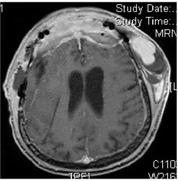
Fig. 1. Axial MRI demonstrating flow voids in the region of the STA.
A B
Fig. 2. (A) Diagnostic angiogram, lateral view, demonstrating a large STA pseudoaneurysm. (B) Diagnostic angiogram, AP view, dem- onstrating a large STA pseudoaneurysm.
sphenoid wing meningioma and right frontal para- sagittal meningioma. The patient recovered well and the immediate perioperative period was uncomplicated.
Three weeks after surgery he presented with a large pulsatile left temporal mass anterior to the surgical in- cision site. A contrasted MRI demonstrated a 7.5 × 5
× 2 cm complex hematoma over the left calvarium with
flow void concerning for a pseudoaneurysm (Fig. 1).
Digital subtraction angiography confirmed STA pseu- doaneurysm (Fig. 2).
Treatment strategies were discussed in detail with the patient. Thrombin injection was deemed inappropriate due to the large size of the pseudoaneurysm. Catheter coil embolization of the pseudoaneurysm was attempted via right femoral artery access but was unsuccessful due to tortuous parent vessels. With local anesthetic, ultrasound guided access of the STA just proximal to the site of the pseudoaneurysm was then performed.
A 4 French catheter was used for access through the scalp distal to the pseudoaneurysm. Thirteen fibered microcoils were delivered sequentially into the STA until the distal and proximal defects were occluded.
A post-coil angiogram demonstrated complete ob- literation of flow through the pseudoaneurysm (Fig.
3). The contents of the pseudoaneurysm were not as- pirated as the decision was made to see if the pseu- doaneurysm would decompress naturally. No coils were placed within the pseudoaneurysm in an at- tempt at obliteration via blockage of both proximal and distal flow. There were no complications of this procedure. At twelve month follow up, MRI demon- strated complete obliteration of the pseudoaneurysm
A B C
Fig. 3. (A) Coiling of the pseudoaneurysm. (B) Angiogram (DSA) after coiling of the pseudoaneurysm. (C) Lateral angiogram of the coils used to occlude the STA.
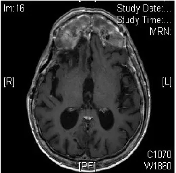
Fig. 4. MRI one year after coiling demonstrating no recurrence of the STA pseudoaneurysm.
and the patient suffered from no cosmetic sequelae (Fig. 4).
DISCUSSION
STA pseudoaneurysms are rare vascular lesions that ordinarily occur weeks to months after blunt trauma.
Pseudoaneurysms are formed by damage to the arte- rial wall leading to extravasation of blood and hema- toma formation in the surrounding soft tissue.14) The frontal branch of STA is particularly vulnerable since
it lies directly above a bony surface and lacks cush- ioning from overlying muscles.1) Clinical examination reveals an enlarging, pulsatile, painless mass in the distribution of STA with or without a headache.
Additionally, STA pseudoaneurysms can be accom- panied by regional pain, auditory and visual dis- turbance and facial palsy.14)15) It is important to treat these lesions due to risk of thromboembolism, rup- ture, bony erosion, prevent cosmetic disfiguration, and potentially alleviate headache.19)
Current treatment modalities include surgical liga- tion and resection, endovascular coil embolization, ul- trasound guided percutaneous thrombin injection, and conservative surveillance. Surgical ligation and ex- cision is safe, highly effective, and the most common treatment option employed. Once the pseudoaneur- ysm is located, the afferent and efferent branches of the artery are ligated, followed by excision of the pseudoaneurysm sac. Thomassen et al. and Van Uden et al. reported 128 and 67 cases of STA pseudoaneur- ysms, respectively, which were successfully treated with surgical excision.18)19) Although highly effective and relatively low risk, complications of surgery in- clude scarring, temporalis muscle wasting, and facial nerve palsy.10)
Recently, alternative treatment options such as en- dovascular coil embolization and ultrasound guided thrombin injection have gained popularity. In coil em- bolization, angiography is used to visualize the STA
pseudoaneurysm, followed by insertion of platinum microcoils to occlude the lesion.5) Advantages of this method include simultaneous performance of diag- nosis and treatment, lack of a surgical scar, decreased risk of scalp infection, and improved recovery time.5) Disadvantages include a 2.8% risk of transient, perma- nent, or reversible neurologic complications secondary dislocation of an aortic or carotid plaque. Additionally, coil embolization does not address issue of mass ef- fect, and may result in poor cosmesis.16)19)20)
Partap et al.11) described direct injection of thrombin into the pseudoaneurysm under ultrasound guidance.
Since then at least five other cases have been successfully treated via this technique, including one case in a pedia- tric patient.10)18) This method is easy to perform, may be done in the outpatient setting without anesthesia, lacks an incisional scar and has immediate therapeutic benefits for narrow neck pseudoaneurysms less than 4 cm in diameter.11) In addition, there is complete resolution of mass effect created by thrombosis after 2-3 months.11) However, ultrasound guided thrombin injection carries a high rate of risk of recanalization,16) occlusion of the underlying artery, and embolization.10) Teh et al.17) de- scribe a case of ischemia of the scalp and seizure fol- lowing ultrasound guided thrombin injection, and rec- ommend avoiding this technique when treating pseu- doaneurysms of arteries supplying vital end organs.
Gulati et al.3) present the only other report of direct ultrasound guided access to the STA for coil emboli- zation of a STA pseudoaneurysm. After an un- successful attempt at manual compression, the pseu- doaneurysm was coiled with three one-centimeter coils, leaving a patent STA and completely occluded aneurysm. Complete obliteration was confirmed on ultrasound and by angiography, however, the size of the mass had only decreased by one-third after one month and was not expected to resolve given the mass effect of the coils.3) In our case, the aneurysm was partially thrombosed but still with a large flow void. Given the size of the pseudoaneurysm, the number of coils required for total occlusion, and con-
sequent risk of skin necrosis and poor cosmetic result, the decision was made to perform a low profile coil- ing of the STA.
We report a case of an iatrogenic STA pseudoaneurysm treated with direct percutaneous ultrasound guided coil embolization of the parent vessel. Post coil angiogram revealed complete occlusion of the pseudoaneurysm.
Additionally, there were no recurrences or any cos- metic defects at the one-year follow up. After a review of the literature, Gulati et al.3) describe the only other case using this approach for STA pseudoaneurysm treatment. However, they performed coil embolization of the pseudoaneurysm resulting in a significant coil burden and permanent cosmetic deformity.
A patient's clinical status, aesthetic preferences, and size and chronicity of the pseudoaneurysm are important factors in formulating the right treatment plan for STA pseudoaneurysms.7) We highlight the use of per- cutaneous ultrasound guided coil embolization as a safe, effective and minimally invasive technique for treating STA pseudoaneurysms. Direct coiling of the STA resulted in a decreased coil burden and diminution of the aneurysm over time. Local, extracranial arterial access avoids the neurologic sequelae sometimes associated with distal access for arteriography. This method is especially useful for poor surgical candidates, patients for whom thrombin injection or catheterization is con- traindicated, pseudoaneurysms of significant size and deformity, and for pseudoaneurysms that are difficult to reach with groin or brachial arterial access.
Disclosure
The authors report no conflict of interest concerning the materials or methods used in this study or the findings specified in this paper.
REFERENCES
1. Ahn HS, Cho BM, Oh SM, Park SH. Traumatic pseu- doaneurysm of the superficial temporal artery in a child:
a case report. Child Nerv Syst. 2010 Jan;26(1):117-20.
2. Angevine PD, Connolly ES Jr. Pseudoaneurysms of the superficial temporal artery secondary to placement of ex-
ternal ventricular drainage catheters. Surg Neurol. 2002 Sep-Oct;58(3-4):258-60.
3. Gulati MS, Gupta H, Sharma S, Kapoor V, Paul S, Berry M. Re: Direct percutaneous coil embolization of a pseu- doaneurysm under color Doppler guidance. Cardiovasc Intervent Radiol. 1999 May-Jun;22(3):265-6.
4. Hakan T, Ersahin M, Somay H, Aker F. Pseudoaneurysm of the superficial temporal artery following revision of a middle cerebral artery aneurysm clipping: case report and review of the literature. Turk Neurosurg. 2011;21(3):430-4.
5. Hong JT, Lee SW, Ihn YK, Son BC, Sung JH, Kim IS, et al.
Traumatic pseudoaneurysm of the superficial temporal artery treated by endovascular coil embolization. Surg Neurol.
2006 Jul;66(1):86-8.
6. Kim JH, Jung YJ, Chang CH. Superficial temporal artery pseudoaneurysm treated with manual compression alone.
J Cerebrovasc Endovasc Neurosurg. 2015 Mar;17(1):49-53.
7. Kim SW, Jong Kim E, Sung KY, Kim JT, Kim YH. Treatment protocol of traumatic pseudoaneurysm of the superficial temporal artery. J Craniofac Surg. 2013 Jan;24(1):295-8.
8. Levisianos I, Sood V. Traumatic aneurysm (pseudoaneurysm) of the superficial temporal artery. Emerg Med J. 2008 Apr;25(4):239-40.
9. Mann GS, Heran MK. Percutaneous thrombin emboliza- tion of a post-traumatic superficial temporal artery pseudoaneurysm. Pediatr Radiol. 2007 Jun;37(6):578-80.
10. Murphy M, Hughes D, Liaquat I, Edmondson R, Bullock P. Giant traumatic pseudoaneurysm of superficial temporal artery: treatment challenges and case review. Br J Neurosurg.
2006 Jun;20(3):159-61.
11. Partap VA, Cassoff J, Glikstein R. US-guided percuta- neous thrombin injection: a new method of repair of su- perficial temporal artery pseudoaneurysm. J Vasc Interv Radiol. 2000 Apr;11(4):461-3.
12. Pourdanesh F, Salehian M, Dehghan P, Dehghani N, Dehghani S. Pseudoaneurysm of the superficial temporal artery following penetrating trauma. J Craniofac Surg. 2013 Jul;24(4):e334-7.
13. Rancic A, Pecoraro F, Nigro G, Simon R, Frauenfelder T, Mayer D. Branch ligatures and blood aspiration for post-traumatic superficial temporal artery pseudoaneur- ysm: surgical technique. Gen Thorac Cardiovasc Surg.
2014 Jan;62(1):68-70.
14. Roman AA, Arsenault AJ, Jackson KD, Price JM. Novel onset of a posttraumatic superficial temporal artery pseudoaneurysm. Case Rep Emerg Med. 2013;2013:369309.
15. Rubio-Palau J, Ferrer-Fuertes A, Garcia-Diez E, Garcia-Linares J, Marti-Pages C, Sieria-Gil R. Traumatic pseudoaneurysm of the superficial temporal artery: a case report and re- view of literature. Oral Surg Oral Med Oral Pathol Oral Radiol. 2014 Feb;117(2):e112-4.
16. Stapleton CJ, Fusco MR, Thomas AJ, Levy El, Ogilvy CS.
Traumatic pseudoaneurysms of the superficial temporal ar- tery: case series, anatomy, and multidisciplinary treatment considerations. J Clin Neurosci. 2014 Sep;21(9):1529-32.
17. Teh LG, Sieunarine K. Thrombin injection for repair of pseudoaneurysms: a case for caution. Australas Radiol.
2003 Mar;47(1):64-6.
18. Thomassen I, Klompenhouwer EG, Willigendael EM, Teijink JA. Treatment of temporal artery pseudoaneurysms. Vascular.
2014 Aug;22(4):274-9.
19. Van Uden DJ, Truijers M, Schipper EE, Zeebregts CJ, Reijnen MM. Superficial temporal artery aneurysm: diagnosis and treatment options. Head Neck. 2013 Apr;35(4):608-14.
20. Willinsky R, Taylor S, TerBrugge K, Farb RI, Tomlinson G, Montanera W. Neurologic complications of cerebral angiog- raphy: prospective analysis of 2,899 procedures and re- view of the literature. Radiology. 2003 May;227(2):522-8.