87 Introduction
Stress induced cardiomyopathy (SCMP) is characterized by a transient left ventricular (LV) dysfunction with akinesis and systolic expansion of apical segments and hyperkinesis or nor- mokinesis of the basal segments. Various emotional or physi- cal stressors have been described as triggering factors of this syndrome. Although the prognosis of SCMP is generally known to be excellent, several fatal complications have been described. Here, we report a rare case of SCMP complicated by LV thrombosis and multiple cerebral infarctions in a pa- tient with essential thrombocythemia (ET).
Case
A 75-year-old female presented with sudden onset of men- tal deepening, hemiparesis, and dysarthria which developed 1 hour ago. She had a history of dural sinus thrombosis and deep vein thrombosis 2 years ago, and the cause of venous thrombosis was ET. She had been treated with 0.5 mg of
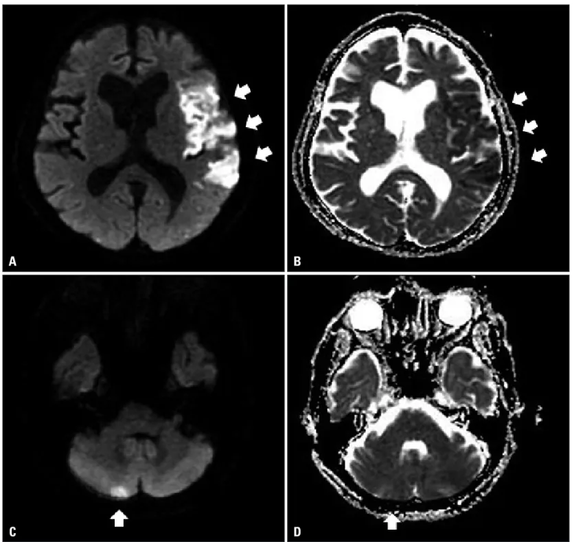
anagrelide and 500 mg of hydroxyurea daily for ET at hema- tologic department for last 2 years. Before 1 day of admission, she had experienced severe emotional stress due to arguments with her husband for economic problems, and thereafter chest tightness developed and persisted. Brain magnetic resonance imaging revealed acute infarctions of left middle cerebral and right posterior inferior cerebellar arterial territories (Fig. 1).
Electrocardiogram (ECG) at admission showed convex ST segment elevation and T-wave inversions in precordial leads with QT prolongation. Laboratory studies showed thrombo- cytosis with platelet count of 665 × 103/mm3 and mildly ele- vated cardiac troponin I with the level of 0.65 ng/mL. Other laboratory findings were unremarkable. Transthoracic echocar- diography (TTE) was performed to evaluate cardiac abnormal- ities relevant to the abnormalities of ECG and cardiac en- zymes, and cardiac source of cerebral embolism. TTE revealed akinesia and systolic bulging of the apical segments with hy- perkinetic basal wall motions, and the estimated ejection frac-
pISSN 1975-4612/ eISSN 2005-9655 Copyright © 2011 Korean Society of Echocardiography www.kse-jcu.org DOI: 10.4250/jcu.2011.19.2.87
CASE REPORT J Cardiovasc Ultrasound 2011;19(2):87-90
Stress Cardiomyopathy Complicated by Left Ventricular Thrombi and
Cerebral Infarctions in a Patient with Essential Thrombocythemia
Seung Hwan Hwang, MD, Kye Hun Kim, MD, Hyun Ju Yoon, MD, Young Joon Hong, MD, Ju Han Kim, MD, YoungKeun Ahn, MD, Myung Ho Jeong, MD, Jeong Gwan Cho, MD, Jong Chun Park, MD and Jung Chaee Kang, MD
Department of Cardiovascular Medicine, Chonnam National University Hospital,
The Research Institute of Medical Sciences, Chonnam National University, Gwangju, Korea
The prognosis of stress induced cardiomyopathy (SCMP) is generally known to be excellent, however, several fatal complications such as cardiac rupture and left ventricular (LV) thrombosis with subsequent embolic complications have been described. We re- port a rare case of SCMP complicated by LV thrombosis and multiple cerebral infarctions in a patient with essential thrombocy- themia. After intravenous anticoagulation with heparin and general managements for heart failure and cerebral infarctions, her neurologic symptoms and the wall motion abnormalities of the LV apex were improved, and the thrombus was disappeared on follow-up echocardiography.
KEY WORDS: Cardiomyopathy · Thrombocythemia · Thromboembolism.
• Received: December 14, 2010 • Revised: May 11, 2011 • Accepted: May 25, 2011
• Address for Correspondence: Kye Hun Kim, The Heart Center of Chonnam National University Hospital, 8 Hak-dong, Dong-gu, Gwangju 501-757, Korea Tel: +82-62-220-6978, Fax: +82-62-227-4760, E-mail: [email protected]
• This is an Open Access article distributed under the terms of the Creative Commons Attribution Non-Commercial License (http://creativecommons.org/licenses/by-nc/3.0) which permits unrestricted non-commercial use, distribution, and reproduction in any medium, provided the original work is properly cited.
Journal of Cardiovascular Ultrasound 19 | June 2011
88
tion was 51.8%. About 3.3 × 2.4 cm sized mural thrombus was observed within the dyskinetic apical segments of the LV (Fig. 2A and B). Coronary angiography was performed to ex- clude acute myocardial infarction (AMI) and revealed no sig- nificant stenotic lesions in both coronary arteries. The most probable diagnosis in the present case was SCMP with apical mural thrombi complicated by multiple cerebral infarctions in the setting of ET. Anticoagulation with intravenous hepa- rin and general managements for heart failure and cerebral in- farctions were done without discontinuation of anagrelide and hydroxyurea. The symptoms and signs of cerebral infarctions including motor and speech disturbances were improved gradually. The wall motion abnormalities of the LV apex were
improved, and the thrombus was disappeared on follow-up TTE (Fig. 2C and D). The patients was discharged with med- ications including anagrelide and hydroxyurea and managed at out-patient clinic without any clinical events.
Discussion
SCMP, also known as apical ballooning or Takotsubo car- diomyopathy, is a syndrome of transient cardiac dysfunction associated with acute emotional or physical stress, but the pathogenesis of SCMP is not well established. Catecholamine- mediated multivessel epicardial or microvascular coronary va- sospasm or direct catecholamine-mediated myocyte injury has been proposed as possible pathophysiological mechanisms.1)
Fig. 1. Diffusion weighted (A and C) and apparent diffusion coefficient (B and D) brain magnetic resonance imaging show acute cerebral infarction in the left middle cerebral artery territory (A and B, arrow) and right posterior inferior cerebellar artery territory (C and D, arrow).
A
C
B
D
Stress Cardiomyopathy and Thromboembolic Events | Seung Hwan Hwang, et al.
89 Because clinical characteristics of SCMP are quite similar to
those of AMI, the differential diagnosis between SCMP and AMI is very difficult and usually requires coronary angiogra- phy.2) Coronary angiography was also performed in the present case to rule out AMI as a cause of LV dysfunction and revealed normal coronary circulation. Provocation test for vasospasm was not performed, because vasoactive drugs including intra- venous nitrates were prescribed already. Therefore, the possi- bility of vasospasm as a cause of wall motion abnormality could not completely rule out in the present case.
Emotional stress, as in most of the previously reported cases, was the most probable predisposing cause of SCMP in the present case, but the role of anagrelide should also be consid- ered. Anagrelide inhibits platelet aggregation through the se- lective inhibition of type III cyclic adenosine monophosphate phosphodiesterase on the megakaryocyte cell lineage, and thus anagrelide used in the treatment of ET to prevent the inci- dence of thromboembolic events. Because it may have positive inotropic and chronotropic activity, and directly induce vaso- spasm of the coronary arteries, several serious cardiovascular side effects including congestive heart failure, arrhythmia, acute coronary syndrome, and SCMP has been described.3)
These side effects of anagrelide developed usually within 1 month of medication and decreased with improved tolerance to treatment over time in the previous report.3)4) Because anagrelide had been prescribed for last two years without any side effects in the present case, it is assumed that the role of anagrelide in the development of SCMP might be negligible in the present case. Therefore, anagrelide and hydroxyurea were prescribed continuously in the present case during admission and after discharge. Because the platelet count of the patient was maintained in acceptable range with the combination therapy of relatively low dose of anagrelide (0.5 mg) and hy- droxyurea, we did not change the dosage of anegrelide. If the platelet count is not controlled within acceptable range, it would be reasonable to adjust the dosage of anegrelide. In case of confirmed acute coronary syndrome, the discontinuation of anagrelide has been recommended.5)
Although the prognosis of SCMP is known to be excellent, various complications, including severe heart failure with pul- monary edema, shock and inhospital death have been report- ed. The present case presented with multiple cerebral infarc- tion presumably caused by the embolization of the LV thrombi, and cerebral infarction associated with LV thrombosis is a rare
Fig. 2. Transthoracic echocardiography at admission reveals akinesia and systolic bulging of apical segments with thrombi formation (A and B, arrow). The wall motion abnormalities of the left ventricular are normalized and the apical thrombi are resolved completely on follow-up echocardiographic examination (C and D).
A
C
B
D
Journal of Cardiovascular Ultrasound 19 | June 2011
90
complication of SCMP. In a recent systematic review of de Gregorio,6) LV thrombosis occurred approximately 5% of the patients with SCMP, and one third of them experienced thromboembolic events. The study of Haghi et al.7) and Mit- suma et al.8) also demonstrated that LV thrombosis and subse- quent embolism was a possible complication of SCMP. Recent meta-analysis showed that LV thrombus is a significant com- plication and systemic embolism is 2nd common causes of mortality in SCMP.9) Therefore, it is suggested that the early initiation of anti-coagulation therapy to prevent LV thrombo- sis and subsequent embolic complications would be a reason- able approach in patients with SCMP.
The LV thrombosis in patients with SCMP is probably caused by low blood flow in the left ventricle as well as LV sys- tolic dysfunction. ET is known to be associated with bleeding or thromboembolic complications. Arterial thromboses occur- ring in coronary, cerebral or peripheral arteries are regarded as more typical than venous thromboembolic events in patients with ET. The present case had a tendency of thrombosis evi- denced by the previous history of dural sinus and deep vein thrombosis associated with ET, and thus ET might play an im- portant additional role in the thrombus formation within the LV in the present case, besides SCMP itself. Baker et al.10) also reported a similar case of LV mural thrombus in a patient with thrombocytosis and agnogenic myeloid metaplasia.
In conclusion, we report a rare case of SCMP complicated by LV thrombosis and multiple cerebral infarctions in a pa- tient with ET. Early initiation of anti-coagulation therapy should be considered in patients with SCMP, especially who had combined coagulation disorders such as ET.
References
1. Abraham J, Mudd JO, Kapur NK, Klein K, Champion HC, Witt- stein IS. Stress cardiomyopathy after intravenous administration of catechol- amines and beta-receptor agonists. J Am Coll Cardiol 2009;53:1320-5.
2. Shin SK, Jin SA, Park YK, Park JH. A case of acute ST-segment eleva- tion myocardial infarction mimicking stress induced cardiomyopathy; dem- onstration of typical echocardiographic finding correlated with unusual distri- bution of left anterior descending coronary artery. J Cardiovasc Ultrasound 2010;18:101-3.
3. Steurer M, Gastl G, Jedrzejczak WW, Pytlik R, Lin W, Schlögl E, Gisslinger H. Anagrelide for thrombocytosis in myeloproliferative disorders:
a prospective study to assess efficacy and adverse event profile. Cancer 2004;101:2239-46.
4. Mazzucconi MG, Redi R, Bernasconi S, Bizzoni L, Dragoni F, Lata- gliata R, Santoro C, Mandelli F. A long-term study of young patients with essential thrombocythemia treated with anagrelide. Haematologica 2004;89:1306-13.
5. Birgegård G. Long-term management of thrombocytosis in essential throm- bocythaemia. Ann Hematol 2009;88:1-10.
6. de Gregorio C. Cardioembolic outcomes in stress-related cardiomyopathy complicated by ventricular thrombus: a systematic review of 26 clinical stud- ies. Int J Cardiol 2010;141:11-7.
7. Haghi D, Papavassiliu T, Heggemann F, Kaden JJ, Borggrefe M, Suselbeck T. Incidence and clinical significance of left ventricular thrombus in tako-tsubo cardiomyopathy assessed with echocardiography. QJM 2008;101:381-6.
8. Mitsuma W, Kodama M, Ito M, Kimura S, Tanaka K, Hoyano M, Hirono S, Aizawa Y. Thromboembolism in Takotsubo cardiomyopathy. Int J Cardiol 2010;139:98-100.
9. Donohue D, Movahed MR. Clinical characteristics, demographics and prognosis of transient left ventricular apical ballooning syndrome. Heart Fail Rev 2005;10:311-6.
10. Baker KM, Hess CE, Ayers CR, Johns DW, Mentzer RM, Wellsons HA, Taylor GJ, Martin RP. Left ventricular mural thrombus in a patient with thrombocytosis and agnogenic myeloid metaplasia. Arch Intern Med 1981;141:1527-9.