ISSN 2234-3806 • eISSN 2234-3814
152 www.annlabmed.org http://dx.doi.org/10.3343/alm.2015.35.1.152 Ann Lab Med 2015;35:152-154
http://dx.doi.org/10.3343/alm.2015.35.1.152
Letter to the Editor
Diagnostic Hematology
The First Case Report of Composite Bone Marrow
Involvement by Simultaneously Developed Peripheral T-Cell Lymphoma, Not Otherwise Specified, and
Diffuse Large B-Cell Lymphoma
Hyun-Ki Kim, M.D.1, Chan-Jeoung Park, M.D.1, Seongsoo Jang, M.D.1, Young-Uk Cho, M.D.1, Sang Hyuk Park, M.D.2, Jene Choi, M.D.3, Chan-Sik Park, M.D.3, Jooryung Huh, M.D.3, Young-Hwa Chung, M.D.4, and Jung-Hee Lee, M.D.4
Departments of Laboratory Medicine1, Pathology3, and Internal Medicine4, University of Ulsan College of Medicine and Asan Medical Center, Seoul;
Department of Laboratory Medicine2, Pusan National University School of Medicine, Pusan National University Hospital, Busan, Korea
Lymphomas of different histological types can occur at the same anatomical site (composite lymphoma) or at different sites, and can arise simultaneously or sequentially [1, 2]. Reports of patients in whom histologically distinct lymphomas occur simultaneously at different sites are rare [3, 4]. Among these rare cases, the si- multaneous occurrence of T- and B-cell lymphomas is extremely rare [5]. To the best of our knowledge, composite involvement of T- and B-cell lymphomas in the bone marrow (BM) have not been reported so far. We report a case of composite BM involvement by T- and B-cell lymphomas.
A 52-yr-old woman presented with abdominal discomfort.
She had a history of hepatitis B-related liver cirrhosis and was taking lamivudine. She had lost 4 kg of weight in the previous 4 months. Laboratory investigation revealed pancytopenia (white blood cells, 1.7 ×10⁹/L [reference range: 4-10 ×109/L]; hemo- globin, 11.0 g/dL [12-16 g/dL]; and platelets, 75×109/L [150- 350×109/L]), elevated levels of liver enzymes (aspartate trans- aminase, 75 IU/L [0-40 IU/L]; alkaline phosphatase, 129 IU/L [40-120 IU/L]; γ-glutamyltransferase, 99 IU/L [8-35 IU/L]), and an increased level of lactate dehydrogenase (315 IU/L [120-250
IU/L]). Dynamic computed tomography revealed an ill-defined, 3-cm lesion in the left lateral segment of the liver and multiple abdominal and mediastinal lymphadenopathies. Esophagogas- troduodenoscopy revealed a 3-cm ulcerofungating mass between the antrum and the lower body of the stomach. A liver core nee- dle biopsy identified a peripheral T-cell lymphoma, not otherwise specified, and an endoscopic biopsy of the stomach identified a diffuse large B-cell lymphoma (DLBL). A BM biopsy revealed BM involvement by DLBL with a nodular infiltration pattern, which was corroborated by positive immunohistochemical staining for CD3 and CD20. CD20-positive lymphocytes were present in nodular aggregates. CD3-positive lymphocytes were present in small lymphocytes surrounding CD20-positive lymphocytes sug- gesting reactive T lymphocytes. A cytogenetic study of BM re- vealed an apparently normal karyotype. The patient was started on the combination chemotherapy regimen including rituximab, cyclophosphamide, doxorubicin, vincristine and prednisolone (R-CHOP).
After 6 cycles of R-CHOP, the liver mass and lymphadenopa- thies had disappeared. However, a follow-up BM biopsy revealed
Received: April 21, 2014 Revision received: June 23, 2014 Accepted: October 13, 2014
Corresponding author: Chan-Jeoung Park
Department of Laboratory Medicine, University of Ulsan College of Medicine and Asan Medical Center, 88 Olympic-ro 43-gil, Songpa-gu, Seoul 138-736, Korea
Tel: +82-2-3010-4508, Fax: +82-2-478-0884, E-mail: cjpark@amc.seoul.kr
© The Korean Society for Laboratory Medicine.
This is an Open Access article distributed under the terms of the Creative Commons Attribution Non-Commercial License (http://creativecommons.org/licenses/by-nc/3.0) which permits unrestricted non-commercial use, distribution, and reproduction in any medium, provided the original work is properly cited.
Kim H-K, et al.
Composite BM involvement by T&B cell lymphomas
http://dx.doi.org/10.3343/alm.2015.35.1.152 www.annlabmed.org 153
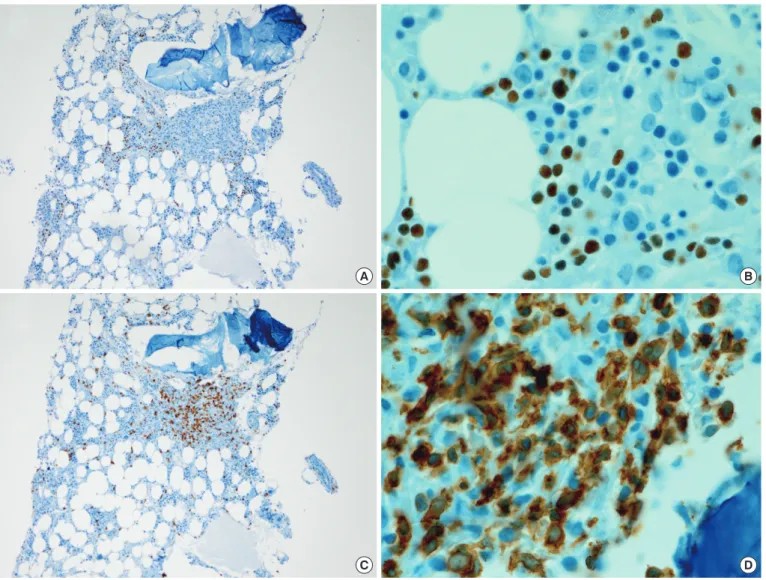
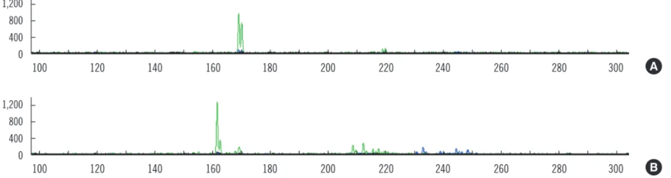
BM involvement by T-cell lymphoma and DLBL. Immunohisto- chemical staining showed medium to large sized PAX5-positive lymphocytes with a marked interstitial infiltration pattern (Fig. 1A, B), and the cells were also CD79a-positive. Peritrabecular and interstitial infiltration of small to medium sized CD3-positive lym- phocytes was also prominent (Fig. 1C, D). Rearrangements of the T-cell receptor gamma locus (TRG) and immunoglobulin heavy locus (IGH) were studied in BM (after the 6th cycle of R-CHOP), liver, and stomach (at diagnosis) specimens using the BIOMED-2 multiplex PCR protocol [6]. A 170-bp monoclonal TRG rearrange- ment was detected in the liver sample (Fig. 2A), while TRG in the BM had a larger 160-bp peak with a smaller 170-bp peak (Fig. 2B). IGH gene rearrangements were not detected in the
stomach sample or in the BM aspirate, and the BM aspirate had a normal karyotype. After the 8th R-CHOP cycle, computed to- mography and positron emission tomography findings suggested involvement of multiple lymph nodes (celiac, splenic hilar, and abdominal paraaortic lymph nodes). The patient died of pneumo- nia 1 yr after diagnosis.
In our TRG rearrangement study, monoclonal TRG rearrange- ments were detected in both liver and BM samples, but the sizes of amplicons were slightly different, that is a finding that may be explained by clonal evolution [7]. We speculate that there is BM involvement on the basis of a subclone from the hepatic T-cell lymphoma. Moreover, a small peak corresponding to a 170-bp PCR product was observed in a BM sample, which suggests that
Fig. 1. Bone marrow biopsy after 6 cycles of rituximab, cyclophosphamide, doxorubicin, vincristine and prednisolone (R-CHOP) combina- tion chemotherapy. (A, B) Neoplastic B lymphocytes infiltrating in an interstitial pattern was immunohistochemically analyzed by anti-PAX5 antibody (A, ×100; B, ×1,000). (C, D) Neoplastic T lymphocytes infiltrating with a peritrabecular or interstitial pattern was immunohisto- chemically analyzed by anti-CD3 antibody (C, ×100; D, ×1,000).
A
C
B
D
Kim H-K, et al.
Composite BM involvement by T&B cell lymphomas
154 www.annlabmed.org http://dx.doi.org/10.3343/alm.2015.35.1.152 the same clone present in the liver sample was also present in
the BM sample. Monoclonal IGH rearrangements were not de- tected in the stomach or BM sample. As monoclonal IGH rear- rangements are detected in approximately 80% of DLBL cases using BIOMED-2 multiplex PCR [8], the fact that rearrangement of IGH was not detected in the stomach sample is not unusual.
T- and B-cell lymphomas simultaneously developed in this pa- tient. Moreover, a follow-up examination suggested BM involve- ment by both these lymphomas. To our knowledge, this is the first report of a patient with BM involvement by both T- and B- cell lymphomas. Factors contributing to the development of syn- chronous primary lymphomas have not been investigated, and a case with involvement of the same site by two different lympho- mas has not been reported; however, primary composite lympho- mas have been reported [9]. The clinical significance of BM in- volvement by both T- and B-cell lymphomas is unknown, although the simultaneous development of more than one histological type of lymphoma is associated with a poor prognosis [1]. The present patient died 1 yr after her diagnosis, suggesting that composite BM involvement may also be associated with a poor prognosis.
Authors’ Disclosures of Potential Conflicts of Interest
No potential conflicts of interest relevant to this article were re- ported.
REFERENCES
1. Tucci A, Motta M, Ungari M, Ruggeri G, Crippa C, Borlenghi E, et al. The development of more than one histologic type of lymphoma in the same patient is frequent and confers a worse prognosis. Haematologica 2005;
90:348-52.
2. Furlan A, Pietrogrande F, Marino F, Menin C, Polato G, Vianello F. Se- quential development of large B cell lymphoma in a patient with periph- eral T-cell lymphoma. Haematologica 2008;93:e6-8.
3. Terada T. One patient with double lymphomas: simultaneous gastric MALT lymphoma and ileal diffuse large B-cell lymphoma. Int J Clin Exp Pathol 2012;5:260-3.
4. Tang Z, Jing W, Lindeman N, Harris NL, Ferry JA. One patient, two lym- phomas. Simultaneous primary gastric marginal zone lymphoma and primary duodenal follicular lymphoma. Arch Pathol Lab Med 2004;128:
1035-8.
5. Cotter FE, Hall PA, Young BD, Lister TA. Simultaneous presentation of T- and B-cell malignant lymphoma with bcl-2 gene involvement. Blood 1989;73:1387-8.
6. van Dongen JJ, Langerak AW, Bruggemann M, Evans PA, Hummel M, Lavender FL, et al. Design and standardization of PCR primers and pro- tocols for detection of clonal immunoglobulin and T-cell receptor gene recombinations in suspect lymphoproliferations: report of the BIOMED-2 Concerted Action BMH4-CT98-3936. Leukemia 2003;17:2257-317.
7. Umino A, Nakagawa M, Utsunomiya A, Tsukasaki K, Taira N, Katayama N, et al. Clonal evolution of adult T-cell leukemia/lymphoma takes place in the lymph nodes. Blood 2011;117:5473-8.
8. Evans PA, Pott Ch, Groenen PJ, Salles G, Davi F, Berger F, et al. Signifi- cantly improved PCR-based clonality testing in B-cell malignancies by use of multiple immunoglobulin gene targets. Report of the BIOMED-2 Concerted Action BHM4-CT98-3936. Leukemia 2007;21:207-14.
9. Wang HW, Yang W, Wang L, Lu YL, Lu JY. Composite diffuse large B- cell lymphoma and classical Hodgkin’s lymphoma of the stomach: case report and literature review. World J Gastroenterol 2013;19:6304-9.
Fig. 2. T cell receptor gamma locus (TRG) rearrangement analyzed by using BIOMED-2 multiplex PCR. Horizontal axis represents amplicon size (base pairs) and vertical axis represents fluorescence intensity. (A) A monoclonal TRG rearrangement was detected at 170 bp in the liver sample. (B) Both a larger peak (160 bp) and a smaller peak (170 bp) were observed in the bone marrow aspirate sample by GeneScan analysis of the PCR results.
1,200 800 400 0
1,200 800 400 0
100 120 140 160 180 200 220 240 260 280 300
100 120 140 160 180 200 220 240 260 280 300 A
B