Korean J Hepatobiliary Pancreat Surg 2016;20:133-136
http://dx.doi.org/10.14701/kjhbps.2016.20.3.133
Case Report
Inferior vena cava stenosis-induced sinusoidal obstructive syndrome after living donor liver transplantation
Batsaikhan Bat-Erdene1, Sergelen Orgoi1, Erdene Sandag1, Ulzii-Orshikh Namkhai2, Bat-Ireedui Badarch2, and Batsaikhan Batsuuri2
1Department of Surgery, Mongolian National University of Medical Science,
2Department of General Surgery, First Central Hospital of Mongolia, Ulaanbaatar, Mongolia
The sinusoidal obstructive syndrome (SOS) is a complication that usually follows hematopoietic stem cell transplantation. It is also known as veno-occlusive disease, which is a rare complication of living donor liver trans- plantation (LDLT). Herein, we reported a 34 year-old female patient presenting SOS after LDLT. Its underlying cause was presumed to be associated with liver abscess and subsequent inferior vena cava stenosis. SOS led to graft failure, thus requiring retransplantation with a deceased donor liver graft. The underlying causes of SOS are complex patho- logic entity with multifactorial etiology. It is likely that its multifactorial etiology includes a decrease of hepatic venous outflow that is caused by graft liver infection and inferior vena cava stenosis. (Korean J Hepatobiliary Pancreat Surg 2016;20:133-136)
Key Words: Graft failure; Stenting; Liver abscess; Retransplantation
Received: May 27, 2016; Revised: June 10, 2016; Accepted: June 20, 2016 Corresponding author: Batsaikhan Bat-Erdene
Department of Surgery, Mongolian National University of Medical Science, S.Zorig Street -3, Ulaanbaatar, Mongolia Tel: +976-99-239-059, Fax: +976-11-321249, E-mail: batsaikhan@mnums.edu.mn
Copyright Ⓒ 2016 by The Korean Association of Hepato-Biliary-Pancreatic Surgery
This is an Open Access article distributed under the terms of the Creative Commons Attribution Non-Commercial License (http://creativecommons.org/
licenses/by-nc/4.0) which permits unrestricted non-commercial use, distribution, and reproduction in any medium, provided the original work is properly cited.
Korean Journal of Hepato-Biliary-Pancreatic Surgery ∙ pISSN: 1738-6349ㆍeISSN: 2288-9213
INTRODUCTION
Sinusoidal obstructive syndrome (SOS) was initially called veno-occlusive disease,1 based on the triad of jaun- dice/hyperbilirubinemia (>2 mg/dl), painful hepatomegaly and ascites/weight gain. It is confirmed by the histologic findings of fibrous obliteration of small hepatic veins by connective tissue and centrilobular hemorrhagic necrosis.2 The most frequent cause of SOS is the use of high-dose chemotherapy in recipients of hematopoietic stem cell transplantation.3-5 SOS is also described after liver trans- plantation (LT), but with relatively rare occurrence of ap- proximately 2%.6 Severe SOS leads to mortality rates of 84-90%.7,8 Development of SOS after LT is reportedly as- sociated with tacrolimus administration.9 Herein, we de- scribed a case of SOS following stenosis of the inferior vena cava after living donor liver transplantation (LDLT).
CASE
A 34-year-old woman with hepatitis B virus-associated liver cirrhosis with model for end-stage liver disease score of 15 underwent LDLT operation using a right lobe graft from a 27-year-old living donor in 2015. The cold and warm ischemic times were 130 minutes and 80 minutes, respectively. Serum total bilirubin increased to 25.4 mg/dl until post-operation day (POD) 7, and Gamma glutamyl transpeptinase (GGT) 248 mg/dl at POD 13. Doppler ul- trasonography indicated the right hepatic vein (RHV) out- flow rate of 37 cm/sec and enhanced computed tomog- raphy (CT) at POD 14 revealed no vascular abnormality.
Thereafter, the patient condition was stabilized.
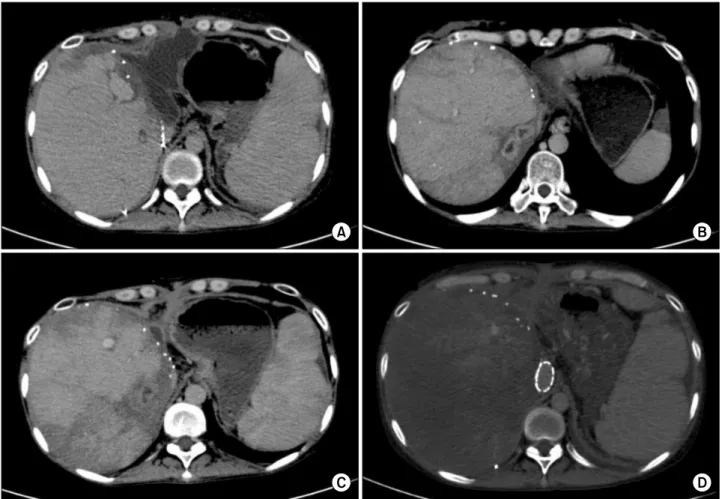
Repeat CT scan at POD24 revealed biloma around the liver graft (Fig. 1A). The laboratory profiles showed de- crease in total bilirubin to 1.6 m/dl, but increase in C-re- active protein (CRP) to 129 mg/dl. Abdominal drainage culture was Escherichia coli-positive; therefore, antibiotic was changed to Meropenem. Thereafter, CRP decreased
134 Korean J Hepatobiliary Pancreat Surg Vol. 20, No. 3, August 2016
Fig. 1. Sequences of computed tomography (CT) findings: (A) CT scan shows biloma around the liver graft; (B) CT scan taken at day 73 shows multifocal liver abscesses; (C) CT scan shows treatment of liver abscess following antibiotics therapy; and (D) CT scan shows that the liver parenchyma was undefined after stenting of the inferior vena cava.
to 3.9 mg/dl and CT scan at POD 33 showed normal liver findings. The patient was discharged from the hospital.
Blood tacrolimus level was maintained around 10-14 ng/ml during the hospitalization.
At POD 73, she complained of high fever and upper abdominal discomfort. Laboratory study showed CRP 47.4 mg/dl and white blood cell count 3890/l, and tacro- limus level 15-20 ng/ml. Doppler ultrasonography showed RHV outflow rate 30 cm/sec; and CT scan showed multi- focal liver abscesses (Fig. 1B). On admission, she re- ceived antibiotic and supportive therapy. The size of liver abscess decreased after 9 days (Fig. 1C) and the patient was discharged at POD 83.
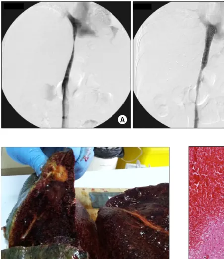
After 6 months, she complained of foot edema and ascites. CT scan revealed stenosis of the retrohepatic in- ferior vena cava (IVC), thus direct angiography was per- formed (Fig. 2A). At that time, the liver function profiles were not impaired: total bilirubin 1.1 mg/dl; aspartate
aminotransferase (AST) 18 IU/L; alanine aminotransferase (ALT) 10 IU/L; GGT 31 IU/L; alkaline phosphatase (ALP) 100 IU/L; and creatinine 0.6 mg/dl. To resolve IVC stenosis, an IVC stent (Hercules Vascular Stent, S&G Bio Tech: Seongnam-si, Korea) was inserted without any noticeable complication (Fig. 2B).
However, after IVC stenting, she started to complain of abdominal discomfort. The liver function was impaired:
total bilirubin 11.5 mg/dl; AST 698 IU/L; ALT 317 IU/L;
GGT 42 IU/L; ALP 120 IU/L; and creatinine 2.35 mg/dl.
Doppler ultrasonography showed significant decrease in RHV flow rate to 13 cm/sec after IVC stenting; and CT scan 9 days after stenting showed non-enhancement of the liver parenchyma (Fig. 1D). The patient’s condition con- tinued to worsen with aggravating jaundice, ascites, and hepatic encephalopathy. Finally, 8 months after LDLT, she underwent retransplantation with a deceased donor liver graft. The patient’s condition was critical (total bilir-
Batsaikhan Bat-Erdene, et al. Inferior vena cava stenosis-induced sinusoidal obstructive syndrome 135
Fig. 2. Direct venography find- ings showing stenosis of the ret- rohepatic inferior vena cava (A) and expansion after inferior vena cava stenting (B).
Fig. 3. Gross photograph of the explant liver graft at the time of retransplantaion. The cut surface of the liver shows tan,
congested and rubbery parenchyma. Fig. 4. Microscopic examination of the explant liver showing findings consistent with sinusoidal obstructive syndrome.
Massive venous congestion is visible; the liver parenchyma was replaced by blood; and a few hepatocytes survived around the portal tracts.
ubin 31.8 mg/dl; AST 42 IU/L; ALT 9 IU/L ; GGT 43 IU/L; ALP 513 IU/L; and creatinine 0.95 mg/dl; INR 3.72). The retransplantation operation was complex and took 15 hour 20 min due to patient’s condition and ab- dominal adhesion. Blood loss was massive and we were unable to control the leakage of blood when closing the abdominal incision. After the operation, she continued in critical condition (total bilirubin 4.7 mg/dl; AST 1555 IU/L; ALT 269 IU/L ; GGT 15 IU/L; ALP 23 IU/L; and creatinine 1.06 mg/dl; INR 1.94). The patient was in- tubated for 9 days post-operatively, and showed gradual improvement. Her blood tacrolimus level was maintained around 10-14 ng/ml during the hospitalization.
The explant liver pathology was consistent with SOS (Figs. 3 and 4). Microscopic examination suggested SOS, characterized by massive venous congestion, replacement of parenchyma with blood, and a few surviving hep-
atocytes around the portal tracts.
DISCUSSION
SOS is characterized by toxic injury of sinusoidal endo- thelial cell, which is revealed as loss of sinusoidal wall.
SOS is occasionally associated with perisinusoidal fib- rosis, centrilobular hepatic vein fibrotic obstruction, nod- ular regenerative hyperplasia, and peliosos.10 The in- cidence of clinical SOS after LT is reported to be 1.9-2.9%.11,12 The clinical signs and symptoms are non-specific after LT, which results in difficult and com- plicated diagnosis. Developing SOS after LT reportedly correlates with acute cellular rejection.5 An in vitro study showed that toxins and drugs that cause hepatic SOS are
136 Korean J Hepatobiliary Pancreat Surg Vol. 20, No. 3, August 2016
more toxic to the hepatic sinusoidal endothelial cells than to the hepatocytes.13,14
Our present case was diagnosed based on explant path- ology after retransplantation. The precedent underlying cause of SOS was possibly graft liver abscess and sub- sequent IVC stenosis. This case was not associated with any episode of acute rejection and we could exclude other potential pathology as well as infectious complication.
Moreover, the explant liver pathology showed 25% mac- rovesicular steatosis. A specific polymorphism of the glu- tathione-S-transferase gene is found more frequently among patients with SOS.15 Thrombotic microangiopathy associated with significantly reduced survival following the allogeneic hematopoetic stem cell transplantation is another risk factor for development of transplantation- associated SOS.16 Also, evidence of clotting abnormalities in the experimental model of hepatic SOS is lacking.17
In particular, cytomegalovirus infection mediated vas- cular injury, results in incidence of hepatic artery throm- bosis after LT. This injury occurs in the sinusoidal endo- thelium, which increases the risk of SOS development.
Positive expression of cytomegalovirus infection was ab- sent in our case.
Tacrolimus is primarily metabolized by the cytochrome P450 (CYP) 3A subfamily in liver microsomes. The zone 3 of the liver acinus has the highest levels of CYP and is the most affected by SOS.18 Hence, we considered Tacrolimus a potential factor for development of SOS af- ter LT in our case. However, its 24-hour trough level was maintained between 10-16 ng/ml, which suggested no as- sociation with SOS development.
In conclusion, the underlying causes of SOS are a com- plex pathologic entity with multifactorial etiology. Its multifactorial etiologies are likely to include a decrease of hepatic venous outflow that is caused by graft liver in- fection and IVC stenosis.
REFERENCES
1. Bras G, Jelliffe DB, Stuart KL. Veno-occlusive disease of liver with nonportal type of cirrhosis, occurring in Jamaica. AMA Arch Pathol 1954;57:285-300.
2. Takamura H, Nakanuma S, Hayashi H, Tajima H, Kakinoki K,
Kitahara M, et al. Severe veno-occlusive disease/sinusoidal ob- struction syndrome after deceased-donor and living-donor liver transplantation. Transplant Proc 2014;46:3523-3535.
3. Dulley FL, Kanfer EJ, Appelbaum FR, Amos D, Hill RS, Buckner CD, et al. Venocclusive disease of the liver after che- moradiotherapy and autologous bone marrow transplantation.
Transplantation 1987;43:870-873.
4. Kumar S, DeLeve LD, Kamath PS, Tefferi A. Hepatic veno-occlu- sive disease (sinusoidal obstruction syndrome) after hematopoietic stem cell transplantation. Mayo Clin Proc 2003;78:589-598.
5. Carreras E. Veno-occlusive disease of the liver after hemopoietic cell transplantation. Eur J Haematol 2000;64:281-291.
6. Marín-Gómez LM, Álamo-Martínez JM, Suárez-Artacho G, Ramírez-Santos J, Bernal-Bellido C, Barrera-Pulido L, et al. Is the sinusoidal obstructive syndrome post-liver transplantation a pathologic entity with a multifactorial etiology? Rev Esp Enferm Dig 2015;107:235-238.
7. Dignan FL, Wynn RF, Hadzic N, Karani J, Quaglia A, Pagliuca A, et al. BCSH/BSBMT guideline: diagnosis and management of veno-occlusive disease (sinusoidal obstruction syndrome) fol- lowing haematopoietic stem cell transplantation. Br J Haematol 2013;163:444-457.
8. Tsirigotis PD, Resnick IB, Avni B, Grisariu S, Stepensky P, Or R, et al. Incidence and risk factors for moderate-to-severe ve- no-occlusive disease of the liver after allogeneic stem cell trans- plantation using a reduced intensity conditioning regimen. Bone Marrow Transplant 2014;49:1389-1392.
9. Shen T, Feng XW, Geng L, Zheng SS. Reversible sinusoidal ob- struction syndrome associated with tacrolimus following liver transplantation. World J Gastroenterol 2015;21:6422-6426.
10. Rubbia-Brandt L. Sinusoidal obstruction syndrome. Clin Liver Dis 2010;14:651-668.
11. Sebagh M, Debette M, Samuel D, Emile JF, Falissard B, Cailliez V, et al. "Silent" presentation of veno-occlusive disease after liv- er transplantation as part of the process of cellular rejection with endothelial predilection. Hepatology 1999;30:1144-1150.
12. Sebagh M, Azoulay D, Roche B, Hoti E, Karam V, Teicher E, et al. Significance of isolated hepatic veno-occlusive dis- ease/sinusoidal obstruction syndrome after liver transplantation.
Liver Transpl 2011;17:798-808.
13. Deleve LD. Dacarbazine toxicity in murine liver cells: a model of hepatic endothelial injury and glutathione defense. J Pharmacol Exp Ther 1994;268:1261-1270.
14. DeLeve LD, Wang X, Kuhlenkamp JF, Kaplowitz N. Toxicity of azathioprine and monocrotaline in murine sinusoidal endothe- lial cells and hepatocytes: the role of glutathione and relevance to hepatic venoocclusive disease. Hepatology 1996;23:589-599.
15. DeLeve LD. Glutathione defense in non-parenchymal cells.
Semin Liver Dis 1998;18:403-413.
16. Daly AS, Hasegawa WS, Lipton JH, Messner HA, Kiss TL.
Transplantation-associated thrombotic microangiopathy is asso- ciated with transplantation from unrelated donors, acute graft-versus-host disease and venoocclusive disease of the liver.
Transfus Apher Sci 2002;27:3-12.
17. DeLeve LD, McCuskey RS, Wang X, Hu L, McCuskey MK, Epstein RB, et al. Characterization of a reproducible rat model of hepatic veno-occlusive disease. Hepatology 1999;29:1779-1791.
18. Nakazawa Y, Chisuwa H, Mita A, Ikegami T, Hashikura Y, Terada M, et al. Life-threatening veno-occlusive disease after liv- ing-related liver transplantation. Transplantation 2003;75:727-730.