182
Correspondence to: Young Kyu Park, Division of Gastroenterologic Surgery, Department of Surgery, Chonnam National University Hwasun Hospital, 160, Ilsim-ri, Hwasun-eup, Hwasun 519-809, Korea. Tel: 061-379-7644, Fax: 061-379-7661, E-mail: parkyk@jnu.
ac.kr
Received March 15, 2010, Accepted April 28, 2010
*These two authors equally contributed to this work.
This work was supported by a research grant (0720570) from the Chonnam National University.
Matrix Metalloproteinase 7 (MMP-7) Expression Predicts the Status of Lymph Node Metastasis in Early Gastric Cancer
1Division of Gastroenterologic Surgery, Department of Surgery, 2Department of Pathology, Chonnam National University Hwasun Hospital, Hwasun, Korea
Oh Jeong, M.D.
1,*, Xue-Feng Zhao, M.D.
1,*, Young Kyu Park, M.D., Ph.D.
1, Jae Hyuk Lee, M.D., Ph.D.
2, Young-Jin Kim, M.D., Ph.D.
1Purpose: Accurate prediction of lymph node (LN) status is of crucial importance for appropriate treatment planning
in early gastric cancer (EGC). Matrix metalloproteinases (MMPs) have been shown to be involved in the pathogenesis of tumor progression and metastasis in gastric carcinoma. In this study, we investigated the association between MMPs expressions and LN metastasis in EGC.Methods: Thirty-four LN positive and 80 LN negative pT1 tumors were immunohistochemically analyzed for
MMP-2, MMP-7, and MMP-9 expression. The relation of MMPs expressions to LN metastasis was analyzed in the univariate and multivariate model.Results: There were 73 men and 41 women with a mean age of 60 years. Among the pathologic characteristics,
larger tumor size, submucosal invasion and lymphatic invasion were factors that are significantly associated with LN metastasis in pT1 tumors. Immunohistochemistry showed significantly higher MMP-7 expression (82.4% vs.54.4%, P=0.005) in LN positive tumors, whereas MMP-9 (85.3% in LN positive vs. 67.5% in LN negative) and MMP-2 (70.6% in LN positive vs. 57.5% in LN negative) expression did not reach statistical significance.
Multivariate analysis revealed that MMP-7 expression (OR 4.915, 95% CI 1.375∼17.573) is an independent predictor of LN metastasis in EGCs, along with lymphatic invasion by tumor cells (OR 10.337, 95% CI 2.785∼
38.360).
Conclusion: Our study shows that MMP-7 expression is significantly associated with LN metastasis in EGC.
MMP-7 expression can be used to predict LN status in EGCs in addition to other pathological parameters.
(J Korean Surg Soc 2011;80:182-188)
Key Words: Matrix metalloproteinases, Lymph node, Neoplasm metastasis, Stomach neoplasm
INTRODUCTION
Accurate prediction of lymph node (LN) status is of crucial importance for appropriate curative treatment planning in early gastric cancer (EGC). EGCs without LN
metastasis can be curatively treated with minimally invasive endoscopic resection such as endoscopic mucosal resection or endoscopic submucosal dissection, whereas LN positive EGCs should be referred to gastrectomy with limited or extended LN dissection.(1) Compared to surgical treat- ment, selected cases of LN negative EGCs treated by endoscopic resection show excellent prognosis, in addition to minimal morbidity and mortality and better postopera- tive quality of life.(2) Therefore, the aggressive surgical approach should be reserved only for EGC patients at risk of LN metastasis.
In an attempt for proper prediction of LN status in EGC, several studies have identified the pathologic
characteristics that are associated with an increased likelihood of LN metastasis,(3) which are currently used as eligibility criteria for endoscopic resection. Recently, with an advancement of molecular biology in gastric cancer, some molecular markers, such as, the PCNA labeling index,(4) p53 expression,(5) erbB-2 expression,(6) DNA ploidy,(7) and VEGF expression,(8) are also emerging as novel markers for predicting LN status in EGC.
Matrix metalloproteinases (MMPs) are a family of enzymes that are responsible for the breakdown of connective tissue proteins in the extracellular matrix. It plays central roles in the tissue remodeling associated with growth, development, and tissue repair under normal physiologic condition.(9) Recently, it has been demons- trated that aberrant MMPs expressions contribute to the pathogenesis of metastasis and tumor progression in several human malignancies. More specifically, the following MMPs have been reported to be involved in the tumor progression and metastasis of gastric carcinoma; mem- brane- type MMP (MT1-MMP),(10) MMP-1,(11) MMP- 2,(12,13) MMP-3,(14) MMP-7,(15,16) and MMP-9.(17) In this study, to investigate the association between MMPs expressions and LN metastasis in EGC, we immuno- histochemically analyzed MMPs expressions in the tumor samples of pT1 gastric cancers.
METHODS 1) Patients
Between May 2004 and April 2006, there were 512 EGC patients treated with gastrectomy and regional LN dissection at Chonnam National University Hwasun Hospital. Of these, 34 (6.6%) cases had LN metastasis at final pathologic examinations and were included in this study. Eighty EGC patients without LN metastasis were collected at random and used as controls. All patients underwent standard gastrectomy including distal or total gastrectomy with limited or extended LN dissection.
Gastric cancer samples were obtained with informed consent from the Chonnam National University Hwasun.
2) Histopathology
After resection, all specimens were fixed in 10% buffered formalin, embedded in paraffin wax, and serially sectioned at 3∼5 mm. Sections were stained with hematoxylin and eosin (H&E) and subjected to pathological examination, at which depth of invasion, lymph node metastasis, histologic type, Lauren’s classification, and lymphovascular permea- tion (as outlined by the Japanese Classification of Gastric Carcinoma) were determined.(18) Degrees of histologic differentiation and tumor stages were determined as detailed in the 6th edition of the TNM Classification System of the UICC.(19) Sections containing greatest tumor areas or the invasive front were selected, and each section was serially sectioned at 4μm for immunohisto- chemistry (IHC).
3) Immunohistochemistry
Tissue sections from formalin-fixed, paraffin-embedded blocks were deparaffinized with xylene, rehydrated through graded ethanol, rinsed with distilled water, and washed with Tris-buffered saline (TBS). Antigen retrieval was performed by boiling for 20 min in citrate buffer (pH 6.0).
Avidin-biotin-peroxidase complex staining using DAB as a chromogen was performed using a standard technique, as described by the manufacturer (Dako, Glostrup, Denmark).
The primary antibodies used were rabbit anti-MMP-2 (1:
100, Santa Cruz Biotechnology, Santa Cruz, CA), rabbit anti-MMP-9 (1:100, Santa Cruz Biotechnology, Santa Cruz, CA), mouse anti-MMP-7 (1:75, Chemicon), mouse anti-CK (1:100, Dako, Glostrup, Denmark), and mouse anti-D2-40 (1:50, Dako, Glostrup, Denmark).
4) Evaluation of immunostaining
Staining for MMP-2, MMP-7, and MMP-9 at the invasive front were evaluated by two independent researchers (JH Lee and YK Park) without knowledge of clinicopathological data, and staining intensities (stains were dark brown in color) were graded as: 0, negative; 1+, weak staining; 2+, moderate; and 3+, strong. MMP immunostaining was deemed positive when moderate to
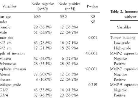
Table 1. The clinicopathological features of pT1 tumors with and without nodal metastasis
Node negative Node positive
Variables (n=80) (n=34) P-value
Mean age 60.0 59.0 NS
Gender NS
Female 29 (36.3%) 12 (35.3%)
Male 51 (63.8%) 22 (64.7%)
Tumor size 0.001
<2 cm 63 (78.8%) 16 (47.1%)
≥2 cm 17 (21.3%) 18 (52.9%)
Depth of invasion <0.001
Mucosa 52 (65.0%) 6 (17.6%)
Submucosa 28 (35.5%) 28 (82.4%)
Lymphatic invasion <0.001
Absent 72 (90.0%) 12 (35.3%)
Present 8 (10.0%) 22 (64.7%)
Histologic grade 0.219
G1/2 43 (53.8%) 14 (41.2%)
G3/4 37 (46.3%) 20 (58.8%)
Table 2. Immunohistochemistries of pT1 gastric cancers with and without lymph node metastasis
Node negative Node positive
Variables (n=80) (n=34) P-value
Tumor budding 0.054
Low-grade 67 (83.8%) 23 (67.6%) High-grade 13 (16.3%) 11 (32.4%)
MMP-2 expression 0.189
Negative 34 (42.5%) 10 (29.4%) Positive 46 (57.5%) 24 (70.6%)
MMP-7 expression 0.005
Negative 36 (45.6%) 6 (17.6%) Positive 43 (54.4%) 28 (82.4%)
MMP-9 expression 0.051
Negative 26 (32.5%) 5 (14.7%) Positive 54 (67.5%) 29 (85.3%) strong membranous and/or cytoplasmic staining was
observed at the invasive front (the percentages of positively stained tumor cells were not counted). Cytokeratin staining was used to improve the visualization of small numbers of tumor cells budding from invasive edges. Tumor budding was classified into four grades; none, minimal, moderate, or severe, as previously described.(20) Degree of tumor budding was then categorized as low-grade (none or minimal) and high-grade (moderate to severe). D2-40 staining was used to differentiate lymphatic vessels and the endothelial cells of blood vessels.
5) Statistical analysis
SPSS version 12.0 (SPSS, Chicago, IL) was used throug- hout. Categorical comparisons of the clinicopathological characteristics of the study groups were carried out using the chi-square or Fisher’s exact test. To determine the significances of associations between variables and nodal metastasis, matched data were subjected to conditional logistic regression analysis. For all statistical tests, P-values of less than 0.05 were considered to be statistically significant.
RESULTS
1) Clinicopathological characteristics associated with lymph node metastasis
The study cohort consisted of 73 men and 41 women of mean age 60 years. No differences were found between the LN positive and LN negative group with respect to age or gender. Pathological examination revealed significant inter-group differences with respect to tumor size, depth of invasion, and lymphatic invasion between LN positive and LN negative group (Table 1). Tumor size was significantly larger in the LN positive group (P=0.001), and there were significantly more submucosal invasion (82.4%
vs. 35.5%, P<0.001) and lymphatic invasion (64.7% vs.
10.0%, P<0.001) in the LN positive group.
2) Immunohistochemistry
Immunohistochemistry showed MMP-7 expression in 28 (82.4%) of the 34 LN positive pT1 tumor samples, which was significantly higher than that in the LN negative group (82.4% vs. 54.4%, P=0.005) (Table 2). MMP-7 expression was observed in the cytoplasm and cell membranes of tumor cells, especially in deeper part of the tumor regions, namely at the invasive front (Fig. 1). MMP-9 expression was also greater in the LN positive group, but with marginal statistical significance (85.3% vs. 67.5%, P=0.051).
Fig. 1. Representative histologic and immunohistochemical staining images of MMP-7 in pT1 gastric cancer with and without lymph node metastasis. Note that MMP-7 was immunostained in carcinoma cells at invasive fronts and in tumor budding cells in pT1 tumor with nodal metastasis (A), whereas no staining was observed in tumor cells at invasive fronts in pT1 tumor without nodal metastasis (B). Cytokeratin (CK) staining was used to improve the visualization of small numbers of tumor cells budding from invasive edges.
The solid line indicates the invasive front. Magnification=200×, scale bars=100μm.
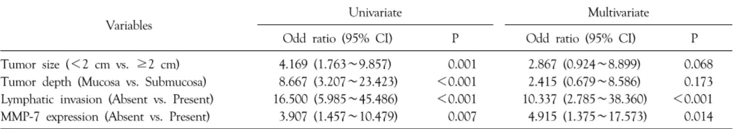
Table 3. Univariate and multivariate analysis of predicting factors for nodal metastasis
Variables Univariate Multivariate
Odd ratio (95% CI) P Odd ratio (95% CI) P
Tumor size (<2 cm vs. ≥2 cm) 4.169 (1.763∼9.857) 0.001 2.867 (0.924∼8.899) 0.068 Tumor depth (Mucosa vs. Submucosa) 8.667 (3.207∼23.423) <0.001 2.415 (0.679∼8.586) 0.173 Lymphatic invasion (Absent vs. Present) 16.500 (5.985∼45.486) <0.001 10.337 (2.785∼38.360) <0.001 MMP-7 expression (Absent vs. Present) 3.907 (1.457∼10.479) 0.007 4.915 (1.375∼17.573) 0.014
However, no significant inter-group difference was found for MMP-2 expression (70.6% in LN positive group vs.
57.5% in LN negative group, P=0.189).
3) Predicting factors for lymph node metastasis
To identify predicting factors for LN metastasis in pT1 tumors, variables found to be significantly associated with LN metastasis in histopathological examinations and IHC, such as, tumor size, submucosal invasion, lymphatic invasion, and MMP-7 expression, were subjected to univariate and multivariate analysis (Table 3). Multivariate analysis was performed using a logistic regression modelwhich incorporated all of these variables, and revealed that MMP-7 expression (OR 4.915, 95% CI 1.375∼17.573) and the presences of lymphatic invasion (OR 10.337, 95%
CI 2.785∼38.360) were independent predictors for LN metastasis in pT1 tumors.
DISCUSSION
Increasing evidence indicates that the extracellular matrix (ECM) of tumors and non-cancerous stromal cells play an important role in tumor progression and metas- tasis.(21) Multistep phenomena involving the proteoloytic
degradations of basement membranes and ECM reduce cellular adhesion and increase the ability of tumor cells to relocate, and these processes are known to be prerequisites of tumor cell invasion and metastasis.(22) Matrix metal- loproteinases (MMPs) are a subfamily of the metzincin proteases, which is one of several families of metalloendo- peptidases. Mechanisms of ECM protein degradation by MMPs regulate various cellular characteristics, such as, cell growth, differentiation, apoptosis, and migration under physiologic condition.(9) Recently, it has been demons- trated that the expressions and activities of MMPs are upregulated in virtually all human cancers.(23)
Minimally invasive endoscopic resection, such as endoscopic mucosal resection or endoscopic submucosal dissection, is a treatment of choice for LN negative EGCs in Asian regions.(1) Compared to surgical treatment, it offers several advantages including minimal morbidity and mortality and better postoperative quality of life as well as excellent long term outcomes when performed in selected patients.(24) Given the increasing use of endoscopic treat- ment for EGC, it is critical that we are able to determine the LN status of EGC for appropriate selection of patients for this treatment. Current indications used for predicting LN status in EGCs are generally based on the histopatholo- gical parameters that suggest the high likelihood of the LN metastasis. EGCs of ulcerative type, undifferentiated histol- ogy, lymphovascular invasion, or submucosal invasion are candidates for surgical treatment due to the high risk of LN metastasis.(25) However, established eligibility criteria for endoscopic resection for EGC are debated, and there is no definitive consensus yet on which patients and/or tumor characteristics are associated with LN metastasis in EGC.(3,26)
Recently, with a better understanding of molecular biology in gastric cancer, several molecular-pathological markers have been investigated to determine their associa- tions with LN metastasis in EGC. PCNA labeling index,(4) p53 expression,(5) erbB-2 expression,(6) DNA ploidy,(7) and VEGF expression,(8) are good examples that have been shown to be associated with LN metastasis in EGC. In this study, although sample size is small, we found that MMP-7
expression is also associated with LN metastasis in EGC.
In our analysis, MMP-7 expression remained as an indepen- dent predicting factor for LN metastasis even after adjusting other pathologic parameters. To our best knowledge, this study is the first to demonstrate the relation of MMP-7 expression to LN metastasis in EGC.
MMP-7 has broad-spectrum proteolytic activity against a variety of ECM substrates, including type IV collagens, proteoglycans, laminin, fibronectin, and casein.(9) Unlike other MMPs, the expression of MMP-7 is predominantly restricted to carcinoma cells in deeply invading tumor cell nests.(16) Yamashita and colleagues(27) showed that its expression levels are directly correlated with vessel invasion and metastasis in human gastric cancer. Aihira and colleagues(28) also showed an association between MMP-7 expression and submucosal invasion and lymph node metastasis in early stage signet ring cell gastric carcinoma.
Similarly, the carcinoma cell-specific expression of MMP-7 and its association with tumor invasiveness and prognosis have also been demonstrated in other malignancies such as pancreatic,(29) esophageal,(30) and colorectal carci- noma.(15) Their findings are consistent with our result indicating that MMP-7 may play an important role in the early stages of tumor invasion and LN metastasis in human gastric cancer.
In the present study, MMP-2 and MMP-9 were found to be more highly expressed in pT1 tumors with LN metastasis, but did not reach statistical significance. In the literature, MMP-2 is one of the most extensively investi- gated MMPs, and has been shown to be significantly associated with tumor invasion, vascular permeation, and lymph node metastasis in gastric cancer.(13) In addition, a significant correlation between MMP-2 expression and prognosis has been suggested.(12) MMP-9 also has been shown to be associated with tumor invasiveness and survival in gastric cancer, and its expression has also been associated with initial gastric cancer invasion.(17) Therefore, considering small sample size of our study, we think we need more studies with larger patients to investigate the association of MMP-2 and MMP-9 with LN metastasis in EGC.
Summarizing, the present study shows that MMP-7 expression is associated with LN metastasis in EGC, which suggests that MMP-7 may have an important role during the early stages of LN metastasis in gastric cancer. Our results suggest that MMP-7 expression can be used as a molecular-pathologic marker for predicting LN metastasis in EGC.
REFERENCES
1) Gotoda T. Endoscopic resection of early gastric cancer. Gastric Cancer 2007;10:1-11.
2) Kim JJ, Lee JH, Jung HY, Lee GH, Cho JY, Ryu CB, et al.
EMR for early gastric cancer in Korea: a multicenter retrospec- tive study. Gastrointest Endosc 2007;66:693-700.
3) Gotoda T, Yanagisawa A, Sasako M, Ono H, Nakanishi Y, Shimoda T, et al. Incidence of lymph node metastasis from early gastric cancer: estimation with a large number of cases at two large centers. Gastric Cancer 2000;3:219-25.
4) Maeda K, Chung YS, Onoda N, Ogawa M, Kato Y, Nitta A, et al. Association of tumor cell proliferation with lymph node metastasis in early gastric cancer. Oncology 1996;53:1-5.
5) Takeno S, Noguchi T, Kikuchi R, Sato T, Uchida Y, Yokoyama S. Analysis of early (pT1) gastric cancer with submucosal invasion: surgical management and possibility to schedule less invasive surgery. Ann Surg Oncol 2001;8:605-10.
6) Yonemura Y, Ninomiya I, Ohoyama S, Fushida S, Kimura H, Tsugawa K, et al. Correlation of c-erbB-2 protein expression and lymph node status in early gastric cancer. Oncology 1992;49:363-7.
7) Korenaga D, Okamura T, Saito A, Baba H, Sugimachi K. DNA ploidy is closely linked to tumor invasion, lymph node metastasis, and prognosis in clinical gastric cancer. Cancer 1988;62:309-13.
8) Onogawa S, Kitadai Y, Amioka T, Kodama M, Cho S, Kuroda T, et al. Expression of vascular endothelial growth factor (VEGF)-C and VEGF-D in early gastric carcinoma: correlation with clinicopathological parameters. Cancer Lett 2005;226:85- 90.
9) Nagase H, Woessner JF Jr. Matrix metalloproteinases. J Biol Chem 1999;274:21491-4.
10) Mori M, Mimori K, Shiraishi T, Fujie T, Baba K, Kusumoto H, et al. Analysis of MT1-MMP and MMP2 expression in human gastric cancers. Int J Cancer 1997;74:316-21.
11) Inoue T, Yashiro M, Nishimura S, Maeda K, Sawada T, Ogawa Y, et al. Matrix metalloproteinase-1 expression is a prognostic factor for patients with advanced gastric cancer. Int J Mol Med 1999;4:73-7.
12) Kubben FJ, Sier CF, van Duijn W, Griffioen G, Hanemaaijer
R, van de Velde CJ, et al. Matrix metalloproteinase-2 is a consistent prognostic factor in gastric cancer. Br J Cancer 2006;
94:1035-40.
13) Nomura H, Fujimoto N, Seiki M, Mai M, Okada Y. Enhanced production of matrix metalloproteinases and activation of matrix metalloproteinase 2 (gelatinase A) in human gastric carcinomas. Int J Cancer 1996;69:9-16.
14) Murray GI, Duncan ME, O'Neil P, McKay JA, Melvin WT, Fothergill JE. Matrix metalloproteinase-1 is associated with poor prognosis in oesophageal cancer. J Pathol 1998;185:256-61.
15) Adachi Y, Yamamoto H, Itoh F, Arimura Y, Nishi M, Endo T, et al. Clinicopathologic and prognostic significance of matrilysin expression at the invasive front in human colorectal cancers. Int J Cancer 2001;95:290-4.
16) Honda M, Mori M, Ueo H, Sugimachi K, Akiyoshi T. Matrix metalloproteinase-7 expression in gastric carcinoma. Gut 1996;
39:444-8.
17) Torii A, Kodera Y, Ito M, Shimizu Y, Hirai T, Yasui K, et al.
Matrix metalloproteinase 9 in mucosally invasive gastric cancer.
Gastric Cancer 1998;1:142-5.
18) Japanese Gastric Cancer Association. Japanese classification of gastric carcinoma-2nd English edition. Gastric Cancer 1998;1:10-24.
19) Sobin L, Witteking C. TNM Classification of Malignant Tumours. 6th ed. Wiley-Liss: New York; 2002.
20) Hase K, Shatney C, Johnson D, Trollope M, Vierra M.
Prognostic value of tumor "budding" in patients with colorectal cancer. Dis Colon Rectum 1993;36:627-35.
21) Stetler-Stevenson WG, Aznavoorian S, Liotta LA. Tumor cell interactions with the extracellular matrix during invasion and metastasis. Annu Rev Cell Biol 1993;9:541-73.
22) Stamenkovic I. Extracellular matrix remodelling: the role of matrix metalloproteinases. J Pathol 2003;200:448-64.
23) Egeblad M, Werb Z. New functions for the matrix metallo- proteinases in cancer progression. Nat Rev Cancer 2002;2:
161-74.
24) Oda I, Saito D, Tada M, Iishi H, Tanabe S, Oyama T, et al.
A multicenter retrospective study of endoscopic resection for early gastric cancer. Gastric Cancer 2006;9:262-70.
25) Nakajima T. Gastric cancer treatment guidelines in Japan.
Gastric Cancer 2002;5:1-5.
26) An JY, Baik YH, Choi MG, Noh JH, Sohn TS, Kim S.
Predictive factors for lymph node metastasis in early gastric cancer with submucosal invasion: analysis of a single institu- tional experience. Ann Surg 2007;246:749-53.
27) Yamashita K, Azumano I, Mai M, Okada Y. Expression and tissue localization of matrix metalloproteinase 7 (matrilysin) in human gastric carcinomas. Implications for vessel invasion and metastasis. Int J Cancer 1998;79:187-94.
28) Aihara R, Mochiki E, Kamiyama Y, Ohno T, Asao T, Kuwano H. Matrilsin expression is a useful marker of submucosal invasion and lymph node metastasis in early stage signet ring
cell carcinoma of the stomach. J Surg Oncol 2006;93:491-7.
29) Yamamoto H, Itoh F, Iku S, Adachi Y, Fukushima H, Sasaki S, et al. Expression of matrix metalloproteinases and tissue inhibitors of metalloproteinases in human pancreatic adenocar- cinomas: clinicopathologic and prognostic significance of
matrilysin expression. J Clin Oncol 2001;19:1118-27.
30) Yamashita K, Mori M, Shiraishi T, Shibuta K, Sugimachi K.
Clinical significance of matrix metalloproteinase-7 expression in esophageal carcinoma. Clin Cancer Res 2000;6:1169-74.